Abstract
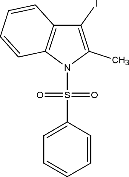
In the title compound, C15H12INO2S, the sulfonyl-bound phenyl ring forms a dihedral angle 82.84 (9)° with the indole ring system. The molecular structure is stabilized by a weak intramolecular C—H⋯O hydrogen bond. The crystal structure exhibits weak intermolecular C—H⋯π interactions and π–π interactions between the indole groups [centroid–centroid distance between the five-membered and six-membered rings of the indole group = 3.7617 (18) Å].
Related literature
For the biological properties of indole derivatives, see: Chai et al. (2006 ▶); Nieto et al. (2005 ▶). For the structures of closely related compounds, see: Chakkaravarthi et al. (2007 ▶, 2008 ▶).
Experimental
Crystal data
C15H12INO2S
M r = 397.22
Monoclinic,

a = 10.7068 (3) Å
b = 16.2670 (4) Å
c = 8.5147 (2) Å
β = 104.540 (1)°
V = 1435.49 (6) Å3
Z = 4
Mo Kα radiation
μ = 2.38 mm−1
T = 295 K
0.30 × 0.24 × 0.20 mm
Data collection
Bruker Kappa APEXII diffractometer
Absorption correction: multi-scan (SADABS; Sheldrick, 1996 ▶) T min = 0.536, T max = 0.648
21249 measured reflections
5276 independent reflections
3696 reflections with I > 2σ(I)
R int = 0.023
Refinement
R[F 2 > 2σ(F 2)] = 0.043
wR(F 2) = 0.144
S = 1.06
5276 reflections
182 parameters
1 restraint
H-atom parameters constrained
Δρmax = 0.94 e Å−3
Δρmin = −1.56 e Å−3
Data collection: APEX2 (Bruker, 2004 ▶); cell refinement: SAINT (Bruker, 2004 ▶); data reduction: SAINT; program(s) used to solve structure: SHELXS97 (Sheldrick, 2008 ▶); program(s) used to refine structure: SHELXL97 (Sheldrick, 2008 ▶); molecular graphics: PLATON (Spek, 2009 ▶); software used to prepare material for publication: SHELXL97.
Supplementary Material
Crystal structure: contains datablocks I, global. DOI: 10.1107/S1600536811004685/gk2346sup1.cif
Structure factors: contains datablocks I. DOI: 10.1107/S1600536811004685/gk2346Isup2.hkl
Additional supplementary materials: crystallographic information; 3D view; checkCIF report
Table 1. Hydrogen-bond geometry (Å, °).
Cg3 is the centroid of the C9–C14 ring.
| D—H⋯A | D—H | H⋯A | D⋯A | D—H⋯A |
|---|---|---|---|---|
| C13—H13⋯O1 | 0.93 | 2.29 | 2.871 (4) | 120 |
| C4—H4⋯Cg3i | 0.93 | 2.65 | 3.517 (5) | 156 |
Symmetry code: (i)  .
.
Acknowledgments
CR wishes to acknowledge AMET University management, India, for their kind support.
supplementary crystallographic information
Comment
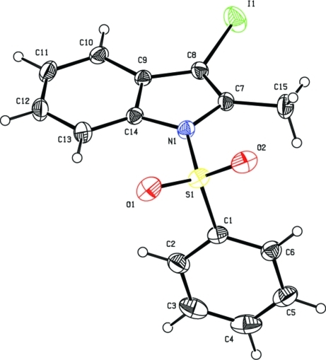
Indole derivatives exhibit antihepatitis B virus (Chai et al., 2006) and antibacterial (Nieto et al., 2005) activities. The geometric parameters of the title molecule (Fig. 1) agree well with the reported similar structures (Chakkaravarthi et al. 2007, 2008).
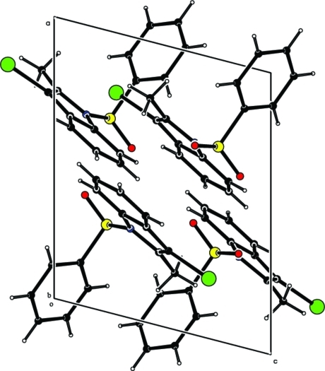
The phenyl ring makes the dihedral angle of 82.84 (9)° with the indole ring system. The sum of the bond angles around N1 [359.4 (2)°] indicates that N1 atom is sp2 hybridized. The molecular structure is stabilized by weak intramolecular C—H···O hydrogen bond. The crystal structure exhibits weak intermolecular C—H···π (Table 1) and π–π interactions [Cg1···Cg3 (1 - x,-y,1 - z) 3.7617 (18) Å; Cg1 and Cg3 are the centroids of the rings N1/C7/C8/C9/C14 and C9—C14, respectively].
Experimental
3-Iodo-2-methylindole (5 g,0.02 mmole) was dissolved in distilled benzene (100 ml).To this, benzenesulfonyl chloride(3.23 ml,0.025 mmol) and 60% aqueous NaOH solution (40 g in 67.0 ml) were added along with tetrabutyl ammonium hydrogensulfate (1.0 g). This two phase system was stirred at room temperature for 2 h. It was then diluted with water (200 ml) and the organic layer was separated. The aqueous layer was extracted with benzene (2x20 ml). The combined organic layer was dried(Na2SO4).The benzene was then removed completely and the crude product was recrystallized from methanol (m.p. 395–397 K).
Refinement
H atoms were positioned geometrically and refined using riding model with C—H = 0.93 Å and Uiso(H) = 1.2Ueq(C) for aromatic C—H and C—H = 0.96 Å and Uiso(H) = 1.5Ueq(C) for CH3. The components of the anisotropic displacement parameters in direction of the bond of I1 and C8 were restrained to be equal within an effective standard deviation of 0.001 using the DELU command in SHELXL (Sheldrick, 2008).
Figures
Fig. 1.
The molecular structure of the title compound with 30% probability displacement ellipsoids for non-H atoms.
Fig. 2.
Crystal packing viewed along the b axis.
Crystal data
| C15H12INO2S | F(000) = 776 |
| Mr = 397.22 | Dx = 1.838 Mg m−3 |
| Monoclinic, P21/c | Mo Kα radiation, λ = 0.71073 Å |
| Hall symbol: -P 2ybc | Cell parameters from 8510 reflections |
| a = 10.7068 (3) Å | θ = 2.5–30.4° |
| b = 16.2670 (4) Å | µ = 2.38 mm−1 |
| c = 8.5147 (2) Å | T = 295 K |
| β = 104.540 (1)° | Block, colourless |
| V = 1435.49 (6) Å3 | 0.30 × 0.24 × 0.20 mm |
| Z = 4 |
Data collection
| Bruker Kappa APEXII diffractometer | 5276 independent reflections |
| Radiation source: fine-focus sealed tube | 3696 reflections with I > 2σ(I) |
| graphite | Rint = 0.023 |
| ω and φ scans | θmax = 32.8°, θmin = 2.5° |
| Absorption correction: multi-scan (SADABS; Sheldrick, 1996) | h = −15→16 |
| Tmin = 0.536, Tmax = 0.648 | k = −23→24 |
| 21249 measured reflections | l = −12→11 |
Refinement
| Refinement on F2 | Primary atom site location: structure-invariant direct methods |
| Least-squares matrix: full | Secondary atom site location: difference Fourier map |
| R[F2 > 2σ(F2)] = 0.043 | Hydrogen site location: inferred from neighbouring sites |
| wR(F2) = 0.144 | H-atom parameters constrained |
| S = 1.06 | w = 1/[σ2(Fo2) + (0.0743P)2 + 0.987P] where P = (Fo2 + 2Fc2)/3 |
| 5276 reflections | (Δ/σ)max < 0.001 |
| 182 parameters | Δρmax = 0.94 e Å−3 |
| 1 restraint | Δρmin = −1.56 e Å−3 |
Fractional atomic coordinates and isotropic or equivalent isotropic displacement parameters (Å2)
| x | y | z | Uiso*/Ueq | ||
| C1 | 0.8489 (3) | 0.14775 (16) | 0.8788 (3) | 0.0350 (5) | |
| C2 | 0.8848 (4) | 0.0951 (2) | 1.0097 (4) | 0.0495 (7) | |
| H2 | 0.8265 | 0.0574 | 1.0324 | 0.059* | |
| C3 | 1.0098 (4) | 0.0998 (3) | 1.1063 (5) | 0.0638 (10) | |
| H3 | 1.0365 | 0.0646 | 1.1944 | 0.077* | |
| C4 | 1.0940 (4) | 0.1562 (3) | 1.0722 (5) | 0.0643 (11) | |
| H4 | 1.1778 | 0.1587 | 1.1375 | 0.077* | |
| C5 | 1.0569 (3) | 0.2092 (3) | 0.9431 (5) | 0.0556 (9) | |
| H5 | 1.1149 | 0.2478 | 0.9227 | 0.067* | |
| C6 | 0.9331 (3) | 0.20492 (19) | 0.8436 (4) | 0.0433 (6) | |
| H6 | 0.9073 | 0.2398 | 0.7549 | 0.052* | |
| C7 | 0.7397 (3) | 0.04833 (16) | 0.5161 (3) | 0.0329 (5) | |
| C8 | 0.7262 (3) | −0.03190 (16) | 0.4724 (3) | 0.0336 (5) | |
| C9 | 0.6619 (2) | −0.07527 (14) | 0.5739 (3) | 0.0297 (4) | |
| C10 | 0.6230 (3) | −0.15718 (17) | 0.5777 (4) | 0.0387 (6) | |
| H10 | 0.6379 | −0.1950 | 0.5025 | 0.046* | |
| C11 | 0.5626 (3) | −0.1805 (2) | 0.6940 (4) | 0.0478 (7) | |
| H11 | 0.5362 | −0.2348 | 0.6978 | 0.057* | |
| C12 | 0.5402 (3) | −0.1249 (2) | 0.8061 (4) | 0.0486 (7) | |
| H12 | 0.5005 | −0.1429 | 0.8852 | 0.058* | |
| C13 | 0.5753 (3) | −0.0428 (2) | 0.8043 (4) | 0.0435 (6) | |
| H13 | 0.5585 | −0.0055 | 0.8792 | 0.052* | |
| C14 | 0.6367 (2) | −0.01869 (15) | 0.6857 (3) | 0.0309 (5) | |
| C15 | 0.7993 (4) | 0.1168 (2) | 0.4432 (5) | 0.0509 (7) | |
| H15A | 0.8230 | 0.0972 | 0.3483 | 0.076* | |
| H15B | 0.8748 | 0.1362 | 0.5208 | 0.076* | |
| H15C | 0.7383 | 0.1609 | 0.4137 | 0.076* | |
| N1 | 0.6822 (2) | 0.05838 (13) | 0.6481 (3) | 0.0335 (4) | |
| O1 | 0.6046 (2) | 0.13114 (15) | 0.8577 (4) | 0.0552 (6) | |
| O2 | 0.6719 (2) | 0.21172 (13) | 0.6496 (3) | 0.0533 (6) | |
| S1 | 0.68958 (6) | 0.14402 (4) | 0.75748 (10) | 0.03797 (16) | |
| I1 | 0.78752 (3) | −0.084909 (16) | 0.28650 (3) | 0.06383 (12) |
Atomic displacement parameters (Å2)
| U11 | U22 | U33 | U12 | U13 | U23 | |
| C1 | 0.0355 (12) | 0.0307 (11) | 0.0379 (13) | 0.0043 (9) | 0.0078 (10) | −0.0078 (10) |
| C2 | 0.0583 (19) | 0.0487 (17) | 0.0391 (15) | 0.0014 (14) | 0.0076 (14) | 0.0020 (12) |
| C3 | 0.071 (3) | 0.070 (2) | 0.0421 (18) | 0.020 (2) | −0.0008 (17) | 0.0018 (17) |
| C4 | 0.0421 (17) | 0.087 (3) | 0.056 (2) | 0.0121 (17) | −0.0013 (15) | −0.025 (2) |
| C5 | 0.0409 (16) | 0.065 (2) | 0.061 (2) | −0.0088 (14) | 0.0131 (14) | −0.0205 (18) |
| C6 | 0.0436 (15) | 0.0406 (14) | 0.0458 (15) | −0.0039 (11) | 0.0117 (12) | −0.0062 (12) |
| C7 | 0.0368 (12) | 0.0290 (11) | 0.0337 (12) | −0.0026 (9) | 0.0107 (9) | 0.0026 (9) |
| C8 | 0.0411 (13) | 0.0293 (11) | 0.0303 (11) | −0.0013 (9) | 0.0088 (9) | 0.0012 (8) |
| C9 | 0.0297 (11) | 0.0280 (11) | 0.0287 (11) | −0.0016 (8) | 0.0025 (8) | 0.0014 (8) |
| C10 | 0.0444 (14) | 0.0295 (12) | 0.0376 (13) | −0.0067 (10) | 0.0021 (11) | −0.0005 (10) |
| C11 | 0.0440 (15) | 0.0395 (15) | 0.0552 (17) | −0.0157 (12) | 0.0036 (13) | 0.0100 (13) |
| C12 | 0.0407 (15) | 0.0578 (19) | 0.0489 (17) | −0.0109 (13) | 0.0143 (12) | 0.0130 (15) |
| C13 | 0.0415 (14) | 0.0501 (16) | 0.0427 (15) | −0.0046 (12) | 0.0178 (12) | 0.0006 (12) |
| C14 | 0.0272 (10) | 0.0321 (11) | 0.0326 (11) | −0.0008 (8) | 0.0058 (9) | 0.0005 (9) |
| C15 | 0.064 (2) | 0.0396 (15) | 0.0548 (18) | −0.0110 (14) | 0.0261 (15) | 0.0073 (14) |
| N1 | 0.0389 (11) | 0.0255 (9) | 0.0373 (11) | −0.0027 (8) | 0.0120 (9) | −0.0033 (8) |
| O1 | 0.0433 (11) | 0.0543 (13) | 0.0750 (17) | 0.0013 (10) | 0.0278 (11) | −0.0227 (12) |
| O2 | 0.0528 (13) | 0.0308 (10) | 0.0659 (15) | 0.0113 (9) | −0.0047 (11) | 0.0013 (10) |
| S1 | 0.0335 (3) | 0.0299 (3) | 0.0491 (4) | 0.0048 (2) | 0.0078 (3) | −0.0079 (3) |
| I1 | 0.0954 (2) | 0.05448 (17) | 0.05233 (17) | 0.00090 (11) | 0.03859 (15) | −0.00527 (9) |
Geometric parameters (Å, °)
| C1—C6 | 1.380 (4) | C9—C14 | 1.399 (4) |
| C1—C2 | 1.381 (4) | C9—C10 | 1.399 (3) |
| C1—S1 | 1.759 (3) | C10—C11 | 1.365 (5) |
| C2—C3 | 1.386 (6) | C10—H10 | 0.9300 |
| C2—H2 | 0.9300 | C11—C12 | 1.379 (5) |
| C3—C4 | 1.367 (7) | C11—H11 | 0.9300 |
| C3—H3 | 0.9300 | C12—C13 | 1.387 (5) |
| C4—C5 | 1.375 (6) | C12—H12 | 0.9300 |
| C4—H4 | 0.9300 | C13—C14 | 1.392 (4) |
| C5—C6 | 1.384 (5) | C13—H13 | 0.9300 |
| C5—H5 | 0.9300 | C14—N1 | 1.411 (3) |
| C6—H6 | 0.9300 | C15—H15A | 0.9600 |
| C7—C8 | 1.355 (4) | C15—H15B | 0.9600 |
| C7—N1 | 1.420 (3) | C15—H15C | 0.9600 |
| C7—C15 | 1.493 (4) | N1—S1 | 1.667 (2) |
| C8—C9 | 1.421 (4) | O1—S1 | 1.411 (3) |
| C8—I1 | 2.050 (3) | O2—S1 | 1.416 (3) |
| C6—C1—C2 | 121.9 (3) | C9—C10—H10 | 120.7 |
| C6—C1—S1 | 119.1 (2) | C10—C11—C12 | 121.0 (3) |
| C2—C1—S1 | 119.0 (2) | C10—C11—H11 | 119.5 |
| C1—C2—C3 | 118.5 (4) | C12—C11—H11 | 119.5 |
| C1—C2—H2 | 120.8 | C11—C12—C13 | 122.0 (3) |
| C3—C2—H2 | 120.8 | C11—C12—H12 | 119.0 |
| C4—C3—C2 | 120.0 (4) | C13—C12—H12 | 119.0 |
| C4—C3—H3 | 120.0 | C12—C13—C14 | 117.2 (3) |
| C2—C3—H3 | 120.0 | C12—C13—H13 | 121.4 |
| C3—C4—C5 | 121.1 (3) | C14—C13—H13 | 121.4 |
| C3—C4—H4 | 119.4 | C13—C14—C9 | 121.0 (2) |
| C5—C4—H4 | 119.4 | C13—C14—N1 | 132.0 (3) |
| C4—C5—C6 | 119.9 (4) | C9—C14—N1 | 107.0 (2) |
| C4—C5—H5 | 120.0 | C7—C15—H15A | 109.5 |
| C6—C5—H5 | 120.0 | C7—C15—H15B | 109.5 |
| C1—C6—C5 | 118.6 (3) | H15A—C15—H15B | 109.5 |
| C1—C6—H6 | 120.7 | C7—C15—H15C | 109.5 |
| C5—C6—H6 | 120.7 | H15A—C15—H15C | 109.5 |
| C8—C7—N1 | 106.9 (2) | H15B—C15—H15C | 109.5 |
| C8—C7—C15 | 129.1 (3) | C14—N1—C7 | 108.7 (2) |
| N1—C7—C15 | 123.9 (3) | C14—N1—S1 | 125.89 (19) |
| C7—C8—C9 | 110.2 (2) | C7—N1—S1 | 124.72 (18) |
| C7—C8—I1 | 125.8 (2) | O1—S1—O2 | 120.43 (16) |
| C9—C8—I1 | 124.00 (18) | O1—S1—N1 | 105.47 (13) |
| C14—C9—C10 | 120.1 (2) | O2—S1—N1 | 107.93 (14) |
| C14—C9—C8 | 107.1 (2) | O1—S1—C1 | 109.09 (15) |
| C10—C9—C8 | 132.8 (3) | O2—S1—C1 | 107.83 (14) |
| C11—C10—C9 | 118.7 (3) | N1—S1—C1 | 105.06 (12) |
| C11—C10—H10 | 120.7 | ||
| C6—C1—C2—C3 | −0.7 (5) | C8—C9—C14—C13 | −179.2 (3) |
| S1—C1—C2—C3 | −178.7 (3) | C10—C9—C14—N1 | −177.8 (2) |
| C1—C2—C3—C4 | 0.7 (6) | C8—C9—C14—N1 | 1.6 (3) |
| C2—C3—C4—C5 | 0.3 (6) | C13—C14—N1—C7 | 179.0 (3) |
| C3—C4—C5—C6 | −1.1 (6) | C9—C14—N1—C7 | −2.0 (3) |
| C2—C1—C6—C5 | −0.1 (5) | C13—C14—N1—S1 | 8.1 (4) |
| S1—C1—C6—C5 | 177.9 (2) | C9—C14—N1—S1 | −172.91 (19) |
| C4—C5—C6—C1 | 1.0 (5) | C8—C7—N1—C14 | 1.6 (3) |
| N1—C7—C8—C9 | −0.5 (3) | C15—C7—N1—C14 | −179.7 (3) |
| C15—C7—C8—C9 | −179.2 (3) | C8—C7—N1—S1 | 172.6 (2) |
| N1—C7—C8—I1 | 179.13 (18) | C15—C7—N1—S1 | −8.7 (4) |
| C15—C7—C8—I1 | 0.5 (5) | C14—N1—S1—O1 | −19.0 (3) |
| C7—C8—C9—C14 | −0.7 (3) | C7—N1—S1—O1 | 171.5 (2) |
| I1—C8—C9—C14 | 179.62 (18) | C14—N1—S1—O2 | −149.0 (2) |
| C7—C8—C9—C10 | 178.6 (3) | C7—N1—S1—O2 | 41.5 (3) |
| I1—C8—C9—C10 | −1.1 (4) | C14—N1—S1—C1 | 96.2 (2) |
| C14—C9—C10—C11 | −1.3 (4) | C7—N1—S1—C1 | −73.3 (2) |
| C8—C9—C10—C11 | 179.5 (3) | C6—C1—S1—O1 | −139.6 (2) |
| C9—C10—C11—C12 | 0.0 (5) | C2—C1—S1—O1 | 38.4 (3) |
| C10—C11—C12—C13 | 1.3 (5) | C6—C1—S1—O2 | −7.2 (3) |
| C11—C12—C13—C14 | −1.1 (5) | C2—C1—S1—O2 | 170.8 (3) |
| C12—C13—C14—C9 | −0.2 (4) | C6—C1—S1—N1 | 107.7 (2) |
| C12—C13—C14—N1 | 178.7 (3) | C2—C1—S1—N1 | −74.2 (3) |
| C10—C9—C14—C13 | 1.4 (4) |
Hydrogen-bond geometry (Å, °)
| Cg3 is the centroid of the C9–C14 ring. |
| D—H···A | D—H | H···A | D···A | D—H···A |
| C13—H13···O1 | 0.93 | 2.29 | 2.871 (4) | 120 |
| C4—H4···Cg3i | 0.93 | 2.65 | 3.517 (5) | 156 |
Symmetry codes: (i) −x+2, −y, −z+2.
Footnotes
Supplementary data and figures for this paper are available from the IUCr electronic archives (Reference: GK2346).
References
- Bruker (2004). APEX2 and SAINT Bruker AXS Inc., Madison, Wisconsin, USA.
- Chai, H., Zhao, C. & Gong, P. (2006). Bioorg. Med. Chem. 14, 911–917. [DOI] [PubMed]
- Chakkaravarthi, G., Dhayalan, V., Mohanakrishnan, A. K. & Manivannan, V. (2008). Acta Cryst. E64, o542. [DOI] [PMC free article] [PubMed]
- Chakkaravarthi, G., Ramesh, N., Mohanakrishnan, A. K. & Manivannan, V. (2007). Acta Cryst. E63, o3564.
- Nieto, M. J., Alovero, F. L., Manzo, R. H. & Mazzieri, M. R. (2005). Eur. J. Med. Chem. 40, 361–369. [DOI] [PubMed]
- Sheldrick, G. M. (1996). SADABS. University of Göttingen, Germany.
- Sheldrick, G. M. (2008). Acta Cryst. A64, 112–122. [DOI] [PubMed]
- Spek, A. L. (2009). Acta Cryst. D65, 148–155. [DOI] [PMC free article] [PubMed]
Associated Data
This section collects any data citations, data availability statements, or supplementary materials included in this article.
Supplementary Materials
Crystal structure: contains datablocks I, global. DOI: 10.1107/S1600536811004685/gk2346sup1.cif
Structure factors: contains datablocks I. DOI: 10.1107/S1600536811004685/gk2346Isup2.hkl
Additional supplementary materials: crystallographic information; 3D view; checkCIF report


