Abstract
The fracture of the medial orbital wall is relatively common in orbital trauma. Titanium mesh is possibly the actual standard material for orbital wall reconstruction. When the floor of the orbit and the medial wall are simultaneously affected, one larger mesh gives better results than two independent meshes that need to be fixated independently. However, large meshes need a wider surgical field. To gain sufficient exposure to the medial and inferior orbital walls simultaneously, we present an approach that combines the transconjunctival and transcaruncular incisions, detaching if needed the inferior oblique muscle and, placing our mesh, repositioning it beside the lacrimal duct. This technique should not entirely displace traditional approaches, but it widens the surgical exposure for middle- and upper-third facial trauma. This alternative has minimum morbidity and can save a great deal of surgery time.
Keywords: Orbit, facial, trauma, transcaruncular
During 2003 to 2007, 10 patients were operated on at our institution using the extended transcaruncular approach (Table 1). Three patients had traumatic injuries of the medial orbital wall, six had combined medial and inferior wall injury, and one had an orbital apex syndrome (OAS). Anatomic restitution with titanium orbital mesh (Synthes, Solothurn, Switzerland) of the medial wall and floor of the orbit was achieved in all except for the OAS patient, in which a posterior ethmoidectomy was performed for decompression of the optic canal. In this case, a 2.4-mm, 30-degree rigid endoscope was used to improve the vision at the orbital apex via the transcaruncular incision. To evaluate postoperative diplopia, Lancaster tests were made on all patients but one who had failed the follow-up process. Digital imaging exophthalmometry was used to assess the position of the ipsilateral globe related to the contralateral globe position.
Table 1.
Patients
| Patient | Sex | Age (Years) | Diagnosis | Transconjunctival Dissection | Endoscope | Detachment Inferior Oblique Muscle | Reconstruction | Enophthalmos (Preoperative/Postoperative) | Postoperative Diplopia | Follow-Up (Months) |
|---|---|---|---|---|---|---|---|---|---|---|
| 1 | F | 25 | Medial wall and floor fracture | Preseptal | No | Yes | 1.5-mm plate and PDS | –1.6/0.0 | No | 23 |
| 2 | F | 21 | Medial wall and floor fracture | Retroseptal | No | No | Titanium mesh and PDS | –1.5/–0.3 | No | 36 |
| 3 | F | 23 | Medial wall and floor fracture | Preseptal | No | Yes | Titanium mesh | –0.4/–0.2 | No | 12 |
| 4 | F | 15 | Medial wall and floor fracture | Retroseptal | No | No | PDS | –1.1/–0.1 | No | 15 |
| 5 | M | 48 | Orbital apex syndrome | Retroseptal | Yes | Yes | Ethmoidectomy | Not assessed | Yes | 6 |
| 6 | F | 27 | Panfacial wall and floor fracture | Preseptal | No | Yes | Titanium mesh | –0.7/0.0 | No | 20 |
| 7 | M | 36 | Medial wall fracture | Retroseptal | No | No | Titanium mesh | –1.1/–0.2 | No | 36 |
| 8 | F | 34 | Strut fracture trapdoor | Preseptal | No | No | Resorbable mesh | –0.5/+ 0.5 | Yes | 13 |
| 9 | F | 25 | Strut fracture trapdoor | Retroseptal | No | No | Resorbable mesh | –0.7/0.0 | No | 30 |
| 10 | F | 33 | Medial wall and floor fracture | Retroseptal | No | Yes | Titanium mesh | –1.7/–0.2 | ? | 5 |
SURGICAL TECHNIQUE
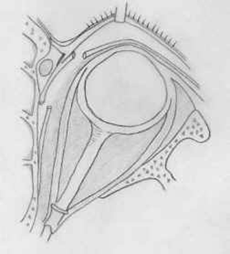
The inferior conjunctival incision must be made as low as possible in the inferior fornix. Retroseptal dissection is preferred, as it prevents having to change the plane of dissection. The orbital periosteum is cut 2 or 3 mm behind the inferior orbital rim. Corneal protectors are used. Retracting the caruncle medially using a Desmarres retractor, the conjunctival incision is made from the inferior to the medial fornix. This can be done using Stevens scissors. Staying posterior to the septum prevents the damage to the lacrimal sac. The posterior lacrimal crest is palpated with a Freer elevator (Fig. 1). Blunt dissection up to the posterior lacrimal crest separates the medial canthal ligament and lacrimal sac anteriorly from the medial rectus muscle and the globe, which are gently rejected laterally. The dissection should be made medially toward the lacrimal crest, not posteriorly, so as not to damage the medial rectus muscle. The orbital periosteum is cut at the level of the posterior lacrimal crest, and the periosteum of the medial wall is elevated.
Figure 1.
Transcaruncular and transconjunctival approach. It is important to stay posterior to the orbital septum in order not to enter the lacrimal sac.
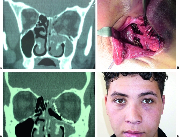
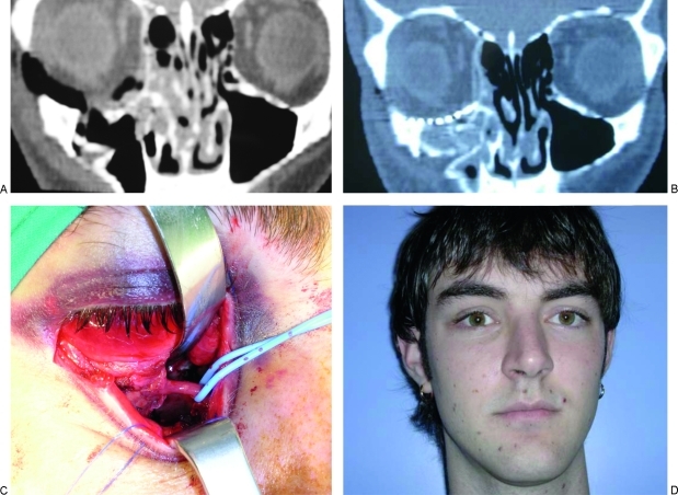
The inferior limit of the transcaruncular dissection is the inferior oblique muscle. This muscle is also the medial end of the transconjunctival dissection (Figs. 2B and 3C). Once the muscle is identified, it is marked with a long suture and detached from its origin to be restored. When the inferior oblique muscle is freed, the surgical field is noticeably extended because all the contents of the orbit can be retracted laterally with no risk of damaging the lacrimal canaliculus. It is important to leave a small cuff of muscle (2 mm) for suture restoration (Fig. 2B,C).
Figure 2.
Case 1. (A) Preoperative computed tomography (CT) scan of a 25-year-old man with fracture of the medial wall and floor of the orbit. (B) Preseptal inferior fornix dissection and transcaruncular dissection was performed. Repositioning of the inferior oblique muscle was performed. (C) Reconstruction with an 0.5-mm orbital mesh was performed up to the superomedial angle of the orbit (postoperative CT scan). (D) Three-month postoperative result.
Figure 3.
Case 2. (A) Preoperative CT scan of a 21-year-old man with fracture of the medial wall and floor of the orbit. (B) Postoperative CT scan showing the reconstruction of the floor of the orbit with titanium mesh and the medial wall with PDS sheets. (C) Retroseptal inferior fornix and transcaruncular dissection without detachment of the inferior oblique muscle. (D) Six-month postoperative view of the patient.
For the reconstructive procedure, 0.5-mm titanium mesh (Synthes) and polydioxanone (PDS) sheets (Ethicon; Johnson & Johnson, Aunea, France) were used. Once the orbital walls are repaired, the inferior obturator muscle, the periorbita, and the septum are closed with absorbable suture material. The transconjunctival incision may be left open if the incision is at the fornix.
RESULTS
Enophthalmos and diplopia were corrected in all patients except one that had a minimal trapdoor fracture with an injured inferior rectus muscle. This patient developed an intramuscular hematoma that led to dystopia and persistent diplopia (Table 1). In the patient with OAS, extraocular movements were recovered but visual perception was not improved.
There were no complications related to the approach. Postoperative aesthetic results were excellent.
DISCUSSION
The access to the medial orbital wall has always been a challenge to the surgeon because of its difficulty. The first orbital approach to the frontal sinus, ethmoidal cells, and sphenoid sinus was described by Bergh in 1886. Lynch1 placed the incision between the medial canthus and the glabellar region. The transorbital approach as described by McCord2 and Anderson and Lindberg,3 which is used for orbital decompression in Graves' disease, leads to extensive dissection of the inferior conjunctival fornix and lateral canthal ligament and leaves cutaneous scars. The transcaruncular approach has mainly been described in ophthalmology journals for the medial decompression of the orbit4 and the repair of the medial canthal ligament5,6,7 and the lacrimal duct.8 More recently, Kennedy et al9 described an endoscopic approach for treating thyroid ophthalmoplegia and decompression of the optic nerve.10,11 These approaches do not leave scars, but the operation takes a long time, and the surgeon's maneuverability is limited to procedures like decompression, removal of small tumors, and drainage of abscesses.12
De Chalain et al13 extended the transconjunctival incision laterally via a lateral canthotomy and a skin incision. Shorr14 added the inferior fornix incision to the transcaruncular incision to improve the surgical field at the maxillo-ethmoidal strut.
Chang15 used this approach for a medial orbitotomy for a traumatic optic nerve decompression with little success but no complications related to the approach.
In contrast with Goldberg et al,16 we prefer the retroseptal dissection. Although it may be technically more difficult because of fat spillage, it provides a safer plane of dissection, thus staying farther away from the lacrimal canaliculi.
Su and Harris17 describe the extended transcaruncular approach added to the inferior fornix incision but without repositioning of the inferior oblique muscle.
The main disadvantage of the pure transcaruncular approach is that the field for working in the medial wall is very limited—only small grafts (15 × 20 mm maximum) can be inserted.
The detachment of the inferior oblique muscles unites the inferior and medial approaches, which gives the surgeon a larger field of vision into the medial wall.18 This should be done when a wide exposure of the medial wall is required, for example, when the repair of the inferomedial buttress is needed. When this strut is damaged, there is no support for our grafts, either bone or allografts.19 Thanks to this wide operating field, larger titanium mesh materials can be inserted from the lateral wall up to the superomedial corner of the orbit (Fig. 3). This approach also provides a wide exposure for obtaining hemostasis of the ethmoidal arteries and the direct suture or plication of the medial palpebral ligament.7,20
Endoscopic-assisted transcaruncular approaches have been recently reported by Chen et al21 for optic nerve decompression. In our case, the 2.4-mm, 30-degree endoscope was used in conjunction with the extended transcaruncular approach to achieve a better view of the orbital apex for posterior medial wall ethmoidectomy in the traumatic decompression of the optic nerve.
In acute cases, preoperative exophthalmometry data are less than expected because of orbital swelling.
CONCLUSION
This extended transcaruncular approach offers a better surgical field than do the inferior transconjunctival approach or the isolated transcaruncular approach to correct the inferomedial strut of the orbit, which is a key support for reconstructing the floor and medial wall in severe trauma. It also provides better vision of the orbital apex and is even clearer if the endoscope is used. This approach can oftentimes avoid coronal incision. Detachment and repositioning of the inferior oblique muscle gives the surgeon a wider field of vision of the medial orbital wall with minimal morbidity. Although it may be technically more difficult because of “fat spillage,” retroseptal dissection provides a safer plane of dissection.
References
- Lynch R C. The technique of radical frontal sinus operation which has given me the best results. Laryngoscope. 1921;31:1–5. [Google Scholar]
- McCord C D. Orbital decompression for Graves' disease exposure through lateral canthal and inferior fornix incision. Ophthalmology. 1981;88:533–541. [PubMed] [Google Scholar]
- Anderson R L, Lindberg J V. Transorbital approach to decompression in Graves' disease. Arch Ophthalmol. 1981;99:120–124. doi: 10.1001/archopht.1981.03930010122016. [DOI] [PubMed] [Google Scholar]
- Graham S M. The transcaruncular approach to the medial orbital wall. Laryngoscope. 2002;112:986–989. doi: 10.1097/00005537-200206000-00009. [DOI] [PubMed] [Google Scholar]
- Francis I C. Transcaruncular medial orbitotomy for stabilization of the posterior limb of the medial canthal tendon. Clin Experiment Ophthalmol. 2001;29:85–89. doi: 10.1046/j.1442-9071.2001.d01-6.x. [DOI] [PubMed] [Google Scholar]
- Fante R G. Transcaruncular approach to the medial canthal tendon-application for lower eyelid laxity. Ophthal Plast Reconstr Surg. 2001;17:16–27. doi: 10.1097/00002341-200101000-00004. [DOI] [PubMed] [Google Scholar]
- Demirci H, Hassan A S, Elner S G, Boehkle C, Elner V M. Comprehensive, combined anterior and transcaruncular orbital approach to medial canthal ligament plication. Ophthal Plast Reconstr Surg. 2007;23:384–388. doi: 10.1097/IOP.0b013e3181469e3c. [DOI] [PubMed] [Google Scholar]
- Lee J S. The treatment of the lacrimal apparatus obstruction with the use of an inner canthal Jones tube insertion via a transcaruncular approach. Ophthalmic Surg Lasers. 2001;32:48–54. [PubMed] [Google Scholar]
- Kennedy D W, Goodstein M L, Miller N R, et al. Endoscopic transnasal orbital decompression. Arch Otolaryngol Head Neck Surg. 1990;116:275–282. doi: 10.1001/archotol.1990.01870030039006. [DOI] [PubMed] [Google Scholar]
- Graham S M, Carter K D. Combined-approach orbital decompression for thyroid-related orbitopathy. Clin Otolaryngol Allied Sci. 1999;24:109–113. doi: 10.1046/j.1365-2273.1999.00219.x. [DOI] [PubMed] [Google Scholar]
- Shorr N, Baylis H I, Goldberg R A, et al. Transcaruncular approach to the medial orbit and apex. Ophthalmology. 2000;107:1459–1463. doi: 10.1016/s0161-6420(00)00241-4. [DOI] [PubMed] [Google Scholar]
- Manning S C. Endoscopic management of medial subperiosteal orbital abcess. Arch Otolaryngol Head Neck Surg. 1993;119:789–791. doi: 10.1001/archotol.1993.01880190085018. [DOI] [PubMed] [Google Scholar]
- de Chalain T M, Cohen S R, Burstein F D. Modification of the transconjunctival lower lid approach to the orbital floor; lateral paracanthal incision. Plast Reconstr Surg. 1994;94:877–880. doi: 10.1097/00006534-199411000-00023. [DOI] [PubMed] [Google Scholar]
- Shorr N, Baylis H I, Goldberg R A, Perry J D. Transcaruncular approach to the medial orbit and orbital apex. Ophthalmology. 2000;107:1459–1463. doi: 10.1016/s0161-6420(00)00241-4. [DOI] [PubMed] [Google Scholar]
- Chang E L, Bernardino C R, Rubin P A. Transcaruncular orbital decompression for management of compressive optic neuropathy in thyroid-related orbitopathy. Plast Reconstr Surg. 2003;112:739–747. doi: 10.1097/01.PRS.0000069708.70121.67. [DOI] [PubMed] [Google Scholar]
- Goldberg R A, Mancini R, Demer J L. The transcaruncular approach: surgical anatomy and technique. Arch Facial Plast Surg. 2007;9:443–447. doi: 10.1001/archfaci.9.6.443. [DOI] [PubMed] [Google Scholar]
- Su G W, Harris G J. Combined inferior and medial surgical approaches and overlapping thin implants for orbital floor and medial wall fractures. Ophthal Plast Reconstr Surg. 2006;22:420–423. doi: 10.1097/01.iop.0000242163.03589.0e. [DOI] [PubMed] [Google Scholar]
- Warwik R. Eugene Wolff's Anatomy of the Eye and Orbit. 7th ed. London, England: HK Lewis; 1976.
- Ellis E, III, Tan Y. Assessment of internal orbit reconstructions for pure blowout fractures: cranial bone grafts versus titanium mesh. J Oral Maxillofac Surg. 2003;61:442–453. doi: 10.1053/joms.2003.50085. [DOI] [PubMed] [Google Scholar]
- Tyers A G. Colour Atlas of Ophthalmic Plastic Surgery. Edinburgh, Scotland: Churchill Livingstone; 1995. pp. 7–8.
- Chen C T, Huang F, Tsay P K, et al. Endoscopically assisted transconjunctival decompression of traumatic optic neuropathy. J Craniofac Surg. 2007;18:19–26. discussion 27–28. doi: 10.1097/01.scs.0000248654.15287.89. [DOI] [PubMed] [Google Scholar]


