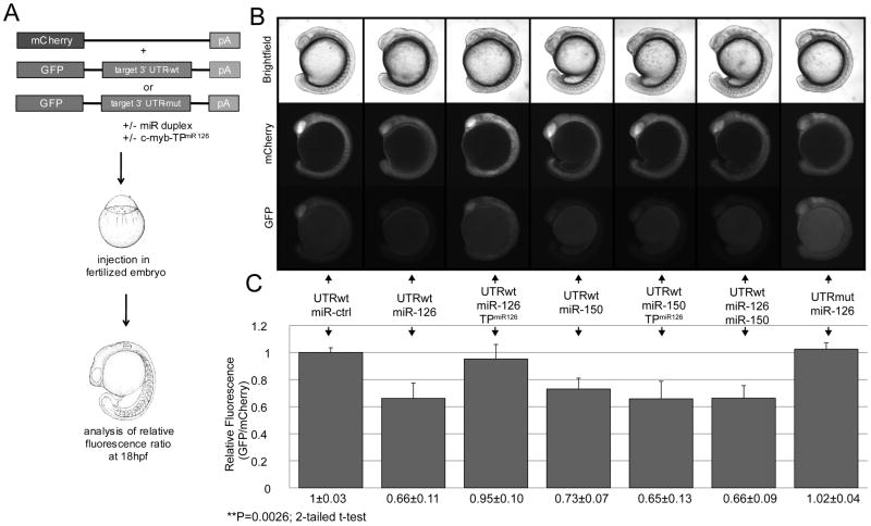
Figure 2. MiR-126 targets the zebrafish c-myb 3′UTR in vivo.
(A) Schematic of the in vivo miRNA/mRNA interaction reporter assay 42. Wild-type or mutated c-myb 3′UTR fused to EGFP mRNA is co-injected into fertilized zebrafish embryos with mCherry mRNA (internal injection control) and miR duplexes or morpholinos. At 18hpf the relative fluorescence intensity levels (GFP/RFP) are measured and normalized to control injections. (B) Upper panel: Live images of co-injected embryos in brightfield, RFP filter and EGFP filter. (C) Lower panel: Quantification of relative fluorescence. Co-injection of wild-type c-myb 3′UTR with miR-126 duplex resulted in a 35% decrease of relative EGFP fluorescence compared to the control miRNA (miR-ctrl). A similar reduction was observed with miR-150, a known regulator of c-myb 18. Conversely, injection of a c-myb 3′UTR containing four point mutations in the putative miR-126 target site or co-injection with a c-myb-TPmiR126 target protector morpholino abolished the regulation of c-myb 3′UTR by miR-126. The c-myb-TPmiR126 target protector morpholino showed no effect on miR-150 mediated c-myb 3′UTR regulation. The combination of miR-126 and miR-150 did not yield stronger regulation than when expressed individually. Injection conditions and relative fluorescence values are indicated. Error bars represent standard deviation.
**P=0.0026 (two-tailed student’s t-test); mCherry, mCherry fluorescent protein; GFP, green fluorescent protein.

