Figure 5.
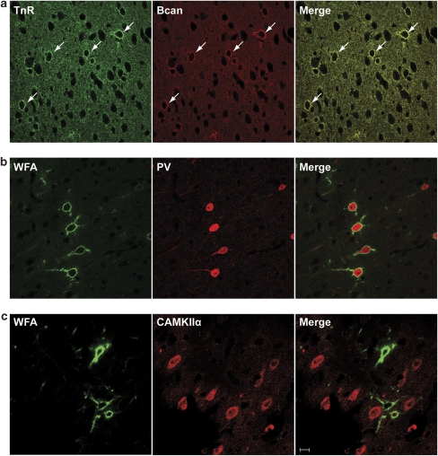
Immunohistochemical identification of ECM proteins in the mPFC. (a) Colocalization of Tnr and Bcan in perineuronal nets in the mPFC. Sections were double-stained using Tnr (green) and Bcan (red) specific antibodies and analyzed by confocal microscopy. Immunoreactivity of Tnr and Bcan is colocalized (merged image) in perineuronal nets (white arrows) surrounding a subset of neurons in the anterior cingulate, prelimbic, and infralimbic area of the mPFC. (b+c) Biotinylated-WFA (green) was used to detect chondroitin sulfate proteoglycan-containing perineuronal nets in combination with a PV-specific antibody (red) or CAMKIIα antibody (red). Throughout all areas of the mPFC, perineuronal nets were associated with the majority of PV-expressing interneurons (b), but never with CAMKII-positive pyramidal neurons (c). Scale bar: 20 μm.

