Activation of PLCγ1 is necessary for angiogenesis in both cell culture and a CAM model of angiogenesis. The data show for the first time that c-Cbl ubiquitin E3 ligase, a recently identified endogenous inhibitor of PLCγ1, plays a critical and novel role in laser-induced choroidal neovascularization, shedding new light on its regulation, and possibly providing a new target for controlling pathologic angiogenesis.
Abstract
Purpose.
Activation of phospholipase Cγ1 (PLCγ1) by vascular endothelial growth factor receptor (VEGFR)-2 is necessary for proliferation and tube formation of endothelial cells in vitro. Previous work has demonstrated that Casitas B-lineage lymphoma (c-Cbl) promotes ubiquitination of PLCγ1 and suppression of its tyrosine phosphorylation. This study was designed to evaluate the importance of PLCγ1 and c-Cbl in experimental choroidal neovascularization (CNV).
Methods.
The role of PLCγ1 was studied in three models of angiogenesis: the endothelial cell culture system, the chorioallantoic membrane (CAM) assay, and the laser-induced CNV model. Endothelial cells were analyzed for the role of PLCγ1 in promoting tube formation. CAMs were incubated with pharmacologic agents that either inhibit or stimulate PLCγ1. CNV was induced in wild-type and c-Cbl–knockout mice, and the progression of CNV was evaluated by fluorescein angiography.
Results.
Activation of PLCγ1 was necessary for tube formation of endothelial cells. PLCγ1 stimulation increased the growth of blood vessels and conversely, PLCγ1 inhibition decreased the growth of blood vessels in the CAM model. CNV lesions in the c-Cbl–knockout mice were significantly greater in number, more confluent, and increased in size with time, compared with those in the control wild-type mice.
Conclusions.
The data show that PLCγ1 plays an important role in angiogenesis. Loss of c-Cbl results in enhanced CNV in the eye. The study also shows that c-Cbl plays an important role in ocular angiogenesis, suggesting that modulation of c-Cbl activity or inhibition of PLCγ1 would be a compelling target for antiangiogenesis therapy.
Angiogenesis, the formation of new blood vessels, is of key importance in a broad array of physiologic and pathologic conditions such as age-related macular degeneration (AMD) and cancer. VEGF orchestrated signaling events in endothelial cells are considered to be one of the earliest and the most prominent signaling events promoting angiogenesis.1–3 A precise physiological balance between endogenous pro- and antiangiogenic regulators control endothelial cell functions, such that endothelial cell growth is normally restrained. However, in pathologic conditions, such as tumor growth and AMD, a shift occurs in the balance of regulators favoring endothelial growth.4,5 Given the prominent role of VEGF receptor (VEGFR)-2 and its associated signaling proteins in angiogenesis,6 regulation of VEGFR-2-associated signaling proteins may represent a critical mechanism for the occurrence of aberrant angiogenesis.
Among the key angiogenic signaling proteins that are activated by VEGFR-2, stimulation of phospholipase (PL)Cγ1 is one of the primary signaling pathways emanating from direct VEGFR-2 activation. Activation of PLCγ1 has been shown to be critically essential for endothelial cell proliferation and tube formation in vitro7 and for vasculature formation during embryonic development in mice.8,9 Our recent work has identified c-Cbl ubiquitin E3-ligase as a negative regulator of angiogenesis.10 The potential function of c-Cbl as an antiangiogenic signaling protein is illustrated by its ability to suppress VEGFR-2-mediated phosphorylation of PLCγ1 and its ability to inhibit VEGFR-2-mediated tube formation by endothelial cells.10 These effects suggest that inhibition of tyrosine phosphorylation of PLCγ1 by c-Cbl is highly important in VEGF-induced cellular responses in endothelial cells.
The critical role of VEGF in the pathogenesis of ocular neovascularization is well recognized. Current treatment for abnormal blood vessel growth in the eye is repetitive intravitreal injections of anti-VEGF antibody (ranibizumab or bevacizumab).11 The repetitive treatments put a substantial burden on the health care system, and the patient is at risk of complications from the invasive procedures, with the possibility of undesirable effects arising from long-term pan-VEGF suppression in the eye. Our study demonstrates that c-Cbl and its target protein, PLCγ1, play important roles in ocular angiogenesis, suggesting that regulation of c-Cbl activity or inhibition of PLCγ1 is a feasible novel target for antiangiogenesis therapy.
Methods
Inhibitors, Antibodies, and Cells
Anti-CD31 antibody was purchased from Abcam (Cambridge, MA); anti-phospho-PLCγ1 antibody from Biosource (Carlsbad, CA); m-3M3FBS (2,4,6-trimethyl-N-[3-(trifluoromethyl)phenyl) and U-73122 from Calbiochem (San Diego, CA); GFX and GÖ6976, potent inhibitors of PKCs, from Biomol (Plymouth Meeting, PA); and PKC peptide inhibitor from Promega (Madison, WI). Porcine aortic endothelial (PAE) cells expressing chimeric VEGFR-2 (CKR), in which the extracellular domain of VEGFR-2 is replaced with CSF-1R and PAE cells expressing palm-PLCγ1,10 were grown in 10% FBS.10,12
Animals
All mice were handled in accordance with the ARVO Statement for the Use of Animals in Ophthalmic and Vision Research and with protocols approved by the Institutional Animal Care and Use Committee of Boston University. Wild-type C57BL mice were purchased from Jackson Laboratories (Bar Harbor, ME). The c-Cbl–knockout mice were generously provided by Jeffrey Chiang (National Cancer Institute, Frederick, MD).13 Eight-week-old mice were used. For all procedures, anesthesia was achieved by intramuscular injection of 50 mg/kg ketamine hydrochloride (Phoenix Pharmaceutical, Inc., St. Joseph, MO) and 10 mg/kg xylazine (Phoenix Pharmaceutical, Inc.), and pupils were dilated with topical 0.5% tropicamide (Alcon, Humacao, Puerto Rico).
Western Blot Analysis
PAE cells expressing chimeric VEGFR-2 (CKR) or constitutive active PLCγ1 were grown in sparse conditions in 10% FBS. The cells were left either resting or stimulated, as indicated in the figure legends. They were then washed twice with H/S buffer (25 mM HEPES [pH 7.4], 150 mM NaCl, and 2 mM Na3VO4) and lysed in lysis (EB) buffer (10 mM Tris-HCl [pH 7.4], 5 mM EDTA, 50 mM NaCl, 50 mM NaF, 1% Triton X-100, 1 mM phenylmethylsulfonyl fluoride, 2 mM Na3VO4, and 20 μg/mL aprotinin). For anti-phospho-PLCγ1 Western blot analysis, the membranes were then incubated in buffer (Block; Roche Applied Science, Indianapolis, IN) containing 10 mM Tris-HCl (pH 7.5), 150 mM NaCl, 10 mg/mL BSA, 10 mg/mL ovalbumin, 0.05% Tween-20, and 0.005% NaN3 and then were incubated with primary antibody diluted in the buffer. The membranes were then washed and incubated with horseradish peroxidase-conjugated goat anti-mouse or anti-rabbit antibodies. Finally, the membranes were washed and developed by chemiluminescence (ECL; Amersham Corp.-GE Health Care, Piscataway, NJ). In some cases, the membranes were stripped by incubating them in a buffer containing 6.25 mM Tris-HCl (pH 6.8), 2% SDS, and 100 mM β-mercaptoethanol in 50°C for 30 minutes and reprobed.
In Vitro Angiogenesis Assay
An Angiogenesis assay was performed essentially according to a published method.14 Briefly, PAE cells were suspended in DMEM containing 1% fetal bovine serum and 0.24% (wt/vol) carboxymethylcellulose (4000 centipoise) in nonadherent, round-bottomed, 96-well plates. After 24 hours, all cells formed a single spheroid per well. The spheroids were cultured for an additional 48 hours before use in the in vitro angiogenesis assay, as described elsewhere.10,14 Sprouting and tubulogenesis were observed under an inverted phase-contrast microscope, and photographs were taken with a digital camera system (Leica, Bannockburn, IL).
Chorioallantoic Membrane Assay (CAM)
Fertilized chicken eggs (Charles River SPAFAS, N. Franklin, CT) were incubated at 99.5°F and 70% humidity for 9 days. A window was drilled (Dremel tools, Racine, WI) in the egg shell, the shell membrane was removed, and the CAM was visualized. Baseline photographs were taken with a microscope equipped with a digital camera (S6D Stereozoom Microscope with DC 300F camera and allied IM50 image management software; Leica). A potent PLCγ1 activator (m-3M3FBS, 50 μM) was applied to eight CAMs and control solution (DMSO with DMEM) to four. A potent PLCγ1 inhibitor (U-73122, 40 μM) was applied to 23 CAMs and control solution (DMSO with DMEM) on 16. These CAMs were photographed after 24 hours of incubation. The images were evaluated with a digital image analysis system (ID 3.5; Eastman Kodak, Rochester, NY). The pharmacologic agents were applied as a drop, which spread once it was on the surface of the CAM. Thus, the vasculature density in the entire surface of the CAM was measured where the initial window was made.
Laser-Induced Choroidal Neovascularization (CNV) in Mice
CNV was induced by a published laser photocoagulation method.15 An ophthalmic dye laser with green light at 532 nm, 0.1-second exposure, 200-mW power, and 50 μm spot size was used (Novus Omni Coherent; Lumenis, Santa Clara, CA). Both eyes of each mouse were treated, and four spots were placed in the peripapillary area approximately 1 to 2 disc diameters from the optic nerve. The procedure was performed with a slit lamp setup with a coverslip used as a contact lens. Spots exhibiting hemorrhagic complications were excluded from further evaluation in the follow-up study. Fourteen lesions were created in the knockout mice and eighteen in the wild-type mice.
Fluorescein Angiography
Fluorescein angiography was performed and photographs obtained (TRC 50IX Megaplus2 ES 3200 retinal camera and Imagenet 2000 with 2.55 software; Topcon, Paramus, NJ). A standard 20-D lens was placed in contact with the fundus camera lens to capture the mouse fundus photographs and angiograms. Fluorescein was administered in the mice via an intraperitoneal injection of 0.1 mL of 2% sodium fluorescein (Akorn, Decatur, IL). Angiograms were performed at 1, 2, 3, and 4 weeks after laser induction. Two masked retina specialists (DH, EA) graded all the angiograms. The angiograms were graded based on a previously described grading scheme: 0 (no leakage), 1 (questionable leakage), 2A (hyperfluorescence increasing in intensity but not in size), and 2B (hyperfluorescence increasing in intensity and size).
Statistical Methods
The difference (delta in the figures) between baseline and the 24 hours after treatment or control was measured for each CAM, and, in addition, the difference between the baseline measurements in the treatment and control groups was measured for each CAM. The statistical significance between the two groups was evaluated using the t-test for independent samples on the two-sample calculator. Statistical significance was calculated by ANOVA (Prism ver. 4.0b; GraphPad, San Diego, CA) and was set at P < 0.05.
Results
Selective Activation of PLCγ1 and Tubulogenesis of Endothelial Cells in Vitro
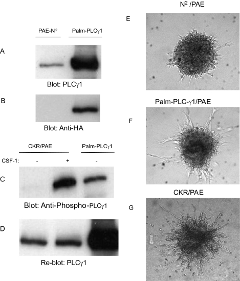
To initially test the hypothesis that PLCγ1 activation is necessary for induction of sprouting/tubulogenesis of endothelial cells, we analyzed the ability of a constitutively active form of PLCγ1 to promote tubulogenesis. A recent study has shown that palmitoylation and myristoylation of PLCγ1 in T-lymphocytes is sufficient for its recompartmentalization to lipid rafts where it undergoes constitutive tyrosine phosphorylation and activation.10 To this end, we used a retroviral system to overexpress a dually acylated form of PLCγ1 with a myristoylation and palmitoylation motif (palm-PLCγ1) in PAE cells. Expression of palm-PLCγ1 in PAE cells was detected with the anti-PLCγ1 and HA antibodies (Figs. 1A, 1B). As expected, palm-PLCγ1 was constitutively tyrosine phosphorylated (Fig. 1C). Nevertheless, its tyrosine phosphorylation was significantly lower than that of the chimeric VEGFR-2 (CKR)-mediated phosphorylation (Fig. 1C). Despite its inefficient tyrosine phosphorylation, overexpression of PLCγ1 in the PAE cells stimulated a modest level of tube formation and sprouting of endothelial cells (Fig. 1F) suggesting that perhaps robust tyrosine phosphorylation of PLCγ1 is necessary for it to fully support tube formation by endothelial cells. The CKR-mediated tube formation by PAE cells was used as the positive control (Fig. 1G).
Figure 1.
Selective activation of PLCγ1 induces tube formation by endothelial cells. An equal number of serum-starved PAE cells expressing an empty vector (PAE-N2) or constitutive active HA-tagged PLCγ1 (palm-PLCγ1) were lysed, and Western blot analysis was performed to determine total PLCγ1 using anti-PLCγ1 (A) or anti-HA (B) antibody. PAE cells expressing chimeric VEGFR-2 (CKR) were unstimulated or stimulated with CSF-1 for 10 minutes, and the cells were lysed. PAE cells expressing palm-PLCγ1 were lysed without stimulation. Cell lysates were subjected to Western blot analysis with a phospho-specific PLCγ1 antibody (C). The same membrane was analyzed for total PLCγ1 levels (D). PAE cells expressing the empty vector PAE-N2, palm-PLCγ1, or chimeric VEGFR-2 (CKR) were subjected to tube formation assay, and photographs were taken after 48 hours (E, F, G).
Effect of Protein Kinase C Inhibitors on VEGFR-2–Induced Tubulogenesis of Endothelial Cells
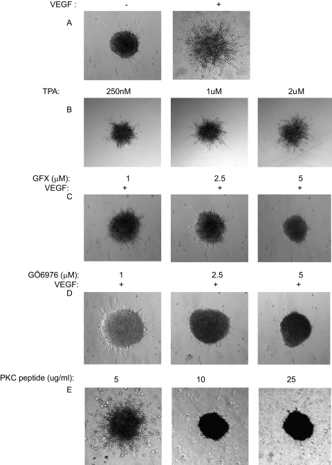
Activation of PLCγ1 by receptor tyrosine kinases (RTKs) leads to hydrolysis of phosphoinositides to generate the second messengers inositol 1,4,5-trisphosphate (IP3) and diacylglycerol (DAG). IP3 increases intracellular Ca2+ levels and DAG activates PKCs and other C1 domain-containing proteins.16. To test whether activation of PKCs contributes to the tubulogenesis of endothelial cells, we analyzed the ability of TPA (12-O-tetradecanoylphorbol 13-acetate), an analog of diacylglycerol, to stimulate tube formation of PAE cells. The results showed that treatment of PAE cells with different concentrations of TPA stimulates tube formation of PAE cells, suggesting that activation of PKCs by PLCγ1 affects its ability to mediate tube formation of endothelial cells (Fig. 2B). To further support the hypothesis that activation of PKCs is important in tube formation by endothelial cells, we attempted to inhibit their activation by a pharmacologic approach with GFX and GÖ6976, potent inhibitors of PKCs. It is known that GFX and GÖ6976 inhibit various isozymes of PKC, including classic PKC (α, βI, βII, and γ), novel PKC (δ, ε, η, and θ), and PKD (protein kinase D). As shown (Figs. 2C, 2D), both GFX and GÖ6976 inhibited VEGFR-2-mediated tubulogenesis of endothelial cells in a dose-dependent manner. Finally, we decided to inhibit activation of PKCs by a myristoylated PKC peptide inhibitor, which specifically inhibits calcium- and phospholipid-dependent PKCs. This inhibitor is based on the pseudosubstrate region of PKC-α and -β.17 Treatment of cells with the PKC peptide inhibitor almost completely inhibited the sprouting and tube formation of the PAE cells (Fig. 2E), further supporting the idea that the PLCγ1/PKC pathway plays a critical role in mediating the angiogenic signaling of VEGF.
Figure 2.
PKC activation is necessary for tube formation of endothelial cells. PAE cells expressing VEGFR-2 (100 ng/mL) were subjected to tube formation assay in the absence or presence of VEGF, as a positive control (A). The same cells were also treated with different concentrations of TPA, a potent PKC activator (B). The same cells were stimulated with VEGF in combination with different concentrations of GFX, a potent inhibitor of PKC (C). The same cells were incubated with VEGF plus GÖ6976, another potent PKC inhibitor (D). PAE cells were also treated with a peptide-derived PKC inhibitor, tube formation was assessed under a light microscope, and photographs were taken with a digital camera (E).
PLCγ1-Induced Growth of Blood Vessels in the CAM Model
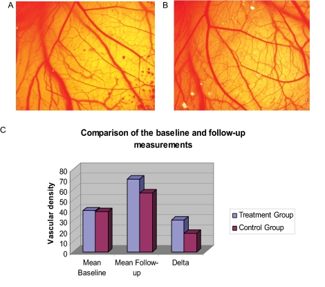
To further analyze the role of PLCγ1 in angiogenesis beyond the cell culture system, we evaluated its role in the CAM assay in which we directly activated PLCγ1 by applying the pharmacologic agent m-3M3FBS, a potent activator of PLCγ1 that stimulates Ca2+ release and inositol phosphate formation in a variety of cell types.18 The results showed that m-3M3FBS stimulated angiogenesis in CAM (Fig. 3B). Further quantification of blood vessel formation showed a significant increase in blood vessel growth in response to m-3M3FBS. The mean change (Fig. 3C, delta) in blood vessel density in the CAM, as measured by the digital image analysis system 24 hours after incubation in the control group was 18 and the mean change (delta) in blood vessel density 24 hours after incubation of the CAM in the treatment group was 30.75. This difference was statistically significant (P = 0.07; Figs. 3A, 3B). The mean baseline blood vessel density in the CAM in the treatment group was 40, and that in the control group was 39. The difference between these two groups was not statistically significant (P = 0.96; Fig. 3C). The vascular density in the CAM model of angiogenesis was significantly higher in the drug (PLCγ1 activator) group than in the control group.
Figure 3.
PLCγ1 activation is necessary for angiogenesis in vivo. CAM treated with PLCγ1 activator (m-3M3FBS). (A) CAM at baseline. (B) A CAM 24 hours after incubation, showing increased vascular density. (C) Difference in vascular density between the baseline and 24-hour measurements after treatment with PLCγ1 activator in each CAM, the difference (delta) between the baseline and 24 hours after control for each CAM. The measurements were higher in the treatment group than in the control group indicating increased vascularity in the PLCγ1 activator–treated group.
Effect of Inhibition of PLCγ1 on the Growth of Blood Vessels in the CAM Angiogenesis Model
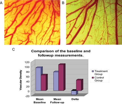
To further evaluate the role of PLCγ1 in angiogenesis, we studied the effect of inhibition of PLCγ1 by a pharmacologic agent, U-73122, in the CAM assay. We found that after 24 hours of incubation with U-73122 in the CAM assay, the mean of the change in blood vessel density in the control group was 43, whereas the mean change in the treatment group was −17. This difference was statistically significant (P = 0.00039; Figs. 4A, 4B). The mean baseline blood vessel density in the treatment group was 94 and that in the control group was 64. The differences between the two groups were not statistically significant (P = 0.122; Fig. 4C, delta). The data show that the vascular density in the CAM model of angiogenesis was significantly higher in the control group than in the group treated with PLCγ1 inhibitor.
Figure 4.
PLCγ1 activation was shown to be necessary for angiogenesis in vivo. CAMs were treated with the PLCγ1 inhibitor (U-73122). (A) CAM at baseline. (B) CAM 24 hours after incubation, showing decreased vascular density compared with baseline photos. (C) Measurement difference (delta) between the baseline and 24 hours after treatment with PLCγ1 inhibitor for each CAM, and the difference between the baseline and 24-hour measurements and the control for each CAM. The measurements were much lower in the treatment group than in the control group, indicating decreased vascularity in the treated group. The difference was statistically significant (P = 0.00039).
Increased Laser-Induced CNV in c-Cbl–knockout Mice
CNV perhaps represents the best example of pathologic angiogenesis. In a previous study, we identified c-Cbl as a negative regulator of PLCγ1.10 Our more recent study indicates that loss of c-Cbl in the mouse model results in elevated PLCγ1 activation, leading to increased endothelial cell proliferation (Meyer RD, et al., manuscript submitted, 2010). To investigate whether loss of c-Cbl, a negative regulator of PLCγ1, also plays role in this pathologic setting, we decided to analyze the biological consequence of loss of c-Cbl in laser-induced CNV.
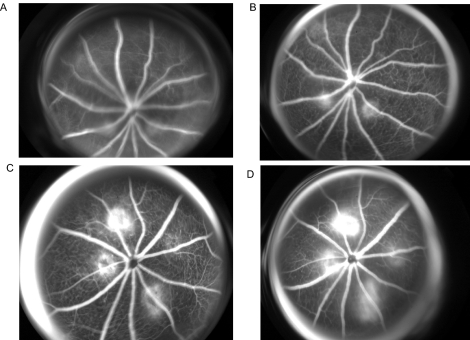
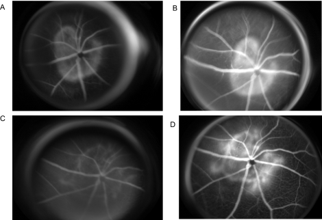
The CNV was induced as described in the Methods section, and the eyes were examined weekly with retinal fluorescein angiography, to detect CNV at the site of laser injury. Fluorescein angiograms performed on eyes of wild-type and knockout mice 2 weeks after laser induction to create CNV showed that CNV had formed in 7 of 18 lesions in the wild-type mice, corresponding to 39% (Figs. 5A, 5B; Table 1). In comparison, CNV formation was noted in 11 (78%) of 14 of the c-Cbl–knockout mice (Figs. 5C, 5D; Table 1). CNV formation in the c-Cbl–knockout mice was not only twice that formed in the wild-type mice, but the leakage from the CNV lesions in the c-Cbl–knockout mice was also robust and larger. As a result, the CNV lesions in the c-Cbl–knockout mice were more confluent than those in the wild-type mice (Fig. 6). In fact 42% (6/14) of the lesions became confluent in the c-Cbl–knockout mice, compared with 11% (2/18 lesions) in the wild-type (Table 1).
Figure 5.
CNV at 2 weeks of follow-up after laser induction of CNV. (A) Early-phase fluorescein angiogram (FA) in wild-type mice showing early hyperfluorescence in two of the four areas treated with the laser. (B) Late-phase FA in wild-type mice showing increasing hyperfluorescence in the two areas, confirming the presence of CNV in two of the four laser-treated areas. (C) Early-phase FA in a knockout mouse eye, showing hyperfluorescence all four areas. (D) Late-phase FA in a knockout mouse eye showing increasing hyperfluorescence all four areas, indicating CNV in all four lasered areas.
Table 1.
CNV in c-Cb1–Knockout Mice versus Wild-Type Mice
| Duration (wk) | Lesions with 2B Grade Leakage (%) | P | |
|---|---|---|---|
| c-Cb1−/− | 1 | 64.3 | 0.03659* |
| Wild-type 1 | 1 | 38.9 | |
| c-Cb1−/− | 2 | 84.6 | 0.0034* |
| Wild-type 1 | 2 | 33.3 | |
| c-Cb1−/− | 4 | 71.4 | 0.03659* |
| Wild-type | 4 | 33.3 |
| Lesions with Confluent Leakage (%) | P | ||
|---|---|---|---|
| c-Cb1−/− | 42.9 | 0.04984* | |
| Wild-type | 11.1 |
The percentage of lesions with 2B grade leakage in the c-Cb1 versus the wild-type mice and the percentage of lesions with confluent leakage are shown at 1, 2, and 4, weeks after laser treatment.
Statistically significant at P < 0.05, by ANOVA.
Figure 6.
Confluent CNV after laser induction in c-Cbl–knockout mouse eyes. (A) Early-phase fluorescein angiogram (FA) at 2 weeks showing hyperfluorescence encircling the optic nerve. (B) Late-phase FA showing increasing hyperfluorescence encircling the optic nerve, indicating that CNV, forming from all four laser spots, have become confluent. (C) Early-phase FA at 4 weeks showing early hyperfluorescence around the optic nerve. (D) Late-phase FA at 4 weeks showing leakage. Note the increasing hyperfluorescence from the borders of CNV seen at 2 weeks indicating continued growth of CNV at 4 weeks after laser induction.
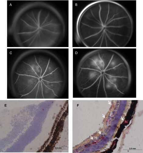
Further follow-up of CNV formation 4 weeks after laser induction, as demonstrated by fluorescein angiogram, showed that CNV in the wild-type mice was still present in 6 (33%) of 18 lesions, with a slight regression from the 2-week post–laser-induction time point. Overall, the CNV leakage was smaller (Figs. 7A, 7B; Table 1). However, unlike what we observed in the wild-type mice, CNV formation in the c-Cbl–knockout mice was still strongly active 4 weeks after laser induction. In fact, CNV was observed in 10 (71%) of 14 lesions, with only minor regression from the 2-week time point. However, we noted that in the c-Cbl–knockout, the CNV leakage was overall more extensive than observed 2 weeks ago (Figs. 7C, 7D; Table 1). Immunohistochemistry of the eye balls of the c-Cbl–knockout mice showed that PLCγ1 phosphorylation and activation were increased substantially compared with that in the wild-type mice (Figs. 7E, 7F). Taken together, the data suggest that loss of c-Cbl results in enhanced ocular angiogenesis in part by increasing activation of PLCγ1.
Figure 7.
CNV at 4 weeks of follow-up after laser induction of CNV. (A) Early-phase FA in a wild-type mouse eye showing no hyperfluorescence. (B) Late-phase FA in a wild-type mouse eye showing no hyperfluorescence, indicating regression of CNV. (C) Early-phase FA in a c-Cbl–knockout mouse eye showing early hyperfluorescence in all four areas. (D) Late-phase FA in a knockout mouse eye showing increasing hyperfluorescence in all four areas. The leakage was larger at 4 weeks than at 2 weeks, indicating continued growth of the CNV at 4 weeks. Phosphorylation of PLCγ1 in c-Cbl–knockout and wild-type mice after 4 weeks of laser induction of CNV is shown (E, F). Immunohistochemistry was performed with the phospho-PLCγ1 antibody, which specifically detects the active form of PLCγ1. (E) A few areas of speckled phospho-PLCγ1 positivity were detected in the layers of the retina in wild mouse eye. (F) Phospho-PLCγ1 positivity (white arrows) was detected between the choroid and RPE and the inner layers of the retina in c-Cbl–knockout mouse eye. Magnification: (E, F) ×400.
Discussion
Aberrant angiogenesis (neovascularization) occurs in several diseases of the eye, including AMD and proliferative diabetic retinopathy. CNV is considered to be the defining characteristic of late-stage wet AMD, where new sprouting vessels penetrate Bruch's membrane, growing into the subretinal space and sometimes into vessels that are derived from the retinal circulation.15 The primary mediator of CNV is VEGF. VEGF binds to and activates an endothelial receptor tyrosine kinase (RTK), vascular endothelial growth factor receptor-2 (VEGFR-2/FLK-1/KDR), as well as other endothelial RTKs, including VEGFR-1 and -3.16 It is thought that VEGF induces vascular leakage and inflammation by promoting the increased production and permeability of capillary endothelial cells. VEGF is considered to play a critical role in the pathogenesis of AMD, and recent therapies targeting VEGF have shown promising results of significant improvement of central vision and reduction of CNV.11
Activation of VEGFR-2 by VEGF family ligands in endothelial cells leads to a wide variety of biological responses that emanate from several key autophosphorylation sites within the cytoplasmic domain of VEGFR-2.16 Activation of PLCγ1 in endothelial cells is considered an important mediator of angiogenic signaling of VEGFR-2.7 The initial evidence linking PLCγ1 to endothelial cell function and angiogenesis in vivo was provided by targeted deletion of PLCγ1, which resulted in early embryonic lethality in zebrafish and mice that was caused by significantly impaired vasculogenesis and erythrogenesis.9
Regulation of angiogenesis is often viewed as a balance between proangiogenic and antiangiogenic factors. Recent studies have shown that PLCγ1, a major signaling substrate of VEGFR-2, undergoes c-Cbl-mediated ubiquitination. Ubiquitination of PLCγ1 blocks its activation by VEGFR-2 and with it VEGF-induced endothelial cell proliferation and angiogenesis.10 Further analysis showed that loss of c-Cbl in mice is associated with elevated PLCγ1 activation and endothelial cell proliferation19 (Meyer RD, et al., manuscript submitted, 2010). However, to date, the biological importance of PLCγ1 in pathologic angiogenesis and the possible link between c-Cbl and PLCγ1 activation in pathologic angiogenesis has remained unknown. In the present study we used various in vitro and in vivo angiogenesis models and demonstrated that PLCγ1 activation plays a pivotal role in angiogenesis. More important, our study for the first time linked c-Cbl to the negative regulation of CNV.
The data presented in this study as well as in a manuscript submitted elsewhere (Meyer RD, et al., manuscript submitted, 2010) strongly implicate c-Cbl as a negative regulator of angiogenesis. Our observation is consistent with the conventional and well-recognized function of c-Cbl as a negative regulator of receptor tyrosine kinase signaling.20 Loss of c-Cbl is dispensable for normal embryonic development and angiogenesis,21 whereas loss of both c-Cbl and Cbl-b is lethal before embryonic day (E)10.5,22 suggesting that the proteins of the Cbl family are compensatory in their ability to regulate the activity of target proteins. Regardless of this possibility, our data show that the absence of c-Cbl led to robust and sustained angiogenesis in the eye. This finding indicates that c-Cbl specifically regulates pathologic angiogenesis and that the unique characteristics of c-Cbl may make it a good target for antiangiogenic therapy.
Although various risk factors have been linked to AMD, the etiology and pathogenesis of AMD remain largely unclear. Similarly, treatment options for AMD patients are limited, further arguing a need for new preventative and therapeutic strategies for this disease. Our present study indicates a vital role for PLCγ1 in pathologic angiogenesis of the eye. Therefore, targeting PLCγ1 may potentially offer a novel treatment for ocular neovascularization which would be more specific and longer-acting than the treatments currently available.
Footnotes
Supported by the Macula Foundation (DH), National Institutes of Health/National Eye Institute Grant R01EY017955-01A2 (NR), and the Massachusetts Lion's Foundation to Department of Ophthalmology (NR/DH).
Disclosure: D. Husain, None; R.D. Meyer, None; M. Mehta, None; W.M. Pfeifer, None; E. Chou, None; G. Navruzbekov, None; E. Ahmed, None; N. Rahimi, None
References
- 1. Carmeliet P. Mechanisms of angiogenesis and arteriogenesis. Nat Med. 2000;6:389–395 [DOI] [PubMed] [Google Scholar]
- 2. Zachary I, Gliki G. Signal transduction mechanisms mediating biological actions of the vascular endothelial growth factor family. Cardiovasc Res. 2001;9:568–581 [DOI] [PubMed] [Google Scholar]
- 3. Yancopoulos GD, Davis S, Gale NW, Rudge JS, Wiegand SJ, Holash J. Vascular-specific growth factors and blood vessel formation. Nature. 2000;407:242–248 [DOI] [PubMed] [Google Scholar]
- 4. Carmeliet P. Angiogenesis in health and disease. Nat Med. 2003;9:653–660 [DOI] [PubMed] [Google Scholar]
- 5. Ferrara N. VEGF and the quest for tumour angiogenesis factors. Nat Rev Cancer. 2002;2:795–803 [DOI] [PubMed] [Google Scholar]
- 6. Sakurai Y, Ohgimoto K, Kataoka Y, Yoshida N, Shibuya M. Essential role of Flk-1 (VEGF receptor 2) tyrosine residue 1173 in vasculogenesis in mice. Proc Natl Acad Sci U S A. 2005;102:1076–1081 [DOI] [PMC free article] [PubMed] [Google Scholar]
- 7. Meyer RD, Latz C, Rahimi N. Recruitment and activation of phospholipase Cgamma1 by vascular endothelial growth factor receptor-2 are required for tubulogenesis and differentiation of endothelial cells. J Biol Chem. 2003;278:16347–16355 [DOI] [PMC free article] [PubMed] [Google Scholar]
- 8. Liao HJ, Kume T, McKay C, Xu MJ, Ihle JN, Carpenter G. Absence of erythrogenesis and vasculogenesis in Plcg1-deficient mice. J Biol Chem;277:9335–9341 [DOI] [PubMed] [Google Scholar]
- 9. Lawson ND, Mugford JW, Diamond BA, Weinstein BM. Phospholipase C gamma-1 is required downstream of vascular endothelial growth factor during arterial development. Genes Dev. 2003;17:1346–1351 [DOI] [PMC free article] [PubMed] [Google Scholar]
- 10. Singh AJ, Meyer RD, Navruzbekov G, et al. A critical role for the E3-ligase activity of c-Cbl in VEGFR-2-mediated PLCgamma1 activation and angiogenesis. Proc Natl Acad Sci U S A. 2007;104:5413–5418 [DOI] [PMC free article] [PubMed] [Google Scholar]
- 11. Bressler NM. Antiangiogenic approaches to age-related macular degeneration today. Ophthalmology. 2009;116:S15–S23 [DOI] [PubMed] [Google Scholar]
- 12. Rahimi N, Dayanir V, Lashkari K. Receptor chimeras indicate that the vascular endothelial growth factor receptor-1 (VEGFR-1) modulates mitogenic activity of VEGFR-2 in endothelial cells. J Biol Chem. 2000;275:16986–16992 [DOI] [PubMed] [Google Scholar]
- 13. Naramura M, Kole HK, Hu RJ, Gu H. Altered thymic positive selection and intracellular signals in Cbl deficient mice. Proc Natl Acad Sci U S A. 1998;95:15547–15552 [DOI] [PMC free article] [PubMed] [Google Scholar]
- 14. Meyer RD, Sacks DB, Rahimi N. IQGAP1-dependent signaling pathway regulates endothelial cell proliferation and angiogenesis. PLoS One. 2008;3:e3848. [DOI] [PMC free article] [PubMed] [Google Scholar]
- 15. Grossniklaus HE, Green WR. Choroidal neovascularization. Am J Ophthalmol. 2004;137:496–503 [DOI] [PubMed] [Google Scholar]
- 16. Rahimi N. Vascular endothelial growth factor receptors: molecular mechanisms of activation and therapeutic potentials. Exp Eye Res. 2006;83:1005–1016 [DOI] [PMC free article] [PubMed] [Google Scholar]
- 17. Eichholtz T, de Bont DB, de Widt J, Liskamp RM, Ploegh HL. A myristoylated pseudosubstrate peptide, a novel protein kinase C inhibitor. J Biol Chem. 1993;268:1982–1986 [PubMed] [Google Scholar]
- 18. Li Y, Jia YC, Li N, Zheng ZY, Wang YZ, Yuan XB. Essential role of TRPC channels in the guidance of nerve growth cones by brain-derived neurotrophic factor. Nature. 2005;434:894–898 [DOI] [PubMed] [Google Scholar]
- 19. Rahimi N. A role for protein ubiquitination in VEGFR-2 signaling and angiogenesis. Biochem Soc Trans. 2009;37:1189–1192 [DOI] [PubMed] [Google Scholar]
- 20. Thien CB, Langdon WY. c-Cbl and Cbl-b ubiquitin ligases: substrate diversity and the negative regulation of signalling responses. Biochem J. 2005;391:153–166 [DOI] [PMC free article] [PubMed] [Google Scholar]
- 21. Murphy MA, Schnall RG, Venter DJ, et al. Tissue hyperplasia and enhanced T-cell signalling via ZAP-70 in c-Cbl-deficient mice. Mol Cell Biol. 1998;18:4872–4882 [DOI] [PMC free article] [PubMed] [Google Scholar]
- 22. Naramura M, Jang IK, Kole H, Huang F, Haines D, Gu H. c-Cbl and Cbl-b regulate T cell responsiveness by promoting ligand-induced TCR down-modulation. Nat Immunol. 2002;3:1192–1199 [DOI] [PubMed] [Google Scholar]


