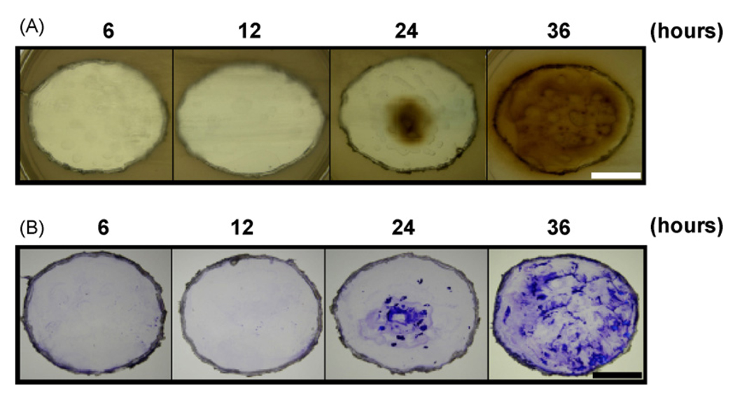
Fig. 1.
Detection of VSC production and biofilm formation of F. nucleatum. F. nucleatum (4 × 10⁹ CFU/well) was cultured on a 6-well nonpyrogenic polystyrene plate for 6, 12, 24 and 36 h. (A) After removing the media, each well containing attached bacteria was excised and pasted onto the top of OHO-C agar plate overnight under anaerobic conditions using Gas-Pak. (B) For detection of biofilm formation, excised wells were removed from OHO-C agar plates and stained with 0.4% (w/v) crystal violet for 1 min. The brown/dark precipitates of lead sulfides and purple crystal violet stain on the 24 and 36 h-cultured plates indicated the VSCs and biofilms, respectively. Bars = 1.5 cm.

