Abstract
Blockade of neurotransmitter release by botulinum neurotoxin type A (BoNTA) underlies the severe neuroparalytic symptoms of human botulism, which can last a few years. The structural basis for this remarkable persistence remains unclear. Herein, recombinant BoNTA was found to match the neurotoxicity of that from Clostridium botulinum, producing persistent cleavage of synaptosomal-associated protein of 25 kDa (SNAP-25) and neuromuscular paralysis. When two leucines near the C terminus of the protease light chain of A (LCA) were mutated, its inhibition of exocytosis was followed by fast recovery of intact SNAP-25 in cerebellar neurons and neuromuscular transmission in vivo. Deletion of 6–7 N terminus residues diminished BoNTA activity but did not alter the longevity of its SNAP-25 cleavage and neuromuscular paralysis. Furthermore, genetically fusing LCE to a BoNTA enzymically inactive mutant (BoTIMA) yielded a novel LCE-BoTIMA protein that targets neurons, and the BoTIMA moiety also delivers and stabilizes the inhibitory LCE, giving a potent and persistent cleavage of SNAP-25 with associated neuromuscular paralysis. Moreover, its neurotropism was extended to sensory neurons normally insensitive to BoNTE. LCE-BoTIMA(AA) with the above-identified dileucine mutated gave transient neuromuscular paralysis similar to BoNTE, reaffirming that these residues are critical for the persistent action of LCE-BoTIMA as well as BoNTA. LCE-BoTIMA inhibited release of calcitonin gene-related peptide from sensory neurons mediated by transient receptor potential vanilloid type 1 and attenuated capsaicin-evoked nociceptive behavior in rats, following intraplantar injection. Thus, a long acting, versatile composite toxin has been developed with therapeutic potential for pain and conditions caused by overactive cholinergic nerves.
Keywords: Botulinum Toxin, Drug Design, Exocytosis, Gene Expression, Neurotransmitters, Protein Chimeras, Protein Motifs, Protein Targeting, SNAREs
Introduction
Botulinum neurotoxins (BoNTs)3 inhibit transmitter release from peripheral cholinergic neurons causing the life-threatening flaccid paralysis underlying botulism (1). The most potent biological substances, their estimated lethal doses (LD50) in humans are between 0.1 and 1 ng/kg. Seven BoNT serotypes (A–G), produced by Clostridium botulinum, are synthesized as pro-form single chain proteins (SC, Mr ∼150,000) and converted by either Clostridial or tissue proteases into fully active dichain (DC) forms, consisting of a protease domain (LC; Mr ∼50,000) linked to a heavy chain (HC; Mr ∼100,000) through disulfide and noncovalent bonds. BoNTA preferentially enters cholinergic nerve endings by binding via the C-terminal half of their HC to a membrane acceptor, a lumenal domain of synaptic vesicle protein 2 (2, 3). On the other hand, type E only binds the glycosylated synaptic vesicle protein 2 A/B isoforms (4), which are sparsely expressed on sensory neurons, explaining its lack of effects on trigeminal ganglionic neurons (TGNs) (5). These toxins undergo acceptor-mediated endocytosis (6, 7) with translocation of the LCs into the cytosol through a channel formed by the N-terminal half of HC, where they cleave and inactivate soluble N-ethylmaleimide sensitive factor attachment protein receptors (SNAREs) that are essential for regulated exocytosis (8). BoNTA, which removes nine residues from the C terminus of SNAP-25, gives a potent and persistent blockade of neurotransmitter release (9, 10). These features underlie its unrivalled success for long term treatment of a wide variety of neurogenic and idiopathic hyperactivity disorders, e.g. dystonias (3–6 months), overactive bladder (6–9 months), and hyperhydrosis (6 months to ≫1 year) (11). BoNTA has also been used successfully to treat some types of chronic pain (12). However, in terms of an analgesic, a BoNTE protease retargeted to sensory neurons has proved much more efficacious in attenuating the firing of delayed spiking neurons elicited by a pain mediator, calcitonin gene-related peptide (CGRP), or activated via transient receptor potential vanilloid type 1 (TRPV1) in brain slices (5). BoNTE protease removes 17 more C-terminal residues from SNAP-25 than BoNTA and thereby even more effectively prevents formation of stable SNARE complexes and causes a more disruptive blockade of neuroexocytosis (5). Accordingly, neuromuscular paralysis induced by BoNTA can be reversed by elevating [Ca2+]i but not in the case of BoNTE treatment (13–15). Although BoNTE blocks neurotransmission more quickly and more potently than BoNTA (16), the clinical applications of BoNTE are restricted by its neuromuscular paralytic action being transient: less than 4 weeks in contrast to more than 4 months for BoNTA (17, 18).
This intriguing difference in the duration of action warrants investigation, especially into a molecular basis for the amazing longevity of BoNTA and the attractive prospect of transferring this advantageous therapeutic feature into new generations engineered for particular therapies. Initially, the more persistent muscle immobilization by BoNTA, compared with BoNTE, was ascribed to a faster turnover of SNAP-25 that had been cleaved by BoNTE (SNAP-25E) rather than BoNTA (SNAP-25A) after observations in humans (18) and mice (15) that injection of BoNTE alongside BoNTA, or even a few days later, accelerated recovery of muscle function. However, other studies in cultured neurons (9, 19) and animals (10) found that BoNTE transiently converts the SNAP-25 and/or SNAP-25A to SNAP-25E, but that SNAP-25A will return after the disappearance of SNAP-25E, suggestive of the A protease outliving the E enzyme. This conclusion was bolstered by a careful pulse-chase analysis, which established that the turnover rate for SNAP-25 is little changed following its cleavage by BoNTA or BoNTE, whereas the half-life of the E protease is much shorter than that of the A (19).
Such observations prompted a search for unique features of BoNTA that may stabilize its protease activity. Expression as GFP-tagged fusion proteins in neuroendocrine cells revealed a difference in subcellular localization of LCA, which concentrated on the cell membrane, compared with LCE that distributed throughout the cytosol (20). Site-directed mutations and deletions pin-pointed two regions involved in membrane association of LCA: eight N-terminal residues and a dileucine motif (EFYKLL) near its C terminus (20). Thus, the authors speculated that interaction with the plasmalemma may contribute to the longevity of the LCA protease. Because it is not possible to ascertain the lifetime of proteins in such an overexpression model, it was not clear whether such findings could be reconciled with the properties of toxins that act in minute amounts in the nerve endings of clinical recipients.
Herein, to address such issues, BoNTA was engineered by incorporating a synthetic gene into a prokaryotic vector and expressed as C-terminal His6-tagged SC proteins in Escherichia coli, creating a recombinant BoNTA that could be evaluated in vivo against its natural counterpart. An additional advantage of this strategy was the exploitation of mutagenesis, which revealed a C-terminal dileucine that was essential for the protease longevity. Furthermore, a BoNTA enzymically inactive mutant (BoTIMA) delivered a fused LCE protease into cholinergic neurons and stabilized its activity, transforming the transient action of BoNTE into a prolonged neuromuscular paralysis. Additionally, BoTIMA transferred LCE into sensory neurons and blocked CGRP release from TGNs and attenuated capsaicin-evoked nociceptive behavior in rats. This novel LCE-BoTIMA “composite” protein inherited the desirable characteristics of two BoNT serotypes, the powerful LCE protease combined with the longevity endowed by LCA offering scope to extend therapeutic applications.
EXPERIMENTAL PROCEDURES
Animals
Female Tyler's Ordinary mice were purchased from Harlan UK; Sprague-Dawley rats were bred in an approved Bioresource Unit at Dublin City University or obtained from Charles River UK and housed at the National University of Ireland, Galway. All of the procedures involving live animals have been approved by the Dublin City University Research Ethics Committee and the Animal Care and Research Ethics Committee at the National University of Ireland, Galway and were carried out under licenses granted by the Minister for Health and Children under the Cruelty to Animals Act, 1876 as amended by European Communities Regulations 2002 and 2005 (license numbers 100/3609 and 100/3613).
Construction of Plasmids
Experimentation on recombinant BoNTs has been approved by the Environmental Protection Agency of Ireland and performed under containment level 2, according to a detailed and strictly enforced safety protocol. Genes encoding BoTIMA, which had a single mutation at the active site (His227 to Tyr) or BoNTE were custom synthesized for optimal expression in E. coli. The BoTIMA gene was subcloned into pET29a vector (Novagen), using suitable restriction enzymes. Furthermore, two thrombin cleavage sites, one in the HC/LC loop as reported earlier for BoNT/G (21) and the other between the C terminus of HCA and His6, were engineered into the genes for the SC protein. The sequence of the resultant construct pET29a-BoTIMA was verified. For generating the WT BoNTA construct, the nucleotides encoding Tyr227 were mutated to those for His, using site-directed mutagenesis with suitable primers. BoNTA constructs encoding N-terminal deletions of residues 2–7(ΔN6), 2–8(ΔN7), and 2–9(ΔN8) were obtained by PCR, using appropriate primers and WT BoNT/A as template, followed by self-ligation; the start codon encoding the methionine (residue 1) was retained in the gene sequence. LCE gene encoding residues 1–411, excluding the disulfide-forming cysteines, was subcloned into the BoTIMA construct at the 5′ end to yield pET29a containing LCE-BoTIMA. Mutated BoNTA(AA428–9) and LCE-BoTIMA(AA428–9) constructs were generated by site-directed mutagenesis. A new BoNTEA chimera was engineered by incorporating N-terminal residues 2–9 of BoNTA into the previously engineered chimera EA (after the first methionine), using appropriate primers and the chimera EA construct (22) as template, followed by self-ligation. The sequence of resultant chimeric gene N8-BoNTEA was confirmed.
Expression, Purification, and Characterization of the Recombinant Proteins
Proteins were produced in the E. coli strain BL21(DE3) using an autoinduction medium (22, 23). BoTIMA, BoNTA, or its variant were purified using immobilized metal affinity chromatography on Talon resin and anion exchange chromatography, as for the previously reported BoNT AE chimera (22). Purified SCs were either stored at −80 °C or nicked with biotinylated thrombin (Novagen, 1 unit/mg of toxin) at 22 °C for 1 h; the protease was removed using streptavidin-agarose (Novagen). To further purify LCE-BoTIMA, the immobilized metal affinity chromatography-separated proteins were exchanged into 20 mm sodium phosphate (pH 6.5) and loaded onto a UNO-S1 column followed by washing with 100 mm NaCl before elution by a stepwise gradient up to 1 m NaCl. The fusion protein began to elute at 0.22 m NaCl; after transfer into 20 mm HEPES and 150 mm NaCl (pH 7.6), it was nicked with biotinylated thrombin as for BoTIMA except incubated for 3 h at 22 °C. N8-BoNT EA protein was produced as for chimera EA (22). The samples were monitored by SDS-PAGE followed by Coomassie staining or Western blotting, using antibodies against C-terminal residues of BoNTA, LCE, LCA, or His6 (22). SC or nicked samples were quantified using the Bradford assay. Note that all of the functional assays were performed on the nicked samples; the protease activities of toxins were measured as before (22).
Neuronal Cultures, Exposure to BoNTs, and Assay of Transmitter Release and SNARE Cleavage
Isolation and culturing of rat cerebellar granule neurons (CGNs) were as described previously (19). At 7 days in vitro, the cells were exposed for 24 h at 37 °C to a series of toxin concentrations. In some cases, the cells were incubated for 5 min with 500 pm BoNTs in HEPES-buffered solution containing 70 mm K+ (stimulation solution) with adjustment for the NaCl concentration (19); after being washed twice and incubated in medium at 37 °C for the periods noted in the figure legends, the cells were harvested. Where specified, the neurons were exposed to 100 pm toxin (0.2-μm filter sterilized) in culture medium for 24 h; unbound toxin was removed by three washes, and then the culture medium was replaced. The cells were harvested in SDS sample buffer at the indicated times.
Culturing of rat TGNs followed by their exposure to BoNTs and assay of transmitter release were as described previously (24). For quantifying SNAP-25 cleavage, the lysates were subjected to SDS-PAGE and Western blotting using an antibody (SMI-81; Covance) that recognizes intact and BoNT-truncated SNAP-25. Monoclonal antibody (HPC-1; Sigma-Aldrich) was used to detect another SNARE protein, syntaxin. A two-dimensional SDS-PAGE method (5, 25) was used to investigate the SDS-resistant SNARE complexes in TGNs.
Assessment of Neuromuscular Paralytic Activity and Lethality of Toxins
Mouse hemi-diaphragms were used to measure neuromuscular paralysis by BoNTs (22). Their durations of action were monitored in vivo by the digit abduction score (DAS) assay (22, 26); to accommodate the potencies of the various toxin preparations, a maximal tolerated dose (TDmax; see Table 1) for each was determined by injection into mouse right gastrocnemius muscle (22). In separate experiments, the leg muscles of mice injected with toxin or saline (control) were dissected at different times, and the presence or absence of cleaved SNAP-25 was visualized by Western blotting (15). Toxin lethalities (see Table 1) were determined by intraperitoneal injection into mice as described before (27). All of the mice used had body weights between 20 and 22 g.
TABLE 1.
Mouse lethalities of purified toxins and variants
The doses are those used in the DAS assay.
| Toxin | Lethality | TDmaxa |
|---|---|---|
| mLD50 units/mgc | pg [mLD50 units]c | |
| Natural (n)BoNTAb | 3 × 10⁸ | 20 [6] |
| Natural (n)BoNTEb | 1 × 10⁸ | 70 [7] |
| BoNTA (WT) | 2 × 10⁸ | 10 [2] |
| BoNTA (ΔN6) | 4 × 10⁶ | 500 [2] |
| BoNTA (AA) | 3 × 10⁴ | 1 × 10⁵ [3] |
| BoNTEAb | 7 × 10⁶ | 1140 [8] |
| N8-BoNTEA | 4 × 10⁵ | 5000 [2] |
| BoTIMA | <3 × 10³ | NA |
| LCE-BoTIMA | 7 × 10⁷ | 8 [0.5] |
| LCE-BoTIMA (AA) | 3 × 10⁴ | 1 × 10⁵ [3] |
a TDmax is the maximal tolerated dose that could be injected intramuscularly without producing systemic symptoms (e.g. complete immobilization of injected leg, lower abdomen contraction, and subsequent general paralysis).
b Data from Ref. 22.
c 1 mLD50 unit is the lowest dose of toxin that killed 50% of a group of four mice within 4 days after intraperitoneal injection.
Immunolabeling and Confocal Microscopy
Morphological examination of motor endings in gastrocnemius muscles of mice was performed 14 days after toxin injection, as above. The muscles were then washed and fixed with 4% (w/v) paraformaldehyde in 0.1 m phosphate buffer (pH 7.4) for 10 min before excision. Dissected preparations were kept in the fixative for 2 h, washed, and mounted in Tissue-Tek O.C.T. medium (Sakura Finetek); 50-μm-thick longitudinal frozen sections were cut on a cryostat. Neurons were visualized with a rabbit polyclonal antibody against the 200-kDa neurofilament protein (Sigma), which was detected with Alexa-488 conjugated secondary IgG (Invitrogen). For localizing end plates, postsynaptic acetylcholine receptors were stained with TRITC-conjugated α-bungarotoxin (Invitrogen). The samples were then mounted with Vectashield (Vector Laboratories) and viewed with a confocal microscope (LSM 710; Zeiss).
Intraplantar Injection of Toxins and Capsaicin: In Vivo Assessment of Nociceptive Behavior
On day 1, adult male Sprague-Dawley rats received intraplantar injection of 20 mLD50 (mouse minimal lethal dose) units/kg of body weight of BoNTA or LCE-BoTIMA or vehicle (0.9% NaCl, 0.5% BSA) in 20 μl into the right hind paw, under brief isoflurane anesthesia. The animals were habituated daily to a behavioral observation arena (5 min) and hot plate apparatus (1 min) for 4 days prior to testing. Local muscle activities were unaffected by toxin at the injected dose. On day 5, the rats received intraplantar injections of a 20-μg capsaicin solution in 20 μl (vehicle: 5% ethanol, 5% Tween 80, and 0.9% NaCl) to the same sites as the toxin. Immediately after injection, capsaicin-induced nociceptive behavior was monitored in a Perspex arena with a camera positioned underneath; animal behaviors were recorded onto DVD and scored off-line with the aid of Ethovision software (Noldus, Wageningen, The Netherlands) by a trained experimenter blinded from the treatments. The duration and frequency of licking/shaking/flinching of the capsaicin-injected paw was measured for 10 min. The hot plate test was performed 50 min post-capsaicin injection, using a hot plate analgesia meter (LE7406 from Harvard Apparatus Ltd., Kent, UK). The rats were enclosed by a Plexiglas open cylinder on the hot plate with temperature maintained at 54.5 ± 1 °C. The time taken (latency, s) to lick the right (injected) hind paw was recorded. A cut-off time of 40 s was set to prevent tissue injury. To remove olfactory cues, testing arenas were cleaned and wiped down with 0.5% acetic acid before and after testing of each rat. After experiments, the rats were killed by stunning and decapitation.
Statistical Analysis and Data Presentation
All of the calculations and graphs were performed using GraphPad Prism 4.0, and the p values were calculated as indicated in the figure legends; p < 0.05 was considered statistically significant.
RESULTS
In Vivo Potency of BoNTA Is Reduced Dramatically by N-terminal Truncation or C-terminal Mutation
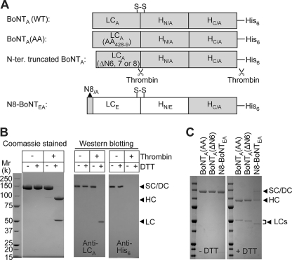
It would be of great interest for both scientists and clinicians if a molecular basis was deciphered for the amazing longevity of BoNTA because this underlies its widespread application in the treatment of various neurological diseases. As noted in the Introduction, there is increasing evidence implicating the protease, rather than other toxin domains, in its prolonged neuroparalytic action (19, 22). To elucidate features that contribute to its stability, a BoNTA construct was generated (Fig. 1A) with a His6 tag to facilitate purification of the expressed protein. Also, sequences were incorporated for selective proteolysis by thrombin (21) to permit subsequent simultaneous activation and removal of the tag (Fig. 1A). BoNTA was highly expressed in E. coli and purified to ∼90% by immobilized metal affinity chromatography, and a pure product was achieved by anion exchange chromatography, with a yield of ∼8 mg/liter of culture (Fig. 1B). The final material gave a single band of Mr ∼150,000 in the absence or presence of DTT, confirming that it was expressed as a SC form (Fig. 1B). Nicking with thrombin completely converted the SC to a disulfide-linked DC, as revealed by the appearance of the HC and LC in the presence of DTT (Fig. 1B). Thrombin simultaneously removed the His6 tag because the latter could not be detected by Western blotting using specific antibodies (Fig. 1B).
FIGURE 1.
Generation of a new BoNTEA chimera, BoNTA, and its deleted or mutated variants. A, representation of engineered constructs. Note that two thrombin recognition sites, one in the HC/LC loop and the other between the C terminus of HCA and His6, were engineered into BoNTA and its variants. S-S denotes the interchain disulfide. HN/A and HC/A represent the N- and C-terminal half of HCA, respectively. B, the purified BoNTA SC was incubated with thrombin (as detailed under “Experimental Procedures”). Aliquots were analyzed by SDS-PAGE in the absence or presence of DTT, followed by Coomassie Blue staining or Western blotting, with the specific antibodies indicated. The stained gel and blot shows that BoNTA was fully nicked by thrombin with complete removal of the tag; also, the interchain disulfide between the LC and HC had been formed. Arrowheads indicate the positions of the SC/DC, HC, and LC. C, SDS-PAGE under reducing (+DTT) or nonreducing conditions and Coomassie staining of nicked N8-BoNTEA, BoNTA, and modified forms.
The potency of this BoNTA, evaluated using a mouse lethality assay, was similar to that for native BoNTA (Table 1). Clearly, incorporation of a thrombin cleavage site in the loop region (21) does not compromise its activity. Such high potency highlights that the toxin produced recombinantly could bind to its acceptor on neuronal membranes, get internalized, and cleave its intracellular target as efficiently as BoNTA purified from C. botulinum. Moreover, this successful recombinant technology provides a means of investigating structural features previously implicated in intracellular membrane association (20) to decipher their roles in the extraordinary longevity of the toxin. Toward this end, a series of N-terminal deletions of BoNTA was generated (Fig. 1A); no soluble intact protein could be obtained if residues 2–9 were deleted (ΔN8). Removal of residues 2–8 (ΔN7) or 2–7 (ΔN6) (Fig. 1C) caused significant (>100-fold) decreases in the expression levels (data not shown). The specific neurotoxicity obtained for ΔN6 was 50-fold lower than for WT BoNTA (Table 1). Deleting seven N-terminal residues (ΔN7) further reduced its specific neurotoxicity (<5 × 10⁵ mLD50 units/mg). The importance of the dileucine was also assessed by mutation to alanines (L428A and L429A) (Fig. 1A) with the resultant plasmid (BoNTA(AA)) transformed into E. coli for expression. This mutated protein was purified as for WT (Fig. 1C), but the specific neurotoxicity of its DC form was ∼4 log units lower (Table 1).
Long Term Cleavage of SNAP-25 by BoNTA and Associated Neuroparalytic Activity Involve a Dileucine in Its LC
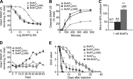
The reduced lethalities of BoNTA variants warranted investigation of their abilities to undergo the multiphasic steps and of the associated durations of action. Therefore, the biological activities of these variants were assessed in vitro using rat cultured CGNs and mouse phrenic nerve diaphragms and in vivo by local injection into mouse gastrocnemius muscle to measure the duration of induced neuromuscular paralysis, using DAS assay (26). In agreement with the in vivo data, BoNTA(ΔN6) was ∼64-fold less active than WT in its cleavage of SNAP-25 in CGNs, with EC50 values being 90 and 1.4 pm, respectively (Fig. 2A). The rate of cellular intoxication by BoNTA (i.e. binding to acceptors, internalization/translocation of protease and cleavage of substrate) was significantly affected by deleting six N-terminal residues, as revealed by the appearance of less cleaved product at the each time point (Fig. 2B). Accordingly, ΔN6 was significantly slower than WT in blocking neuromuscular transmission (Fig. 2C); however, this deletion did not alter its duration of action in CGNs or in vivo (Fig. 2, D and E). Although 100 pm of ΔN6 only cleaved ∼60% of SNAP-25 after 24 h, the ratio of intact to truncated substrate remained fairly constant for 56 days (Fig. 2D); also, injection of mouse gastrocnemius muscle with a maximum tolerated dose (TDmax; Table 1) that does not produce systemic symptoms induced neuromuscular paralysis in vivo of similar duration to that for WT; both lasted over 30 days (Fig. 2E). Likewise, deleting seven N-terminal residues (ΔN7) did not affect the duration of action significantly (data not shown). Because intact protein could not be expressed if the eight N-terminal residues from BoNTA are removed, an alternative strategy was adopted to transfer these residues into a short acting chimera BoNTEA, to test whether its duration of action could be extended. For this purpose, a chimera EA variant construct (N8-BoNTEA) was engineered that encodes the acceptor-binding domain of BoNTA, but the protease and translocation domains of BoNTE with eight N-terminal residues of BoNTA fused to its N terminus (Fig. 1A). Its expression in E. coli, purification, and activation were achieved as for chimera EA (Fig. 1C) (22). Adding these eight extra residues from BoNTA reduced its specific neurotoxicity compared with BoNTEA chimera (Table 1), and it failed to extend the duration of action with respect to the unaltered chimera EA. Injecting the indicated TDmax of N8-BoNTEA induced neuromuscular paralysis that lasted 5 days, similar to BoNTEA (Fig. 2E). Thus, these N-terminal residues of BoNTA seem essential for maintaining the full activity but not for the longevity of its action.
FIGURE 2.
Effects of deleting or mutating residues in BoNTA on its functional properties and duration of action. A, rat CGNs were incubated with each toxin for 24 h before analyzing SNAP-25 cleavage. The proportions of intact substrate remaining were calculated as a percentage of the total (i.e. sum of the intact and cleaved SNAP-25). B, CGNs, exposed to 0.5 nm of each toxin for 5 min at 37 °C in 70 mm K+ stimulation buffer (22), were analyzed immediately as in A or maintained in culture and processed at the indicated times. C, mouse hemi-diaphragms were incubated at 35 °C with 1 nm of each toxin, and the paralysis times were recorded. ***, p < 0.001 versus WT BoNTA, using one-way analysis of variance and post-hoc Student-Newman-Keul's test. D, after incubation of CGNs at 37 °C for 24 h with 100 pm of the toxins indicated, the cells were analyzed for SNAP-25 immediately at day 0 or at the times noted, as above. E, right gastrocnemius muscles of mice were injected with the indicated TDmax of each toxin (Table 1), and the extent of muscle weakening was determined (means ± S.E., n ≥ 5) over time by monitoring DAS (22, 26), BoNTA(AA) versus WT BoNTA using two-way analysis of variance (F(1,119) = 28.28, p < 0.0001), and post-hoc Bonferroni test for comparison of individual time points (*, p < 0.05; **, p < 0.01; ***, p < 0.001). All of the values in A–D are the means ± S.E. (n ≥ 3); in some cases, the error bars are encompassed by the symbols.
Despite a significantly reduced LD50 (Table 1), the dileucine-mutated BoNTA(AA) proved equipotent to WT in entering CGNs and cleaving SNAP-25 (Fig. 2A) with their rates of cellular intoxication being nearly the same (Fig. 2B). Furthermore, BoNTA(AA) blocked neuromuscular transmission as rapidly as WT BoNTA (Fig. 2C; p > 0.05). Clearly, mutating these two leucines does not change the binding/internalization, translocation, or SNAP-25 cleavage; hence, the low mLD50 value may result from faster inactivation in vivo, observed in lethality or DAS assays over several days unlike diaphragm paralysis in 1–2 h. A striking difference was unveiled by the proportion of intact SNAP-25 remaining over time in CGNs preincubated with each toxin, reflecting dissimilarity between the lifetimes of the protease in BoNTA(AA) and WT. Whereas the amount of BoNTA-cleaved substrate remained constant for over 49 days with only a slight decrease on day 63, cells treated with the AA mutant showed a relatively fast recovery of full-length SNAP-25 (Fig. 2D). In vivo experiments confirmed this significant shortening of the action of the toxin upon introducing the AA mutation (Fig. 2E). Injecting a TDmax of the mutant into the hind leg of mice gave full inhibition of muscle movement (DAS = 4) within 24 h, but this persisted for only 2 days; with recovery to DAS = 1 within 10 days, although slight paralysis persisted up to 23 days before DAS = 0 was observed (Fig. 2E). Thus, the potency of the toxin and the duration of muscle paralysis decreased upon mutating the dileucine, with 10,000-fold more protein than BoNTA being required to induce paralysis.
BoTIMA Can Deliver LCE Intraneuronally to Induce Prolonged Neuroparalysis in Mice Because of Its Lifetime Being Extended
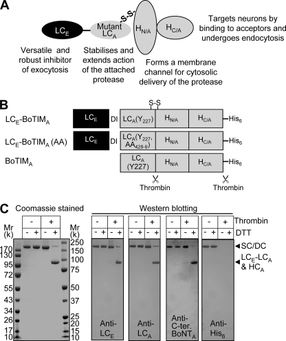
The intriguing question of whether the pronounced longevity of LCA in vivo (see Introduction) could be transferred to transiently acting LCE was addressed by attaching the latter to protease-inactive BoTIMA. This involved designing the composite protein, LCE-BoTIMA (Fig. 3A) whose multiple domains endow synergistic activities: (a) targeting neurons by binding to the synaptic vesicle protein 2 acceptor via the C-terminal half of HCA and undergoing endocytosis; (b) translocation of LCs by the N-terminal half of HCA; and (c) stabilization of LCE protease by enzymically inactive LCA. As a prerequisite, a BoTIMA-His6 construct (Fig. 3B) containing a mutation (H227Y) in the protease active site was generated for ligation to the LCE gene (see below). To act as a control, the BoTIMA-His6 was expressed in E. coli and purified (see “Experimental Procedures”); it behaved as a SC of Mr ∼150,000, which could be converted to the DC form by thrombin (supplemental Fig. S1A).
FIGURE 3.
Synergistic functional properties endowed on a novel multidomain BoNT-based protease. A, illustration of our innovative strategy for compounding advantageous features from types A and E BoNT into a single protein, capable of performing the sequential steps depicted for optimized pharmacological action. B, schematic of the engineered constructs encoding LCE-BoTIMA, LCE-BoTIMA(AA), and BoTIMA. In the composite proteins, two extra residues (DI) were introduced between the pair of LCs. S-S denotes the interchain disulfide. Note that all of the proteins contain two thrombin cleavage sites for simultaneous nicking and tag removal. C, SDS-PAGE, in the absence and presence of DTT, of purified LCE-BoTIMA (see “Experimental Procedures” and supplemental Fig. S1, B and C) showing complete nicking by thrombin with removal of His6; also, the interchain disulfide between the LCA (with LCE attached) and HCA had been formed.
To obtain the required composite toxin, a nucleotide sequence encoding LCE was ligated to the BoTIMA gene to create LCE-BoTIMA (Fig. 3B). Expression in E. coli and purification of this SC protein were accomplished with a yield of ∼1 mg/liter of culture (supplemental Fig. S1, B and C), giving an expected Mr ∼200,000 in the absence or presence of DTT (Fig. 3C). A single band appeared in SDS-PAGE reducing gels after conversion by thrombin to the DC, which also removes the His6 tag (Fig. 3C) at the introduced consensus sequences (Fig. 3B). This band represents LCE-LCA(H227Y) and HCA, which have similar sizes (∼100,000); these bands could not be resolved by SDS-PAGE, but the presence of both was confirmed by Western blotting using specific antibodies (Fig. 3C).
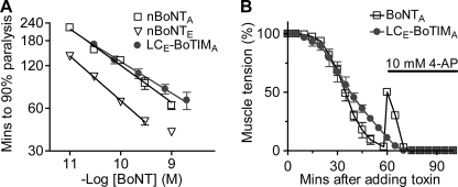
Notably, LCE-BoTIMA cleaved a synthetic SNAP-25 peptide (see “Experimental Procedures”) at the same scissile bond as BoNTE, giving EC50 values (nm) of 1.79 (± 0.6, n = 5) and 1.97 (± 0.6, n = 6), respectively. As expected, the H227Y mutation in the LC of BoTIMA abolished its protease activity. An apparent lack of interference with the protease of LCE, by being coupled to BoTIMA and expressed as a fusion protein, permitted assessment of the performance of LCE-BoTIMA in acceptor-mediated endocytosis and translocation. The capability of BoTIMA to transfer LCE into the motor nerve terminals was demonstrated by potent paralysis of mouse hemi-diaphragm by LCE-BoTIMA (Fig. 4A). Time to neuromuscular paralysis relates to concentration by a power function, as shown by a linear plot on double log axes (Fig. 4A); this revealed that LCE-BoTIMA is almost equipotent to natural type A toxin although somewhat less effective than the more quickly acting type E toxin (Fig. 4A). In contrast, BoTIMA failed to paralyze mouse hemi-diaphragm at 10 nm (Fig. 4 legend), confirming the effectiveness of the H227Y mutation. To reaffirm a successful delivery of LCE via BoTIMA and to establish the expected functional consequence of SNAP-25 being cleaved by LCE, the paralyzed neuromuscular junctions were exposed to 4-aminopyridine. This K+ channel blocker causes nerve terminal depolarization, which is known to transiently overcome the blockade caused by BoNTA, but not BoNTE because of its larger truncation of SNAP-25 (15). As expected, 4-aminopyridine transiently reversed the paralysis of diaphragm induced by BoNTA, but it had no effect on the neuromuscular blockade in the tissue treated with LCE-BoTIMA (Fig. 4B). This lack of reversal of the paralysis confirms the activity of delivered BoNTE protease. Such efficient delivery by BoTIMA of active LCE into motor nerve endings in situ unveiled the surprising but extremely useful capacity of the channel formed by translocation domain of BoNTA (N-terminal half of HCA) to translocate two different covalently linked LCs across the vesicle membrane into the cytoplasm to cleave SNAP-25 (see “Discussion”). These data accord with LCE-BoTIMA having a specific neurotoxicity similar to that for natural BoNTE (Table 1), highlighting the efficiency of the fusion protein in performing the multiple steps of the intoxication process.
FIGURE 4.
LCE-BoTIMA potently blocks neuromuscular transmission, an effect not reversed by 4-aminopyridine, unlike that of BoNTA. A, mouse hemi-diaphragms were incubated with each toxin, and the paralysis times were recorded. The data were fitted with power functions (solid lines) as described (16); BoNTE at 1 nm deviated from this relationship because of approaching saturation. BoTIMA (10 nm) did not cause any paralysis within 3 h. B, mouse diaphragms were incubated with 1 nm BoNTA or LCE-BoTIMA. After reaching ∼90% blockade of initial muscle tension, 10 mm 4-aminopyridine (4-AP) was added to the bath. All of the values are the means ± S.E. (n ≥ 3). In some cases, the error bars are encompassed by the symbols. nBoNT and BoNT represent natural and recombinant toxin, respectively.
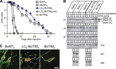
Injection of mouse gastrocnemius muscle with indicated TDmax (Table 1) of LCE-BoTIMA within 24 h induced a full paralysis (DAS = 4) that lasted for ∼30 days, similar to the effect induced by an equivalent amount of BoNTA (Fig. 5A). This long lasting action apparently arises from stabilization of the fusion protein because BoNTE alone gave a much shorter paralysis (<5 days). BoTIMA lacking LCE failed to produce any paralysis even at an over 100-fold higher dose than LCE-BoTIMA (Fig. 5A), consistent with the mutation H227Y having abolished the ability of its LCA to cleave SNAP-25 in the in vitro assay (noted above).
FIGURE 5.
Long lasting neuromuscular paralysis with persistent SNAP-25 cleavage and sprouting of motor nerve terminals induced by LCE-BoTIMA, unlike the short-lived effect of BoNTE. A, right gastrocnemius muscles of mice were injected with the TDmax of each toxin (Table 1), and the extent of muscle weakening was determined (means ± S.E., n = 10) over time by monitoring DAS (22, 26). B, SNAP-25 cleavage after different times in nerve muscle samples from mice injected with BoNTA, BoNTE, LCE-BoTIMA, BoTIMA, or saline. C, LCE-BoTIMA and BoNTA caused nerve sprouting (arrowheads) from original motor terminals at day 14 after injection into mice, unlike BoTIMA or BoNTE (data not shown). Green, staining of neurofilament 200; red, postsynaptic labeling of acetylcholine receptors by α-bungarotoxin. Note that 1 ng of BoTIMA was used in each panel. nBoNT and BoNT represent natural and recombinant toxin, respectively.
LCE-BoTIMA Reverts to a Short-lived Intraneuronally Acting Enzyme Following Alanine Substitution of the Dileucine
If LCE-BoTIMA really did gain longevity from the stabilizing influence of protease-inactive LCA, mutating its two leucines should also cause a loss of this acquired property. Therefore, a LCE-BoTIMA(AA) mutant construct was engineered (Fig. 3B). Mutation of LL to AA in LCE-BoTIMA protein did not reduce the expression or change the purification, but not surprisingly, the resultant mutant exhibited a dramatic decrease in specific toxicity (Table 1), like that described previously for BoNTA(AA). Nevertheless, the duration of action of LCE-BoTIMA(AA) injected into the leg muscle was shortened to a lifetime similar to that observed for BoNTE (Fig. 5A), affirming that the dileucine is as critical for the persistent action of LCE-BoTIMA as it is for BoNTA.
To establish that the prolonged neuroparalysis relates to longevity in the motor nerve endings of the “specially delivered and stabilized” LCE, the presence of SNAP-25 cleaved by three toxins was monitored by Western blotting at various times after injection (Fig. 5B). Cleavage of SNAP-25 by each was noted in the injected limb (with only traces in the noninjected leg) within 1 day, coinciding with the onset of the inability to spread the toes (DAS = 4). In contrast to the product of the long lasting BoNTA, SNAP-25E could not be detected on day 7 or later (Fig. 5B), consistent with the DAS recovery within 5 days of BoNTE-injected muscles (Fig. 5A). On the other hand, SNAP-25E produced by LCE-BoTIMA was visualized up to day 28 (Fig. 5B). Therefore, the extended activity of LCE-BoTIMA is due to stabilization of the LCE protease by BoTIMA within the nerve terminals. Furthermore, synapse remodeling in vivo (Fig. 5C) resulted from the unprecedented persistent production of SNAP-25E at motor endings after exposure to LCE-BoTIMA. New sprouts emerged from the original nerve terminal and projected thin branches (see arrowheads), similar to those observed in end plates paralyzed by BoNTA (Fig. 5C); the aforementioned sprouting could only have been induced by the prolonged generation of SNAP-25E by LCE-BoTIMA, because BoTIMA (even at a 100-fold larger dose) (Fig. 5C) or BoNTE (data not shown; see Ref. 15) failed to induce any sprouts. This provides corroborative evidence for the ability of BoTIMA to substantially extend the normal short period of paralysis by LCE.
LCE-BoTIMA Blocks the Release of a Pain-mediating Peptide from Sensory Neurons
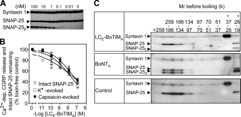
The effects of LCE-BoTIMA were examined in rat cultured TGNs. Because capsaicin activates TRPV1, mainly expressed on primary afferent sensory C-fibers, the capability of this new toxin to block the release of pain mediators evoked by capsaicin should highlight its anti-nociceptive potential. CGRP exocytosis elicited by capsaicin requires residues 181–197 of SNAP-25 but not the nine C-terminal amino acids removed by BoNTA (5). Thus, it was possible to demonstrate the ability of BoTIMA to retarget LCE into cultured sensory neurons. Incubation of TGNs with LCE-BoTIMA gave a dose-dependent cleavage of SNAP-25 (Fig. 6, A and B) and accordingly blocked the release of CGRP evoked by K+ or capsaicin (Fig. 6B). Consistent with our previous data on chimera EA (5), the SNAP-25E produced proved unable to form SDS-resistant stable SNARE complexes (Fig. 6C), which underlies the blockade of CGRP release even when triggered by capsaicin.
FIGURE 6.
Inhibition of CGRP release from sensory neurons by LCE-BoTIMA: alteration of SNARE complex formation. A and B, rat TGNs cultured for 6 days were exposed to LCE-BoTIMA overnight at 37 °C; then release of CGRP was assayed over 30 min (24). The cells were solubilized in SDS sample buffer, and equal volumes were subjected to SDS-PAGE and Western blotting; the percentage of substrate remaining intact was calculated. A, immunoblot showing the cleavage by toxin of SNAP-25 but not syntaxin 1. B, dose-response curves for SNAP-25 cleavage and toxin-induced blockade of CGRP release evoked by 60 mm K+ or 1 μm capsaicin. All of the values are the means ± S.E. (n ≥ 3); in some cases, the error bars are encompassed by the symbols. C, after overnight incubation of TGNs with 100 nm toxin at 37 °C, the cells were stimulated with 1 μm capsaicin for 30 min before solubilization in SDS sample buffer without boiling and SDS-PAGE. Gel sections containing the proteins, separated according to their Mr or that of complexes as indicated, were extracted by boiling in SDS buffer before a second electrophoresis and Western blotting using antibodies as before (5, 25). Note that LCE-BoTIMA-truncated SNAP-25 was absent from the SDS-resistant SNARE complexes, unlike intact and BoNTA-cleaved SNAP-25. *, samples were diluted 5-fold before running in these lanes.
Inhibition of Capsaicin-induced Nociceptive Behavior in Rats
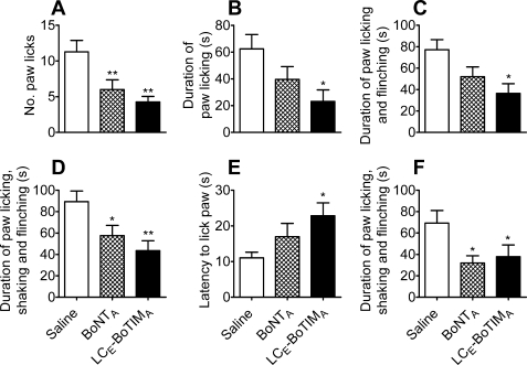
The putative therapeutic value of LCE-BoTIMA was assessed from its effects on the nociceptive behavior evoked in rats by the TRPV1 agonist, capsaicin, a validated model of hypersensitivity/algesia (28, 29). LCE-BoTIMA and, for comparison, BoNTA or saline were injected into the plantar surface of the right hind paw 4 days before administering capsaicin into the same site. Local muscle activities were unaffected by the toxins at the injected dose. Capsaicin application to saline-preinjected control animals immediately evoked nociceptive behavior that was characterized by licking, shaking, and flinching of the paw. This pattern peaked at 1–3 min post-capsaicin and lasted not more than 10 min. Scoring, analysis, and comparison of the number of right hind paw licks over 0–10 min in the rats injected with LCE-BoTIMA (n = 8) or BoNTA (n = 8) revealed a significant reduction compared with vehicle-treated animals (n = 7) (Fig. 7A). The duration of licking, shaking, and flinching was also decreased significantly by both toxins (Fig. 7D), with LCE-BoTIMA causing a larger change than BoNTA at the dose chosen. It is noteworthy that the animals given LCE-BoTIMA, but not those injected with BoNTA, displayed significantly shorter duration of right hind paw licking or licking and flinching (Fig. 7, B and C). Measuring thermal withdrawal latencies of the hind paw in the hot plate test showed that the latency to lick the right hind paw was increased significantly by LCE-BoTIMA but not BoNTA (Fig. 7E). Thus, this new toxin, engineered to acquire multiple synergistic activities, seems more efficacious than BoNTA in attenuating capsaicin-induced nociceptive behavior at the concentration used, with the restricted number of animals permitted for experimentation under our license. After 18 days, the same rats received a second challenge with capsaicin; nociceptive behavior analysis established that LCE-BoTIMA still shortened the duration of right hind paw licking, shaking, and flinching (Fig. 7F) with a similar effect as seen in BoNTA-injected rats.
FIGURE 7.
Effects of LCE-BoTIMA and BoNTA (WT) on capsaicin-evoked nociceptive behavior in rats. The vehicle or the test toxins indicated were administered by intraplantar injection into the right hind paw 4 days (A–E) or 18 days (F) before measuring nociceptive behavior (A–D and F) and hot plate latency (54.5 ± 1 °C, cut-off time 40 s) (E) at 0–10 or 50 min, respectively, following capsaicin injection into the right hind paw (see “Experimental Procedures”). The data are the means ± S.E. (n = 7–8). *, p < 0.05; **, p < 0.01 versus vehicle-pretreated group using one-way analysis of variance and post-hoc Student-Newman-Keul's test.
DISCUSSION
Mutation of a Dileucine in the Protease LC of BoNTA Dramatically Shortens Its Duration of Action and Lowers Systemic Neurotoxicity without Reducing Neuromuscular Paralysis in Vitro
The extreme longevity of BoNTA is key to its successful clinical exploitation, prompting investigations on the molecular basis for this remarkable feature. It has been suggested that membrane interaction might be an important factor; this prediction was based on GFP-LCA overexpressed in neuroendocrine cells preferentially associating with the plasmalemma, whereas a GFP fusion with the short acting LCE distributes throughout the cytosol (Ref. 20; see also Addendum). Although deletion analysis and site-directed mutagenesis implicated eight N-terminal residues and a C-terminal dileucine motif in the interaction of LCA with the plasmalemma, persistent expression of the transgene precluded any correlation with stability of the enzyme in the in vitro model used. To circumvent this limitation, the present study expressed a His6-tagged BoNTA in E. coli as a low activity pro-enzyme that could be purified rapidly and with high yield by a simple two-step chromatographic protocol. Importantly, a fully functional BoNTA was obtained upon controlled nicking of the pro-form by thrombin (21), at a susceptible site introduced into an exposed loop between two disulfide-bridged cysteines, as shown by its lethality in mice being equivalent to that of BoNTA extracted from C. botulinum. Availability of recombinant BoNTA allowed longevity studies in vivo, as well as mutational analysis to be performed with appropriate evaluation of known amounts of BoNTA in physiologically relevant systems. Our results highlight that removing six (or seven) residues from the N terminus of BoNTA lowers the expression level and specific lethality. Nevertheless, these residues are not required for maintaining the longevity of enzymic activity that induces neuromuscular paralysis. On the other hand, a double point mutation in a C-terminal dileucine motif not only reduced specific lethality but also significantly shortened its neuroparalytic effect in vivo (Fig. 2E), indicating that these residues are critical for a persistent action. Likewise, the reappearance of intact SNAP-25 occurred much more quickly in cultured CGNs exposed to BoNTA (AA) rather than WT. Clearly, this dileucine found in BoNTA, but not other serotypes, plays very important roles in stabilizing the LCA protease (also see later). Similar dileucine motifs ((D/E)XXXL(L/I)) are present in many synaptic membrane proteins (20) and are thought to be involved in their sorting and retention during membrane cycling at the synapse, possibly via interaction with adaptor protein complex 2 (30). Consistent with this notion, mutation of these leucines in GFP-LCA resulted in less association with the plasma membrane, compared with WT control, upon overexpression in neuroendocrine cells (20). An additional or alternative possibility is that the dileucine somehow protects the LCA from degradation via proteases or the ubiquitination pathway (31, 32).
Fusion of LCE to BoTIMA Extends Its Duration of Action and Provides a Means for Delivering a Stabilized E Protease into Neurons
Curiously, another long lasting neurotoxin BoNTC1 lacks a dileucine motif in its LC, and GFP-LCC1 does not associate with the plasma membrane when expressed in PC-12 cells (20); this indicates that another unknown mechanism must underlie the longevity of this serotype. Therefore, to confirm the stabilizing influence of the LCA dileucine, a short sequence encompassing this region was transferred from the latter into the LC of short acting BoNTE. Unfortunately, the resultant fusion protein could not be expressed. As an alternative strategy, the entire LCE was fused to a proteolytically inactive mutant of BoNTA (BoTIMA) to create the composite chimera: LCE-BoTIMA. Remarkably, its biological activities were comparable with those of natural BoNTA and WT, indicating that the LCE is effectively translocated into the cytosol through a membrane pore created by BoTIMA. Application of the elegant and sensitive measurement of single channels formed in membranes by BoNTs (33, 34) should yield insights into how two linked LCs can be translocated via the pore-forming domain of the N-terminal half of HCA. Consistent with our observation, other studies have shown that BoNTD can translocate a completely unrelated enzyme fused to its LC, dihydrofolate reductase, albeit with a significant reduction in its catalytic activity (35).
Importantly, injection of LCE-BoTIMA into the hind leg of mice caused a long term neuromuscular paralysis equivalent in duration to that of BoNTA (>30 days) and thus was associated with the persistent presence of an E-type cleavage product of SNAP-25 in nerves innervating the immobilized muscles. In contrast, BoTIMA failed to induce any functional block, despite injection of much greater amounts of protein, with no evidence of SNAP-25 proteolysis. Also, the TDmax of BoNTE caused a short-lived paralysis (< 5 days) that correlated with the rapid disappearance of E-cleaved SNAP-25. Clearly, the protease activity of LCE is stabilized by fusion to BoTIMA.
Prolonged Paralysis by LCE-BoTIMA Induces Nerve Terminal Remodeling
Injecting BoNTA into skeletal muscle induces extensive nerve terminal sprouts that grow until synaptic activity is restored to the parent terminal, after which they retract back until only the original motor terminal remains (15). In contrast, nerve endings treated with BoNTE recover before any sprouting is observed (15). Notably, long term muscle inactivation by LCE-BoTIMA led to the appearance of terminal sprouts at day 14 similar in length and complexity as observed in end plates paralyzed by BoNTA (Fig. 5C). Thus, this synapse remodeling in vivo is a response to prolonged neuromuscular paralysis, irrespective of the site at which SNAP-25 has been truncated or, it can be deduced, any subtle differences between the neurophysiological outcomes of treatment by A or E.
The Dileucine in BoTIMA Is Essential for Conferring Stability onto LCE
The importance of the dileucine in BoTIMA for the stabilization of fused LCE was unequivocally demonstrated by the loss of the acquired protease durability upon substituting these two residues with alanines; consequently, neuromuscular paralysis reverted to a short duration (7 days) only slightly longer than that observed with BoNTE (<5 days). Presumably, the AA mutation endowed an increased susceptibility to tissue proteases or ubiquitination (as suggested above) culminating in accelerated inactivation; such an outcome could also underlie the greatly reduced neurotoxicity of LCE-BoTIMA following mutation of the dileucine. What can be deduced with more certainty is that N-terminal degradation is not solely responsible for the fast decay of BoNTE activity because this region is not altered in either LCE-BoTIMA or LCE-BoTIMA(AA). Instead, the dileucine may protect LCA from C-terminal degradation, and fusion to BoTIMA could similarly prevent such processing at the C terminus of LCE.
Whatever the reasons for the induced stability of LCE by BoTIMA, the advantageous outcome is a novel composite BoNT chimera with properties that combine the most useful features of BoNT A and E, namely, (a) the persistence of A being a dominant trait that can be transferred to another protease and (b) the production by E of a more severely disabled form of SNAP-25 than that produced by A. The physiological consequence of the latter is a more severe block by E of neurotransmission at the neuromuscular junction (15) and of pain mediator release from sensory neurons (5) in vitro.
LCE-BoTIMA Has Anti-nociceptive Potential
LCE-BoTIMA offers the potential to be more effective in clinical applications where BoNTA only provides partial pain relief or in just a subset of patients (such as with headache (36)) or where BoNTE has been found to be more effective than A but gives only short term therapy (such as with epilepsy (37)). To this end, further experiments were performed in a well characterized and widely employed animal model that involves activation by capsaicin of TRPV1 on C-fibers, a prime target for chronic pain therapy (38). The induction by capsaicin of hyperexcitability and excessive secretion of pain and inflammatory mediators, mainly CGRP and substance P in the nervous system, as well altered sensitivity to mechanical and thermal stimuli, are features shared with some chronic pain states (39). Thus, the ability of the newly engineered fusion LCE-BoTIMA to increase the latency of the capsaicin-injected hind paw to respond to noxious heat and to inhibit the evoked nociceptive behavior highlights scope for analgesic applications. This outcome accords with the far superior performance of LCE-BoTIMA (Fig. 6B) relative to BoNTA (24) in blocking TRPV1-mediated exocytosis of CGRP from TGNs.
Supplementary Material
Acknowledgments
We thank Dr. Astrid Sasse and Liam Ryan for purifying some of the recombinant proteins and performing protease assays and Catherine Hagedorn for help with the animal experiments. Dr. L. Smith kindly provided the antibodies specific for the C and N termini of BoNTA.
Addendum
Following the date of this submission, a paper was published (Tsai, Y. C., Maditz, R., Kuo, C. L., Fishman, P. S., Shoemaker, C. B., Oyler, G. A., and Weissman, A. M. (2010) Proc. Natl. Acad. Sci. U.S.A. 107, 16554–16559) reporting that YFP-LCE and RFP-LCA show similar distribution patterns when expressed in N18 neuroblastoma cells, but LCE is degraded more rapidly than LCA via ubiquitin-dependent pathways. Thus, our demonstrated influence of L428L429 in LCA for maintaining the longevity of BoNTA or LCE-BoTIMA may be attributable to protection from this degradation pathway.
This work was supported in part by a research professorship award from Science Foundation Ireland (to J. O. D.) and a grant to D. P. F., plus a studentship to W.M.O. from the Irish Research Council for Science, Engineering and Technology, and the Programme for Research in Third-Level Institutions Cycle four (PRTLI 4) Grant from the Irish Higher Education Authority under the neuroscience section of “Target-driven Therapeutics and Theranostics.”

The on-line version of this article (available at http://www.jbc.org) contains supplemental Fig. S1.
- BoNT
- botulinum neurotoxin
- BoTIMA
- BoNTA enzymically inactive mutant
- CGN
- cerebellar granule neuron
- CGRP
- calcitonin gene-related peptide
- DAS
- digit abduction score
- DC
- dichain
- HC
- heavy chain
- HN
- N-terminal half of HC
- LC
- light chain
- SC
- single chain
- SNARE
- soluble N-ethylmaleimide-sensitive factor attachment protein receptor
- SNAP-25
- synaptosomal-associated protein of 25 kDa
- TGN
- trigeminal ganglionic neuron
- TRPV1
- transient receptor potential vanilloid type 1
- TRITC
- tetramethylrhodamine isothiocyanate.
REFERENCES
- 1. Dolly J. O., Lawrence G. W., Meng J., Wang J., Ovsepian S. V. (2009) Curr. Opin. Pharmacol. 9, 326–335 [DOI] [PubMed] [Google Scholar]
- 2. Dong M., Yeh F., Tepp W. H., Dean C., Johnson E. A., Janz R., Chapman E. R. (2006) Science 312, 592–596 [DOI] [PubMed] [Google Scholar]
- 3. Mahrhold S., Rummel A., Bigalke H., Davletov B., Binz T. (2006) FEBS Lett. 580, 2011–2014 [DOI] [PubMed] [Google Scholar]
- 4. Dong M., Liu H., Tepp W. H., Johnson E. A., Janz R., Chapman E. R. (2008) Mol. Biol. Cell 19, 5226–5237 [DOI] [PMC free article] [PubMed] [Google Scholar]
- 5. Meng J., Ovsepian S. V., Wang J., Pickering M., Sasse A., Aoki K. R., Lawrence G. W., Dolly J. O. (2009) J. Neurosci. 29, 4981–4992 [DOI] [PMC free article] [PubMed] [Google Scholar]
- 6. Dolly J. O., Black J., Williams R. S., Melling J. (1984) Nature 307, 457–460 [DOI] [PubMed] [Google Scholar]
- 7. Black J. D., Dolly J. O. (1986) J. Cell Biol. 103, 535–544 [DOI] [PMC free article] [PubMed] [Google Scholar]
- 8. Dolly J. O., Lawrence G. (2007) in Clinical Uses of Botulinum Toxins (Ward A. B., Barnes M. B. eds) pp. 9–26, Cambridge University Press, Cambridge [Google Scholar]
- 9. Keller J. E., Neale E. A., Oyler G., Adler M. (1999) FEBS Lett. 456, 137–142 [DOI] [PubMed] [Google Scholar]
- 10. Adler M., Keller J. E., Sheridan R. E., Deshpande S. S. (2001) Toxicon 39, 233–243 [DOI] [PubMed] [Google Scholar]
- 11. Ward A. B., Barnes M. P. (2007) Clinical Uses of Botulinum Toxins, pp. 1–384, Cambridge University Press, Cambridge [Google Scholar]
- 12. Dodick D. W., Turkel C. C., DeGryse R. E., Aurora S. K., Silberstein S. D., Lipton R. B., Diener H. C., Brin M. F. (2010) Headache 50, 921–936 [DOI] [PubMed] [Google Scholar]
- 13. Cull-Candy S. G., Lundh H., Thesleff S. (1976) J. Physiol. 260, 177–203 [DOI] [PMC free article] [PubMed] [Google Scholar]
- 14. Molgó J., Lundh H., Thesleff S. (1980) Eur. J. Pharmacol. 61, 25–34 [DOI] [PubMed] [Google Scholar]
- 15. Meunier F. A., Lisk G., Sesardic D., Dolly J. O. (2003) Mol. Cell Neurosci. 22, 454–466 [DOI] [PubMed] [Google Scholar]
- 16. Lawrence G., Wang J., Chion C. K., Aoki K. R., Dolly J. O. (2007) J. Pharmacol. Exp. Ther. 320, 410–418 [DOI] [PubMed] [Google Scholar]
- 17. Sloop R. R., Cole B. A., Escutin R. O. (1997) Neurology 49, 189–194 [DOI] [PubMed] [Google Scholar]
- 18. Eleopra R., Tugnoli V., Rossetto O., De Grandis D., Montecucco C. (1998) Neurosci. Lett. 256, 135–138 [DOI] [PubMed] [Google Scholar]
- 19. Foran P. G., Mohammed N., Lisk G. O., Nagwaney S., Lawrence G. W., Johnson E., Smith L., Aoki K. R., Dolly J. O. (2003) J. Biol. Chem. 278, 1363–1371 [DOI] [PubMed] [Google Scholar]
- 20. Fernández-Salas E., Steward L. E., Ho H., Garay P. E., Sun S. W., Gilmore M. A., Ordas J. V., Wang J., Francis J., Aoki K. R. (2004) Proc. Natl. Acad. Sci. U.S.A. 101, 3208–3213 [DOI] [PMC free article] [PubMed] [Google Scholar]
- 21. Rummel A., Karnath T., Henke T., Bigalke H., Binz T. (2004) J. Biol. Chem. 279, 30865–30870 [DOI] [PubMed] [Google Scholar]
- 22. Wang J., Meng J., Lawrence G. W., Zurawski T. H., Sasse A., Bodeker M. O., Gilmore M. A., Fernández-Salas E., Francis J., Steward L. E., Aoki K. R., Dolly J. O. (2008) J. Biol. Chem. 283, 16993–17002 [DOI] [PubMed] [Google Scholar]
- 23. Studier F. W. (2005) Protein Expression Purif. 41, 207–234 [DOI] [PubMed] [Google Scholar]
- 24. Meng J., Wang J., Lawrence G., Dolly J. O. (2007) J. Cell Sci. 120, 2864–2874 [DOI] [PubMed] [Google Scholar]
- 25. Lawrence G. W., Dolly J. O. (2002) J. Cell Sci. 115, 2791–2800 [DOI] [PubMed] [Google Scholar]
- 26. Aoki K. R. (2001) Toxicon 39, 1815–1820 [DOI] [PubMed] [Google Scholar]
- 27. Maisey E. A., Wadsworth J. D., Poulain B., Shone C. C., Melling J., Gibbs P., Tauc L., Dolly J. O. (1988) Eur. J. Biochem. 177, 683–691 [DOI] [PubMed] [Google Scholar]
- 28. Honda K., Kitagawa J., Sessle B. J., Kondo M., Tsuboi Y., Yonehara Y., Iwata K. (2008) Mol. Pain 4, 59 [DOI] [PMC free article] [PubMed] [Google Scholar]
- 29. Kim H. Y., Wang J., Lee I., Kim H. K., Chung K., Chung J. M. (2009) Pain 145, 332–340 [DOI] [PMC free article] [PubMed] [Google Scholar]
- 30. Kim S. H., Ryan T. A. (2009) J. Biol. Chem. 284, 32803–32812 [DOI] [PMC free article] [PubMed] [Google Scholar]
- 31. Stoka V., Turk B., Schendel S. L., Kim T. H., Cirman T., Snipas S. J., Ellerby L. M., Bredesen D., Freeze H., Abrahamson M., Bromme D., Krajewski S., Reed J. C., Yin X. M., Turk V., Salvesen G. S. (2001) J. Biol. Chem. 276, 3149–3157 [DOI] [PubMed] [Google Scholar]
- 32. Shi X., Curran J. E., Liao Z., Gordon R. K. (2009) J. Cell. Biochem. 108, 660–667 [DOI] [PubMed] [Google Scholar]
- 33. Fischer A., Montal M. (2007) Proc. Natl. Acad. Sci. U.S.A. 104, 10447–10452 [DOI] [PMC free article] [PubMed] [Google Scholar]
- 34. Montal M. (2010) Annu. Rev. Biochem. 79, 591–617 [DOI] [PubMed] [Google Scholar]
- 35. Bade S., Rummel A., Reisinger C., Karnath T., Ahnert-Hilger G., Bigalke H., Binz T. (2004) J. Neurochem. 91, 1461–1472 [DOI] [PubMed] [Google Scholar]
- 36. Jakubowski M., McAllister P. J., Bajwa Z. H., Ward T. N., Smith P., Burstein R. (2006) Pain 125, 286–295 [DOI] [PMC free article] [PubMed] [Google Scholar]
- 37. Costantin L., Bozzi Y., Richichi C., Viegi A., Antonucci F., Funicello M., Gobbi M., Mennini T., Rossetto O., Montecucco C., Maffei L., Vezzani A., Caleo M. (2005) J. Neurosci. 25, 1943–1951 [DOI] [PMC free article] [PubMed] [Google Scholar]
- 38. Christoph T., Gillen C., Mika J., Grünweller A., Schäfer M. K., Schiene K., Frank R., Jostock R., Bahrenberg G., Weihe E., Erdmann V. A., Kurreck J. (2007) Neurochem. Int. 50, 281–290 [DOI] [PubMed] [Google Scholar]
- 39. Witting N., Svensson P., Gottrup H., Arendt-Nielsen L., Jensen T. S. (2000) Pain 84, 407–412 [DOI] [PubMed] [Google Scholar]
Associated Data
This section collects any data citations, data availability statements, or supplementary materials included in this article.


