Abstract
In Saccharomyces cerevisiae, Ash1p is a specific repressor of transcription that localizes exclusively to daughter cell nuclei through the asymmetric localization of ASH1 mRNA. This localization requires four cis-acting localization elements located in the ASH1 mRNA, five trans-acting factors, one of which is a myosin, and the actin cytoskeleton. The RNA-binding proteins that interact with these cis-elements remained to be identified. Starting with the 3′ most localization element of ASH1 mRNA in the three-hybrid assay, element E3, we isolated a clone corresponding to the C-terminus of She3p. We also found that She3p and She2p interact, and this interaction is essential for the binding of She3p with element E3 in vivo. Moreover, She2p was observed to bind the E3 RNA directly in vitro and each of the ASH1 cis-acting localization elements requires She2p for their localization function. By tethering a She3p–MS2 fusion protein to a reporter RNA containing MS2 binding sites, we observed that She2p is dispensable for She3p–MS2-dependent RNA localization.
Keywords: ASH1/mRNA localization/RNA-binding proteins/Saccharomyces cerevisiae/three-hybrid assay
Introduction
The process of cellular differentiation during development of multicellular organisms requires the asymmetric sorting of cell fate determinants between sister cells. One mechanism used for the asymmetric segregation of cell fate determinants is to localize specifically the mRNAs encoding the determinants to one of the sister cells. The currently emerging mechanism for the localization of RNA involves the presence of specific cis-acting localization elements within the localized mRNA. The cis-acting elements are recognized by RNA-binding proteins that physically interact with molecular motors that transport the ribonucleoprotein complex along cytoskeletal filaments to the site of localization (Nasmyth and Jansen, 1997; Oleynikov and Singer, 1998; Schnorrer et al., 2000).
The asymmetric sorting of Ash1p in the yeast Saccharomyces cerevisiae is a model system for studying the asymmetric segregation of cell fate determinants through RNA localization. Ash1p is a cell-type specific transcriptional repressor of the HO endonuclease (Bobola et al., 1996; Sil and Herskowitz, 1996). At the end of mitosis, Ash1p is found in the nucleus of daughter cells where it represses HO transcription and ultimately prevents mating-type switching in these cells (Bobola et al., 1996; Sil and Herskowitz, 1996). This transcriptional regulation of HO expression restricts mating-type switching to mother cells (Bobola et al., 1996; Sil and Herskowitz, 1996). The asymmetric distribution of Ash1p results from the localization of ASH1 mRNA to the distal tip of daughter cells during anaphase (Long et al., 1997; Takizawa et al., 1997).
ASH1 mRNA contains four cis-acting localization elements: E1, E2A, E2B and E3 (Chartrand et al., 1999; Gonzalez et al., 1999). Each of these elements is sufficient to localize a heterologous reporter mRNA to daughter cells. The E3 element is located predominantly in the 3′ untranslated region (3′-UTR) whereas the remaining three elements are found in the ASH1 open reading frame (ORF). The E1 and E3 elements are predicted to fold into structures containing stem–loops. Mutations predicted to disrupt the stem–loop structure of either element prevent RNA localization activity, while compensatory mutations that re-establish the secondary structure restore RNA localization activity (Chartrand et al., 1999; Gonzalez et al., 1999). For the E3 element, specific RNA sequences have not been demonstrated to be important for RNA localization activity; rather, RNA localization activity for E3 is strictly dependent on secondary structure (Chartrand et al., 1999; Gonzalez et al., 1999). In contrast, in E1, specific RNA sequences important for RNA localization activity have been identified (Chartrand et al., 1999).
Five genes, SHE1–5, originally found to be essential for proper sorting of Ash1p, are also required for ASH1 mRNA localization (Jansen et al., 1996; Long et al., 1997; Takizawa et al., 1997). Several observations indicate that actomyosin-based transport is important for ASH1 mRNA localization (Long et al., 1997; Takizawa et al., 1997). A functional actin cytoskeleton was found to be essential for ASH1 mRNA localization and SHE5/BNI1 is required for proper organization of the actin cytoskeleton (Kohno et al., 1996; Evangelista et al., 1997). SHE4 is also hypothesized to be required for proper organization of the actin cytoskeleton (Wendland et al., 1996). Furthermore, SHE1/MYO4 encodes a type V myosin, which co-localizes with ASH1 mRNA at the tip of daughter cells (Haarer et al., 1994; Münchow et al., 1999; Takizawa and Vale, 2000). Myo4p was also found to be associated with ASH1 mRNA in co-immunoprecipitation experiments (Münchow et al., 1999; Takizawa and Vale, 2000). A live cell assay has shown particles containing Myo4p and ASH1 mRNA rapidly moving from mother cells to daughter cells, suggesting a direct role for Myo4p in ASH1 mRNA transport (Bertrand et al., 1998; Beach et al., 1999).
While the role of myosin in the localization of ASH1 mRNA has apparently been determined, a direct interaction between ASH1 mRNA and Myo4p has not been established. On the contrary, new studies suggest that the association of Myo4p with ASH1 mRNA requires She2p and She3p (Münchow et al., 1999; Takizawa and Vale, 2000). By two-hybrid analysis, Myo4p and She3p have been shown to interact (Münchow et al., 1999). ASH1 mRNA can be co-immunoprecipitated in a complex containing Myo4p, She2p and She3p. Co-immunoprecipitation of ASH1 mRNA with Myo4p and She3p is dependent on She2p (Münchow et al., 1999; Takizawa and Vale, 2000); however, these experiments provide no definite function for either She2p or She3p and do not address what additional unknown proteins may be co-immunoprecipitating with the complex.
Using molecular genetics and biochemical assays, we demonstrate directly that She2p is an RNA-binding protein that interacts specifically with individual ASH1 mRNA localization elements and functions to recruit the Myo4p–She3p complex to ASH1 mRNA.
Results
Interaction of She3p with an ASH1 mRNA localization element
Our approach was to identify specific RNA-binding proteins for ASH1 cis-acting localization elements and to use these RNA-binding proteins to characterize protein–protein interactions required for ASH1 mRNA localization. We initiated an in vivo approach for the identification of RNA-binding proteins using the three-hybrid assay (SenGupta et al., 1996). In this system, formation of an RNA–protein complex in yeast results in transcriptional activation of the reporter genes HIS3 and lacZ.
Plasmid pRL80, expressing a fusion RNA containing two copies of the MS2 coat protein stem–loop motif and one copy of the 127 nucleotide (nt) ASH1 3′-UTR localization element (E3), was transformed into the three-hybrid host yeast strain YBZ-1. A yeast cDNA library fused to the Gal4 transcriptional activation domain (Gal4p-AD) was screened for proteins that could interact with the ASH1 E3 localization element. From 500 000 total yeast transformants, we isolated one clone corresponding to the C-terminus (amino acids 236–425) of She3p. SHE3 encodes a protein of 47 kDa with no significant homology to other known proteins (Jansen et al., 1996). In subsequent three-hybrid screens, She3p was repeatedly isolated, but none of these clones contained the entire She3p ORF.
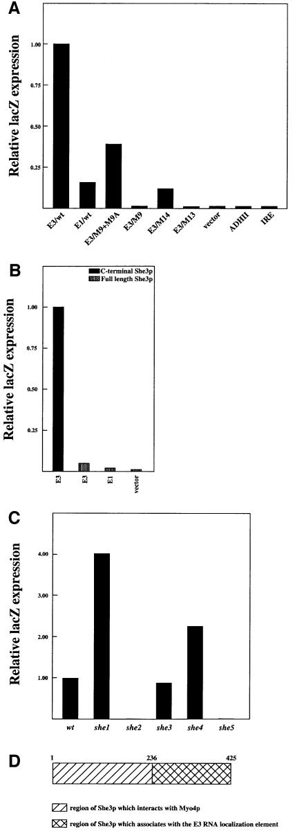
The binding specificity of the SHE3 C-terminal clone was investigated by three-hybrid analysis using mutants in the E3 element (M9, M13, M14) that are predicted to disrupt the secondary structure of element E3 and are defective for RNA localization (Chartrand et al., 1999). We observed in the three-hybrid assay that She3p C-terminal-dependent β-galactosidase expression was lower in mutants M9, M13 and M14 compared with the wild-type E3 element (Figure 1A). In contrast, an E3 double mutant (M9 + M9A), predicted to restore the wild-type stem–loop structure and capable of RNA localization, resulted in higher She3p C-terminal-dependent β-galactosidase expression (Figure 1A) compared with the M9, M13 and M14 mutations (Chartrand et al., 1999). When other yeast 3′-UTRs (ADHII) or double-stranded RNAs, like the iron-responsive element (IRE) (Klausner et al., 1993; Melefors and Hentze, 1993), were investigated for She3p C-terminal-dependent β-galactosidase expression, we observed expression levels similar to the vector control (Figure 1A). These results indicate that the She3p interaction is specific for the E3 localization element. These data suggest that the interaction of the C-terminus of She3p with element E3 is apparently dependent on a specific secondary structure of the E3 element. The sum of this work demonstrates a correlation between mRNA localization activity of E3 and the interaction of She3p with E3.
Fig. 1. By three-hybrid analysis, the C-terminal section of She3p preferentially interacts with the E3 ASH1 RNA localization element. (A) Strain YBZ-1 transformed with a plasmid expressing Gal4p-AD fused to the C-terminus of She3p along with a plasmid expressing one of the indicated MS2 fusion RNAs. Liquid cultures were grown for each strain, and the cells harvested by centrifugation. The cells were permeabilized and β-galactosidase expression levels were determined. β-galactosidase expression levels were normalized to cells expressing the MS2–E3 fusion RNA. (B) Strain YBZ-1 transformed with either a plasmid expressing the C-terminus of She3p fused to Gal4p-AD or full-length She3p fused to Gal4p-AD along with plasmids expressing the indicated MS2 fusion RNAs. Liquid cultures were grown for each strain and used for β-galactosidase assays. β-galactosidase expression levels were normalized to cells expressing the She3p C-terminus fused to Gal4p-AD and the MS2–E3 fusion RNA. (C) Yeast strain YBZ-1 deleted of myo4, she2, she3, she4 or she5. The strains were subsequently transformed with a plasmid expressing full-length She3p fused to Gal4p-AD along with a plasmid expressing the MS2–E3 fusion RNA. Transformants were grown in liquid culture and used for β-galactosidase assays. β-galactosidase levels were normalized to the wild-type strain. (D) Schematic representation of She3p. Amino acids 1–236 of She3p contain a domain required for the association with Myo4p, while amino acids 236–425 contain a domain required for the in vivo association with the E3 ASH1 RNA localization element.
The observation that She3p is required for ASH1 mRNA localization suggests that She3p should interact with each of the ASH1 cis-acting localization elements. We tested the ability of the C-terminus of She3p to interact with the E1 element by three-hybrid analysis. We observed that She3p C-terminal-dependent β-galactosidase expression was higher with element E3 than element E1 (Figure 1A). The different levels of β-galactosidase expression between E1 and E3 could result from different expression levels of the three-hybrid RNA substrates. However, data obtained previously with the three-hybrid assay indicate that the expression levels of these two fusion RNAs are comparable (data not shown). Therefore, we conclude that the association between the C-terminus of She3p and elements E1 and E3 somehow differs.
The association between the C-terminus of She3p and the various cis-acting elements may differ from full-length She3p. Therefore, we investigated the interaction of the full-length She3p with the elements E1 and E3 (Figure 1B). Full-length She3p was cloned into the appropriate three-hybrid vector and the level of β-galactosidase expression was determined for full-length She3p with either E1 or E3 fusion RNAs. We observed only a slight increase in β-galactosidase expression over the vector control (Figure 1B). Furthermore, the C-terminus of She3p displayed higher β-galactosidase expression levels with E3 compared with full-length She3p (Figure 1B). These results suggested that the N-terminus of She3p negatively affected the association between She3p and the cis-acting elements as assayed by the three-hybrid system.
One possibility could be that full-length She3p was bound to a cytoplasmic factor preventing She3p from entering the nucleus in the three-hybrid assay. Since She3p interacts with Myo4p in the two-hybrid assay (Münchow et al., 1999), this interaction could directly or indirectly affect the ability of full-length She3p to associate with the three-hybrid RNA. We tested the interaction of full-length She3p with element E3 in a myo4 strain and observed a 4-fold increase in β-galactosidase expression levels compared with the MYO4 strain (Figure 1C). This result indicates that Myo4p prevented full-length She3p from associating with the cis-acting localization elements in the three-hybrid system. Presumably, Myo4p associates with cytoplasmic actin and full-length She3p, through a domain of She3p between amino acids 1 and 235, making She3p unavailable for associating with nuclear RNA in the three-hybrid assay. In summary, She3p appears to contain at least two important domains: one responsible for interaction with Myo4p and a second independent domain required for an association with ASH1 mRNA cis-acting localization sequences (Figure 1D).
The in vivo interaction of She3p with the E3 localization sequence is dependent on She2p
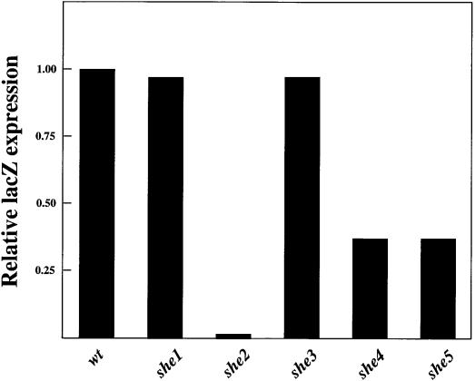
The in vivo association between ASH1 mRNA, Myo4p and She3p is dependent on She2p (Münchow et al., 1999; Takizawa and Vale, 2000). We sought to determine whether the in vivo association of ASH1 mRNA and the C-terminus of She3p was dependent on She2p or any of the other She proteins. Deletions of SHE1–5 were created in strain YBZ-1, and C-terminal She3p–E3-dependent β-galactosidase expression levels were determined (Figure 2). We observed that deletion of MYO4 or SHE3 had no significant effect on β-galactosidase expression, and deletion of SHE4 or SHE5 resulted in a modest reduction in β-galactosidase activity (Figure 2). In contrast, deletion of SHE2 resulted in a dramatic reduction in β-galactosidase expression, indicating that the in vivo association between She3p and E3 requires She2p. By western and northern blotting for the three-hybrid components we eliminated the possible trivial explanation that the abundance of the three-hybrid components is reduced in the she2 strain compared with the wild-type control (data not shown). These results suggest that She2p has a direct role in recruiting She3p to ASH1 mRNA localization sequences.
Fig. 2. She3p C-terminal-dependent lacZ expression assayed by three-hybrid analysis is dependent on She2p. Yeast strain YBZ-1 deleted of myo4, she2, she3, she4 and she5. These strains were subsequently transformed with a plasmid expressing the C-terminus She3p fused to Gal4p-AD and MS2–E3 fusion RNA. The transformants were grown in liquid culture and processed for β-galactosidase assays. β-galactosidase expression levels were normalized to the wild-type strain.
Association of She2p and She3p
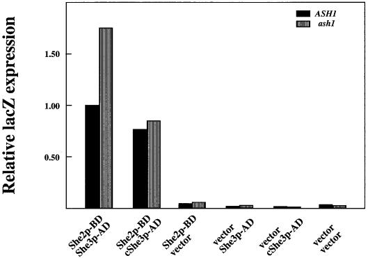
Since the in vivo association between She3p and the E3 localization element was dependent on She2p, we reasoned that She2p and She3p should also associate in vivo. We investigated the association between She2p and She3p by two-hybrid analysis (Fields and Song, 1989). She2p was fused to the Gal4 DNA-binding domain (Gal4p-BD). This She2p bait was tested against the entire ORF for She3p or the C-terminus of She3p when either was fused to Gal4p-AD. We only observed activation of lacZ expression when yeast was simultaneously transformed with She2p–Gal4p-AD and either She3p–Gal4p-BD construct (Figure 3). From these results we conclude that She2p and She3p interact either directly or indirectly, and the domain of She3p responsible for the interaction with She2p resides between amino acids 236 and 425 of She3p. This is the same region of She3p responsible for the in vivo association with the E3 localization element.
Fig. 3. By the two-hybrid assay, She2p and She3p interact. Yeast strains PJ69-4a (ASH1) and YLM445 (ash1) were transformed in combinations with the following plasmids: She2p fused to Gal4p-BD (She2p–BD), full-length She3p fused to Gal4p-AD (She3p–AD), the C-terminus of She3p fused to Gal4p-AD (cShe3p–AD), the vector corresponding to Gal4p-BD (pGBDU-c2) or the vector corresponding to Gal4p-AD (pACT2). Transformants were grown in liquid culture and processed for β-galactosidase assays. β-galactosidase expression levels were normalized to strain PJ69-4a transformed with the plasmid expressing She2p fused to Gal4p-BD and the plasmid expressing full-length She3p fused to Gal4p-AD.
The interaction between She2p and She3p could occur indirectly through ASH1 mRNA tethering the two proteins together. We deleted ASH1 from the two-hybrid strain and assayed She2p–She3p-dependent β-galactosidase expression (Figure 3). In the absence of ASH1 mRNA we measured a modest increase in β-galactosidase expression, providing evidence for a direct association between She2p and She3p.
She2p RNA-binding activity
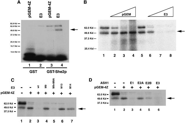
The region of She3p required for the association with ASH1 RNA localization elements was identical to that required for the association with She2p; we reasoned that She2p could be the RNA-binding protein that recruits She3p to ASH1 RNA localization elements. We tested She2p RNA-binding activity directly by expressing and purifying She2p from Escherichia coli as a glutathione S-transferase (GST) fusion protein. Purified GST–She2p was tested for RNA-binding activity by a UV cross-linking assay. When 32P-labeled E3 was incubated with recombinant GST–She2p followed by UV cross-linking, SDS–PAGE and autoradiography, we observed a product corresponding closely to the molecular weight of the GST–She2p fusion protein (49.6 kDa) (Figure 4A). We did not observe this product in the presence of 32P-labeled pGEM-4Z RNA (Figure 4A). Furthermore, the GST–She2p UV cross-linked product was specifically competed with 1000× excess unlabeled E3 and not with 1000× excess unlabeled pGEM-4Z RNA sequences (Figure 4B). We have further observed that 5000× excess pGEM-4Z RNA will not compete the She2p–E3 UV cross-linked product (data not shown).
Fig. 4. She2p RNA-binding activity as assayed by a UV cross-linking assay. (A) One nanogram of 32P-labeled E3 RNA (lanes 2 and 4) or pGEM-4Z RNA (lanes 1 and 3) was incubated with 1.0 µg of purified GST–She2p (lanes 3 and 4) or GST (lanes1 and 2) corresponding to an equivalent volume of GST–She2p used for UV cross-linking. The GST–She2p–E3 complex migrates at 49.6 kDa. The arrow indicates the position of the GST–She2p–E3 complex. (B) Specific cross-linking of GST–She2p to E3. UV cross-linking was performed with GST–She2p and 32P-labeled E3 in the absence (lanes 1 and 5) or presence of 100× (lane 2), 500× (lane 3) and 1000× (lane 4) pGEM-4Z RNA, as well as 100× (lane 6), 500× (lane 7) and 1000× (lane 8) E3 RNA. (C) Competition of GST–She2p–E3 UV cross-linked product with mutant E3 elements. GST–She2p was incubated with 32P-labeled E3 in the absence of pGEM and E3 RNA (lane 1) or 1000× pGEM RNA (lane 2), 1000× pGEM/1000× E3 (lane3), 1000× pGEM/1000× E3/M9 (lane 4), 1000× pGEM/1000× E3/M9 + M9A (lane 5), 1000× pGEM/1000× E3/M13 (lane 6) or 1000× pGEM/1000× E3/M14 (lane 7). (D) Competition of GST–She2p–E3 UV cross-linked product with ASH1 cis-acting localization elements. GST–She2p was incubated with 32P-labeled E3 in the absence of pGEM and E3 RNA (lane 1), 1000× pGEM RNA (lane 2), 1000× pGEM/1000× E1 (lane3), 1000× pGEM/1000× E2A (lane 4), 1000× pGEM/1000× E2B (lane 5) or 1000× pGEM/1000× E3 (lane 6).
The specificity of the She2p–E3 interaction was further examined using mutations in the E3 localization element. Mutations in the E3 element (M9, M13 and M14) previously shown to be defective in RNA localization did not compete the She2p–E3 UV cross-linked product, while a mutation in E3 (M9 + M9A) that supports RNA localization did compete the She2p–E3 UV cross-linked product (Figure 4C). These results demonstrate that She2p is capable of directly and specifically binding the E3 localization element.
We investigated by UV cross-linking whether She2p could interact with each of the cis-acting localization elements using recombinant GST–She2p, 32P-labeled E3 and excess unlabeled RNA for each element. We observed that 1000× excess unlabeled RNA corresponding to either ASH1 element E1 or E3 effectively competed the She2p–E3 UV cross-linked product (Figure 4D). However, a 1000-fold excess of elements E2A and E2B did not compete as effectively as E1 or E3 (Figure 4D). From these results we conclude that, in vitro, She2p is able to bind each of the cis-acting localization elements, but apparently with different affinities.
She2p is essential for the localization function of the four ASH1 mRNA cis-acting elements
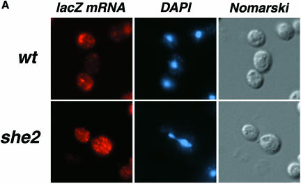
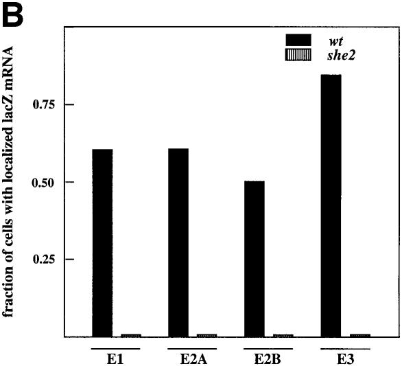
Analogous to She3p, She2p is required for the localization of full-length ASH1 mRNA (Long et al., 1997; Takizawa et al., 1997), but the requirement for She2p by each of the cis-acting localization elements remained to be determined. The She2p RNA-binding data predict that RNA localization directed by each of the cis-acting elements should be dependent on She2p. To test this prediction, a wild-type strain and a she2 strain were transformed with each of the lacZ mRNA reporter plasmids: lacZ-ASH1-E1, lacZ-ASH1-E2A, lacZ-ASH1-E2B and lacZ-ASH1-E3. The intracellular distribution of the lacZ RNA expressed from each of these plasmids was determined by FISH. For all of these reporter constructs we observed that the lacZ mRNA was localized to daughter cells in the wild-type strain and delocalized between mother and daughter cells in the she2 strain (Figure 5). From these results we conclude that the activity of each ASH1 cis-acting localization element requires She2p.
Fig. 5. RNA localization activity for each of the cis-acting localization elements is dependent on She2p. (A) Wild-type (wt) and she2 yeast strains were transformed with a plasmid containing the galactose-inducible lacZ-E3 cassette, and the cells were processed for fluorescent in situ hybridization (FISH). (B) Fraction of budding cells with localized lacZ mRNA for each of the cis-acting localization elements in wild-type and she2 cells. Cells expressing each of the reporter constructs (lacZ-E1, lacZ-E2A, lacZ-E2B and lacZ-E3) were grown in galactose-containing media and processed for in situ hybridization. LacZ RNA was categorized as localized or delocalized from cells displaying FISH signal.
She2p functions to recruit Myo4p–She3p to ASH1 RNA localization elements
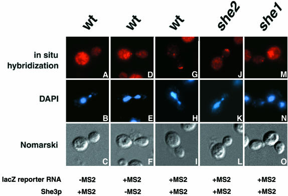
Our work, in addition to the work of others, implies that She2p’s sole function in ASH1 mRNA localization is to interface the Myo4p–She3p complex with ASH1 mRNA. If this is the only function of She2p in Myo4p–She3p-dependent RNA localization, She2p should be dispensable for RNA localization if She3p is given a high affinity RNA-binding domain. We tested this hypothesis by fusing She3p to the MS2 phage RNA-binding coat protein (Fouts et al., 1997) and monitoring the localization of a lacZ reporter RNA containing six MS2 binding sites in its 3′-UTR (Bertrand et al., 1998). In wild-type cells, we observed that lacZ mRNA was localized to the tip of daughter cells dependent on the presence of MS2 binding sites in the reporter mRNA as well as the expression of She3p–MS2 (Figure 6, compare A–C and D–F with G–I). Furthermore, localization of lacZ-MS2 by She3p–MS2 was dependent on Myo4p (Figure 6M–O). However, we observed in she2 cells that She3p–MS2 was still able to localize lacZ-MS2 RNA to daughter cells (Figure 6J–L). These results imply that the sole function of She2p in She3p-dependent RNA localization is to recruit Myo4p–She3p to cis-acting localization elements.
Fig. 6. RNA localization directed by She3p–MS2 is independent of She2p. (A–C) Representative images for in situ hybridization to lacZ mRNA, DAPI staining and Nomarski optics of wild-type cells expressing the She3p–MS2 fusion protein and the lacZ reporter RNA devoid of MS2 binding sites. (D–F) Images for wild-type cells expressing She3p and the lacZ-MS2 reporter RNA. (G–I) Images for wild-type cells expressing the She3p–MS2 fusion protein and the lacZ-MS2 reporter mRNA. Images for she2 (J–L) and she1 (M–O) cells, respectively, expressing the She3p–MS2 fusion protein and the lacZ-MS2 reporter mRNA.
Discussion
We have identified and characterized a new RNA-binding protein required for ASH1 mRNA localization in the yeast S.cerevisiae. Previously, it has been shown that Myo4p, She3p and ASH1 mRNA are associated in a complex containing She2p (Bertrand et al., 1998; Münchow et al., 1999; Takizawa and Vale, 2000); however, a definitive role for She2p and She3p in this complex remained to be determined. Here we show for the first time that She2p can directly and specifically bind ASH1 cis-acting localization elements. These results strongly suggest that She2p is the RNA-binding protein that interfaces ASH1 mRNA to She3p and the transport motor, Myo4p.
Using a segment of ASH1 mRNA (E3) shown to be sufficient for localization of mRNA to the bud in a three-hybrid assay, we found that She3p, a cytoplasmic protein required for ASH1 mRNA localization, associated with this RNA (Jansen et al., 1996; Long et al., 1997; Takizawa et al., 1997). In this in vivo assay, we observed that She3p–E3-dependent β-galactosidase expression was dependent on a functional E3 element, since mutations in E3 (M9, M13 and M14) defective for RNA localization displayed significantly lower levels of β-galactosidase expression. Interestingly, the She3p clone isolated from the three-hybrid assay contained only a C-terminal portion of She3p. Full-length She3p was not isolated in the three-hybrid assay, and when full-length She3p was tested in the assay, β-galactosidase expression was greatly reduced compared with that observed for the C-terminus of She3p. We and others (R.-P.Jansen, personal communication) have found that full-length She3p, via its N-terminal region, is retained in the cytoplasm through protein–protein interactions with Myo4p. Since the three-hybrid analysis requires that the RNA–protein complex be imported into the nucleus for transcriptional activation to occur, anchoring of full-length She3p in the cytoplasm prevents activation of reporter gene expression.
However, the three-hybrid results do not prove that She3p directly binds the E3 localization element. Using recombinant She3p in a mobility shift assay, binding of She3p with element E3 RNA was not comparable in affinity to She2p, suggesting that She3p has low, if any, intrinsic RNA-binding activity. We favor the model whereby She3p interacts in vivo with an RNA-binding protein, She2p, specific for the ASH1 mRNA localization sequences.
Like She3p, She2p is essential for the localization of ASH1 mRNA (Long et al., 1997; Takizawa et al., 1997). She2p is also known to be essential for the association of Myo4p and She3p with ASH1 mRNA (Münchow et al., 1999; Takizawa and Vale, 2000). Moreover, She2p was found to be essential for the proper formation of a green fluorescent protein (GFP)–ASH1 mRNA localization particle, the ‘locasome’ (Bertrand et al., 1998). Our results also show that the three-hybrid interaction of She3p with element E3 is dependent on She2p, and that She2p interacts with a domain in the C-terminus of She3p in the absence of ASH1 mRNA. The possibility remains that She2p and She3p could be tethered together in vivo by an unidentified mRNA. These data indicate that She2p could possess RNA-binding activity and in vivo bind ASH1 mRNA localization elements and recruit the Myo4p– She3p complex to ASH1 mRNA.
Using recombinant She2 protein in a UV cross-linking assay, we observed specific binding of She2p with element E3 RNA, indicating that She2p is an RNA-binding protein. She2p’s binding to element E3 is affected by mutations in the secondary structure of this element. Furthermore, elements E1 and E3 successfully compete the element E3 for binding to She2p, suggesting that She2p recognizes diverse RNA secondary structures.
Furthermore, we have shown that each of the cis-acting elements requires She2p for RNA localization activity. This result supports the conclusion that an identical RNA localization complex assembles on each of the cis-acting localization elements. This conclusion also implies that each of the cis-acting localization elements performs redundant functions and the presence of multiple cis-acting elements in ASH1 mRNA simply serves to increase the efficiency of ASH1 RNA localization to daughter cells.
Model for ASH1 mRNA localization
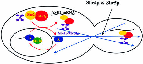
Combining this study with previous work, we propose the following model for ASH1 mRNA localization (Figure 7). ASH1 mRNA is identified in the nucleus by Loc1p, a nuclear RNA-binding protein required for ASH1 mRNA localization (R.M.Long, W.Gu, G.Gonsalvez, R.H.Singer and P.Chartrand, submitted). We propose that Loc1p’s role in the nuclear export of ASH1 mRNA is to increase the efficiency of ASH1 mRNA recognition by the cytoplasmic localization machinery. A component of the cytoplasmic localization machinery, perhaps She2p, might shuttle between the cytoplasm and nucleus to receive ASH1 mRNA from Loc1p. Following transport through the nuclear pore, ASH1 mRNA would be rapidly identified and bound by the cytoplasmic RNA localization machinery: She1p, She2p and She3p.
Fig. 7. Model for the localization of ASH1 mRNA. In the nucleus, ASH1 mRNA is identified by Loc1p in association with nucleocytoplasmic shuttling proteins. Following transport through the nuclear pores, ASH1 mRNA is rapidly assembled into a transport particle through the RNA-binding activity of She2p. The She2p–ASH1 mRNA complex associates with Myo4p–She3p to transport ASH1 mRNA to the bud tip by movement on actin cables. Once at the bud tip, ASH1 mRNA will be anchored through a mechanism that remains to be determined.
She2p’s sole function appears to be to associate ASH1 mRNA indirectly to the motor protein Myo4p, essential for the proper localization of ASH1 mRNA (Long et al., 1997; Takizawa et al., 1997) and an ASH1 mRNA–GFP particle (Bertrand et al., 1998; Beach et al., 1999). Myo4p and She3p have been found to immunoprecipitate with ASH1 mRNA. (Münchow et al., 1999; Takizawa and Vale, 2000). This ASH1 mRNA–Myo4p association requires both She2p and She3p. Moreover, Myo4p and She3p can interact in a two-hybrid interaction assay and co-immunoprecipitate (Münchow et al., 1999; Takizawa and Vale, 2000). We postulate that She2p functions to recruit the Myo4p–She3p complex to the four localization elements in the ASH1 mRNA sequence. Altogether, these results point to a role of She3p in bridging the interaction of ASH1 mRNA with the transport mechanism, specifically Myo4p. Our results support this hypothesis by providing direct evidence of RNA-dependent interactions between She2p and ASH1 mRNA. Furthermore, She2p is unnecessary for She3p-dependent RNA localization when She3p is fused to the MS2 RNA-binding protein. This observation is consistent with the hypothesis that She2p’s sole function in ASH1 mRNA localization is to engage Myo4p–She3p with ASH1 mRNA.
Finally, this RNA localization complex would transport and localize the ASH1 mRNA to the distal tip of daughter cells using polarized actin filaments. Organization of the actin cytoskeleton requires She5p and probably She4p. It remains to be determined how ASH1 mRNA is anchored at the bud tip upon reaching its destination.
Materials and methods
Growth media, yeast strains and plasmids
Yeast cells were grown in either synthetic growth media lacking the nutrients indicated or rich media (Rose et al., 1990). Yeast strains used in this study are listed in Table I. Yeast gene disruption cassettes were created by PCR amplification of the loxP-KAN-loxP construct in plasmid pUG6 and primers specific for the gene of interest (Güldener et al., 1996).
Table I. Yeast strains used in this study.
| Strain | Genotype | Source |
|---|---|---|
| YBZ-1 | Mata, ura3-52, leu2-3,112, his3-200, trp1-1, ade2, LYS2::(LexAop)-lacZ, LexA-MS2 -MS2 coat (N55K) | M.Wickens |
| PJ69-4a | Mata, trp1-901, leu2-3,112, ura3-52, his3-200, gal4, gal80, GAL2-ADE2, LYS2::GAL1-HIS3, met2::GAL7-lacZ | James et al. (1996) |
| K699 | Mata, ura3, leu2-3,112, his3-11, trp1-1, ade2-1, ho, can1-100 | Jansen et al. (1996) |
| YLM195 | K699 she1::KAN | this study |
| YLM197 | K699 she2::KAN | this study |
| YLM445 | PJ69-4a ash1::KAN | this study |
| YLM584 | YBZ-1 she1::KAN | this study |
| YLM585 | YBZ-1 she2::KAN | this study |
| YLM586 | YBZ-1 she3::KAN | this study |
| YLM587 | YBZ-1 she4::KAN | this study |
| YLM588 | YBZ-1 she5::KAN | this study |
Yeast cells transformed with a KAN disruption cassette were plated on YEPD plates containing 200 µg/ml G418. The original selection plates were replica plated to fresh YEPD + G418 plates, and positive colonies were isolated. Genomic DNA was extracted from the candidate colonies using the DNA-Pure Yeast Genomic Kit (CPG Inc.). Specific disruptions were confirmed by PCR analysis of genomic DNA.
Plasmids used for these studies were constructed using standard techniques. The DNA fragments containing the sequences of elements E1 (153 bp), E2A (153 bp) and E3 (127 bp from 2741 to 2867) were cloned in the SmaI site of plasmid pIIIA/MS2-2, generating plasmids pXR196, pXR197 and pRL80, respectively. The specific mutants in element E3 (mutants M9, M9 + 9A, M13 and M14; Chartrand et al., 1999) were also cloned into the SmaI site of pIIIA/MS2-2 (see Table II).
Table II. Plasmids used in this study.
| Plasmid | Features | Source |
|---|---|---|
| pACT2 | yeast vector for expressing Gal4p activation domain fusion proteins | Li et al. (1994) |
| pGBDU-c2 | yeast vector for expression Gal4p DNA binding domain fusion proteins | James et al. (1996) |
| pGEX-2T | Escherichia coli vector for generating GST-tagged fusion proteins | Pharmacia |
| YEplac195 | yeast multicopy shuttle plasmid marked with URA3 | Gietz and Sugino (1998) |
| YEplac195-lacZ-ADHII | LacZ coding sequence with ADHII 3′-UTR cloned in plasmid YEplac195 | Chartrand et al. (1999) |
| YEplac195-lacZ-MS2-ADHII | Yeplac195-lacZ-ADHII with six MS2 stem–loop motifs between lacZ and ADHII | Bertrand et al. (1998) |
| YEplac112 | yeast multicopy shuttle plasmid marked with LEU2 | Gietz and Sugino (1998) |
| pGEM-3Z | vector for in vitro transcription | Promega |
| pGEM-4Z | vector for in vitro transcription | Promega |
| pUG6 | plasmid for generating KAN disruption cassettes by PCR | J.H.Hegemann |
| pIIIA/MS2-2 | three-hybrid vector for the expression of MS2 fusion RNAs | M.Wickens |
| pIII/IRE-MS2 | three-hybrid plasmid expressing MS2–IRE fusion RNA | M.Wickens |
| pEP13 | pACT2 containing entire She3p ORF | this work |
| pRL80 | pIIIA/MS2-2 containing the127 nt SmaI E3 ASH1 localization element | this work |
| pRL128 | Yeplac112 containing SHE3 | this work |
| pRL141 | Yeplac112 expressing She3p–MS2 | this work |
| pRL168 | pGEM-3Z containing element E3 (118 nt) | this work |
| pRL172 | pGEX-2T containing the ORF corresponding to She2p | this work |
| pRL176 | pGEM-3Z containing element E1 (153 nt) | this work |
| pRL177 | pGEM-3Z containing element E2A (154 nt) | this work |
| pRL179 | pGEM-3Z containing element E2B (249 nt) | this work |
| pRL180 | pGEM-3Z containing elements E1, E2A, E2B and E3 (1868 nt) | this work |
| pRL174 | pGBDU-c2 containing She2p ORF | this work |
| pRL194 | element E3-mutant M9 (118 nt) cloned in pGEM-3Z | this work |
| pRL195 | element E3-mutant M9 + 9A (118 nt) cloned in pGEM-3Z | this work |
| pRL196 | element E3-mutant M13 (118 nt) cloned in pGEM-3Z | this work |
| pRL197 | element E3-mutant M13 (118 nt) cloned in pGEM-3Z | this work |
| pRL204 | pIIIA/MS-2 containing 130 bp from the ADHII 3′-UTR | this work |
| pRL205 | pACT plasmid containing She3p clone (amino acids 236–425) isolated through three-hybrid analysis | this work |
| pXR64 | Yeplac195-lacZ-ADHII containing element E3 (118 nt) | Chartrand et al. (1999) |
| pXR113 | Yeplac195-lacZ-ADHII containing element E1 (150 nt) | Chartrand et al. (1999) |
| pXR137 | Yeplac195-lacZ-ADHII containing element E2A (150 nt) | Chartrand et al. (1999) |
| pXR150 | Yeplac195-lacZ-ADHII containing element E2B (275 nt) | Chartrand et al. (1999) |
| pXR192 | element E3-mutant M9 (118 nt) cloned in pIIIA/MS2-2 | this work |
| pXR193 | element E3-mutant M9 + 9A (118 nt) cloned in pIIIA/MS2-2 | this work |
| pXR194 | element E3-mutant M13 (118 nt) cloned in pIIIA/MS2-2 | this work |
| pXR195 | element E3-mutant M14 (118 nt) cloned in pIIIA/MS2-2 | this work |
| pXR196 | element E1 (150 nt) cloned in pIIIA/MS2-2 | this work |
| pXR197 | element E2A (150 nt) cloned in pIIIA/MS2-2 | this work |
Plasmids pEP13 and pRL174 were generated by cloning the ORFs corresponding to She3p and She2p into plasmids pACT2 and pGBDU-c2, respectively.
Plasmid pRL172 was created by cloning the ORF corresponding to She2p into plasmid pGEX-2T, respectively.
Plasmids pRL168, pRL176, pRL177, pRL179 and pRL180 correspond to plasmid pGEM-3Z containing the indicated ASH1 fragments cloned as BamHI–PstI fragments. Plasmid pRL168 corresponds to nt 1750–1867 (E3 localization element); pRL176 corresponds to nt 598–750 (E1 localization element); pRL177 corresponds to nt 1044–1197 (E2A localization element); pRL179 corresponds to nt 1198–1446 (E2B); and pRL180 corresponds to nt 1–1867 (ASH1 ORF plus E3 element). Plasmids pRL194, pRL195, pRL196 and pRL197 contain the M9, M9 + M9A, M13 and M14 mutation element E3, respectively.
Plasmids pRL128 and pRL141 express She3p and She3p–MS2, respectively. Plasmid pRL128 was constructed by subcloning PCR products of SHE3 into YEplac112, resulting in a BamHI restriction site immediately preceding the SHE3 termination codon. A PCR product corresponding to the MS2 RNA-binding protein was subcloned into plasmid pRL128 as a BamHI fragment, generating plasmid pRL141.
Three-hybrid screen
The components for the three-hybrid selection were generously provided by the laboratory of M.Wickens, and the selection was performed essentially as described elsewhere (SenGupta et al., 1996; Zhang et al., 1997). HIS3 and lacZ serve as reporter genes for the three-hybrid assay. Briefly, yeast strain YBZ-1 containing plasmid pRL80 was transformed with an S.cerevisiae cDNA activation library prepared in the S.Elledge laboratory (Li et al., 1994) and obtained from the American Type Culture Collection (ATCC). Transformants were selected on media lacking leucine and histidine and containing 6.67 mg/l adenine and 1 mM 3-aminotriazole (3-AT). The three-hybrid assay contains a screen for RNA dependence. The plasmid expressing the fusion RNA also contains URA3 and ADE2. When the yeast strain YBZ-1 is transformed with this plasmid and grown under non-selective conditions, this plasmid can be lost from the yeast cells, resulting in red and red/white sectored colonies. When grown on the selection media lacking leucine and histidine and containing 6.67 mg/l adenine and 1 mM 3-AT, colonies that remain completely white represent candidates that require the fusion RNA for HIS3 expression. Approximately 5 × 10⁵ total yeast transformants were screened. After 1 week at 30°C, 49 all white colonies were observed. From these 49 colonies 23 were observed to express β-galactosidase. By selection for the loss of the fusion RNA plasmid by plating on media containing 5-fluoroorotic acid, only 9 out of 23 expressed HIS3 and β-galactosidase dependent on the hybrid RNA. The cDNA inserts for these clones were determined, and one of the clones corresponded to She3p. In a subsequent three-hybrid screen with 2.7 × 10⁶ transformants, one of the clones meeting all criteria corresponded to She3p.
Two-hybrid analysis
Yeast strain PJ69-4a was transformed with the following combinations of plasmids: pRL174 pEP13, pRL174 pACT2, pGBDU-C2 pEP13 and pGBDU-C2 pACT2. Yeast transformants were assayed for HIS3 reporter gene expression by assaying growth on synthetic 2% glucose, 1 mM 3-AT media lacking leucine, uracil and histidine. LacZ expression was determined using O-nitrophenyl-β-d-galactopyranoside (ONPG).
LacZ expression
Qualitative β-galactosidase expression levels were determined by X-gal filter assay and quantitative β-galactosidase expression levels were determined by liquid assay using ONPG (Guarente, 1983). The normalized β-galactosidase expression levels represent the average of three independent experiments.
In vitro RNA transcription
For UV cross-linking assays, plasmids pRL168, pRL176, pRL179, pRL194, pRL195, pRL196 and pRL197 were linearized by digestion with HindIII, while plasmid pRL177 was linearized with PstI. In vitro transcription reactions using linearized pRL168, pRL176, pRL177, pRL179, pRL180, pRL194, pRL195, pRL196 and pRL197 were performed using T7 RNA polymerase. 32P-labeled RNAs were purified by PAGE on 6% denaturing gels. In the case of synthesis of unlabeled RNA, purification was achieved by DNase digestion, phenol/chloroform extraction and ethanol precipitation.
GST–She2p expression and purification
A single colony of E.coli DH5α containing either pGEX-2T or pRL172 was inoculated into 50 ml of LB + amp, and the culture was grown overnight at 30°C. The overnight cultures were diluted 1:50 into fresh LB + amp medium prewarmed to 30°C, and cultures were grown to an OD600 of 0.500. Cultures were induced with 1 mM isopropyl-β-d-thiogalactopyranoside and incubated at 30°C for an additional 2 h. Cells were harvested by centrifugation and resuspended in 1× phosphate-buffered saline containing 2 µg/ml aprotinin, 2 µg/ml leupeptin, 1 µg/ml pepstatin and 175 µg/ml phenylmethylsulfonyl fluoride. Extracts were prepared by passing the cell suspension three times through a French press, and the soluble fraction recovered after centrifugation at 31 000 g for 15 min at 4°C. The soluble fraction was aliquoted and frozen at –80°C. GST and GST–She2p were batch purified using GST–Sepharose according to the supplier’s (Pharmacia) recommendations.
UV cross-linking assays
For UV cross-linking assays, recombinant protein and 32P-labeled RNA were incubated for 10 min at 30°C in 20 mM HEPES pH 7.9, 33 mM KCl, 3.2 mM MgCl2, 6% glycerol and 4 mM dithiothreitol in a 25 µl reaction volume containing the indicated amounts of protein. For competition studies, unlabeled RNAs (amounts are indicated in the figure legends) were added just prior to the addition of labeled RNAs to the recombinant proteins. Subsequently, reaction mixtures were UV cross-linked for 20 min at 4°C using a 254 nm UV lamp from Spectroline, model number EF-180C, at a distance of 9 cm. Subsequently, RNase A was added to 0.75 mg/ml, and reactions incubated at 37°C for 15 min. Following RNase treatment, 5 µl of 6× sample buffer were added and the reactions were analyzed by SDS–PAGE. Gels were stained briefly with Coomassie Blue, dried and autoradiography performed.
In situ hybridization
Yeast cells were grown in the appropriate medium to early or mid-log phase, fixed with formaldehyde and spheroplasted as described previously (Long et al., 1995). For in situ hybridization, yeast spheroplasts were hybridized with a pool of Cy3-conjugated lacZ DNA oligonucleotide probes following the protocol described elsewhere (Long et al., 1995).
Acknowledgments
Acknowledgements
We would like to thank Ralf-Peter Jansen for communicating results prior to publication. We would also like to thank Marvin Wickens for assistance as well as providing the yeast strains and plasmids for the three-hybrid assay. This work was supported by NIH grant GM57071 to R.H.S., Canadian FCAR and CIHR Fellowships to P.C. and Training Grant (2-T32-CA09475) to W.G. R.M.L. was supported by NIH grants F32 HD08088 and GM60392, the Pew Scholars Program in the Biomedical Sciences, the March of Dimes and the Medical College of Wisconsin Research Affairs Committee.
References
- Beach D.L., Salmon,E.D. and Bloom,K. (1999) Localization and anchoring of mRNA in budding yeast. Curr. Biol., 9, 569–578. [DOI] [PubMed] [Google Scholar]
- Bertrand E., Chartrand,P., Schaefer,M., Shenoy,S.M., Singer,R.H. and Long,R.M. (1998) Localization of ASH1 mRNA particles in living yeast. Mol. Cell, 2, 437–445. [DOI] [PubMed] [Google Scholar]
- Bobola N., Jansen,R.-P., Shin,T.H. and Nasmyth,K. (1996) Asymmetric accumulation of Ash1p in postanaphase nuclei depends on a myosin and restricts yeast mating-type switching to mother cells. Cell, 84, 699–709. [DOI] [PubMed] [Google Scholar]
- Chartrand P., Meng X., Singer,R.H. and Long,R.M. (1999) Structural elements required for the localization of ASH1 mRNA and of a green fluorescent protein reporter particle in vivo. Curr. Biol., 9, 333–336. [DOI] [PubMed] [Google Scholar]
- Evangelista M., Blundell,K., Longtine,M.S., Chow,C.J., Adames,N., Pringle,J.R., Peter,M. and Boone,C. (1997) Bni1p, a yeast formin linking Cdc42p and the actin cytoskeleton during polarized morphogenesis. Science, 276, 118–122. [DOI] [PubMed] [Google Scholar]
- Fields S. and Song,O. (1989) A novel genetic system to detect protein–protein interactions. Nature, 340, 245–246. [DOI] [PubMed] [Google Scholar]
- Fouts D., True,H. and Celander,D. (1997) Functional recognition of fragmented operator site by R17/MS2 coat protein, a translational repressor. Nucleic Acids Res., 25, 4464–4473. [DOI] [PMC free article] [PubMed] [Google Scholar]
- Gietz R.D. and Sugino,A. (1988) New yeast–Escherichia coli shuttle vectors constructed with in vitro mutagenized yeast genes lacking six-base pair restriction sites. Gene, 74, 527–534. [DOI] [PubMed] [Google Scholar]
- Gonzalez I., Buonomo,S.B., Nasmyth,K. and von Ahsen,U. (1999) ASH1 mRNA localization in yeast involves multiple secondary structural elements and Ash1 protein translation. Curr. Biol., 9, 337–340. [DOI] [PubMed] [Google Scholar]
- Guarente L. (1983) Yeast promoters and lacZ fusions designed to study expression of cloned genes in yeast. Methods Enzymol., 101, 181–191. [DOI] [PubMed] [Google Scholar]
- Güldener U., Heck,S., Fiedler,T., Beinhauer,J. and Hegemann,J.H. (1996) A new efficient gene disruption cassette for repeated use in budding yeast. Nucleic Acids Res., 24, 2519–2524. [DOI] [PMC free article] [PubMed] [Google Scholar]
- Haarer R.K., Petzold,A., Lillie,S.H. and Brown,S.S. (1994) Identification of MYO4, a second class V myosin gene in yeast. J. Cell Sci., 107, 1055–1064. [DOI] [PubMed] [Google Scholar]
- James P., Halladay,J. and Craig,E.A. (1996) Genomic libraries and a host strain designed for highly efficient two-hybrid selection in yeast. Genetics, 144, 1425–1436. [DOI] [PMC free article] [PubMed] [Google Scholar]
- Jansen R.-P., Dowser,C., Michaelis,C., Galova,M. and Nasmyth,K. (1996) Mother cell-specific HO expression in budding yeast depends on the unconventional myosin Myo4p and other cytoplasmic proteins. Cell, 84, 687–697. [DOI] [PubMed] [Google Scholar]
- Klausner R.D., Rouault,T.A. and Harford,J.B. (1993) Regulating the fate of mRNA: the control of cellular iron metabolism. Cell, 72, 19–28. [DOI] [PubMed] [Google Scholar]
- Kohno H. et al. (1996) Bni1p implicated in cytoskeletal control is a putative target of Rho1p small GTP binding protein in Saccharomyces cerevisiae. EMBO J., 15, 6060–6068. [PMC free article] [PubMed] [Google Scholar]
- Li L., Elledge,S.J., Peterson,C.A., Bales,E.S. and Legerski,R.J. (1994) Specific association between the human DNA repair proteins XPA and ERCC1. Proc. Natl Acad. Sci. USA, 91, 5012–5016. [DOI] [PMC free article] [PubMed] [Google Scholar]
- Long R.M., Elliott,D.J., Stutz,F., Rosbash,M. and Singer,R.H. (1995) Spatial consequences of defective processing of specific yeast mRNAs revealed by fluorescent in situ hybridization. RNA, 1, 1071–1078. [PMC free article] [PubMed] [Google Scholar]
- Long R.M., Singer,R.H., Meng,X., Gonzalez,I., Nasmyth,K. and Jansen,R.-P. (1997) Mating type switching in yeast controlled by asymmetric localization of ASH1 mRNA. Science, 277, 383–387. [DOI] [PubMed] [Google Scholar]
- Melefors O. and Hentze,M.W. (1993) Translational regulation by mRNA/protein interactions in eukaryotic cells: ferritin and beyond. BioEssays, 15, 85–90. [DOI] [PubMed] [Google Scholar]
- Münchow S., Sauter,C. and Jansen,R.-P. (1999) Association of the class V myosin Myo4p with a localised messenger RNA in budding yeast depends on She proteins. J. Cell Sci., 112, 1511–1518. [DOI] [PubMed] [Google Scholar]
- Nasmyth K. and Jansen,R.-P. (1997) The cytoskeleton in mRNA localization and cell differentiation Curr. Opin. Cell Biol., 3, 396–400. [DOI] [PubMed] [Google Scholar]
- Oleynikov Y. and Singer,R.H. (1998) RNA localization: different zipcodes, same postman? Trends Cell Biol., 8, 381–383. [DOI] [PubMed] [Google Scholar]
- Rose M.D., Winston,F. and Hieter,P. (1990) Methods in Yeast Genetics. A Laboratory Course Manual. Cold Spring Harbor Laboratory Press, Cold Spring Harbor, NY. [Google Scholar]
- Schnorrer F., Bohmann,K. and Nusslein-Volhard,C. (2000) The molecular motor dynein is involved in targeting swallow and bicoid mRNA to the anterior pole of Drosophila oocytes. Nature Cell Biol., 2, 185–190. [DOI] [PubMed] [Google Scholar]
- SenGupta D.J., Zhang,B., Kraemer,B., Pochart,P., Field,S. and Wickens,M. (1996) A three-hybrid system to detect RNA–protein interactions in vivo. Proc. Natl Acad. Sci. USA, 93, 8496–8501. [DOI] [PMC free article] [PubMed] [Google Scholar]
- Sil A. and Herskowitz,I. (1996) Identification of asymmetrically localized determinant, Ash1p, required for lineage-specific transcription of the yeast HO gene. Cell, 84, 711–722. [DOI] [PubMed] [Google Scholar]
- Takizawa P. and Vale,R. (2000) The myosin motor, Myo4p, binds Ash1 mRNA via the adapter protein, She3p. Proc. Natl Acad. Sci. USA, 97, 5273–5278. [DOI] [PMC free article] [PubMed] [Google Scholar]
- Takizawa P., Sil,A., Swedlow,J., Herskowitz,I. and Vale,R. (1997) Actin-dependent localization of an RNA encoding a cell-fate determinant in yeast. Nature, 389, 90–93. [DOI] [PubMed] [Google Scholar]
- Wendland B., McCaffery,J.M.N., Qin,X. and Emr,S.D. (1996) A novel fluorescence-activated cell sorter-based screen for yeast endocytosin mutants identifies a yeast homologue of mammalian eps15. J. Cell Biol., 135, 1485–1500. [DOI] [PMC free article] [PubMed] [Google Scholar]
- Zhang B., Gallegos,M., Puoti,A., Durkin,E., Field,S., Kimble,J. and Wickens,M.P. (1997) A conserved RNA-binding protein that regulates sexual fates in the C.elegans hermaphrodite germ line. Nature, 390, 477–484. [DOI] [PubMed] [Google Scholar]


