
Figure 6.
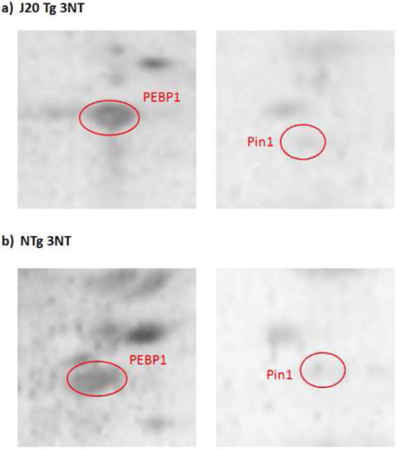
Representative 2D-Western blot images of nitrated PEBP-1 and Pin-1 proteins (images have been zoomed in to focus on these spots) in a) J20 Tg and b) NTg mice. Proteins were probed with anti-3NT antibody.
Official websites use .gov
A
.gov website belongs to an official
government organization in the United States.
Secure .gov websites use HTTPS
A lock (
) or https:// means you've safely
connected to the .gov website. Share sensitive
information only on official, secure websites.
Representative 2D-Western blot images of nitrated PEBP-1 and Pin-1 proteins (images have been zoomed in to focus on these spots) in a) J20 Tg and b) NTg mice. Proteins were probed with anti-3NT antibody.