Abstract
Using a spectrophotometric assay that measures the hyperchromicity that accompanies the unwinding of a DNA duplex, we have identified an ATP-independent step in the unwinding of a herpes simplex virus type 1 (HSV-1) origin of replication, Oris, by a complex of the HSV-1 origin binding protein (UL9 protein) and the HSV-1 single-strand DNA binding protein (ICP8). The sequence unwound is the 18-bp A + T-rich segment that links the two high-affinity UL9 protein binding sites, boxes I and II of Oris. P1 nuclease sensitivity of Oris and single-strand DNA-dependent ATPase measurements of the UL9 protein indicate that, at 37°C, the A + T-rich segment is sufficiently single stranded to permit the binding of ICP8. Binding of the UL9 protein to boxes I and II then results in the formation of the UL9 protein–ICP8 complex, that can, in the absence of ATP, promote unwinding of the A + T-rich segment. On addition of ATP, the helicase activity of the UL9 protein–ICP8 complex can unwind boxes I and II, permitting access of the replication machinery to the Oris sequences.
The herpes simplex virus type 1 (HSV-1) origin binding protein (UL9 protein) is a DNA helicase that, when complexed with the HSV-1 single-strand DNA binding protein (ICP8), can unwind an HSV-1 origin (Oris) to initiate viral DNA replication (1). Measurement of the helicase activity of the UL9 protein has typically made use of nondenaturing polyacrylamide gel electrophoresis (PAGE) to separate the single-stranded DNA products from the duplex DNA substrate (2). This assay, although unambiguous, cannot measure the early events in the unwinding reaction that are necessary to an understanding of its mechanism.
We describe here an optical assay that depends on the hyperchromic effect that results from the rupture of hydrogen bonds as the DNA duplex is unwound. This assay, which can monitor the continuous unwinding of Oris, has revealed that the initial phase of unwinding of Oris consists of the UL9 protein and ICP8-dependent, but ATP-independent unwinding of the A + T-rich sequence linking the two inverted repeats, boxes I and II. On addition of ATP, the UL9 protein–ICP8 complex can then promote the bidirectional unwinding of the entire Oris sequence (3, 4).
Materials and Methods
Nucleotides and Enzymes.
[γ-32P]ATP (6000 Ci/mmol) and proteinase K were obtained from DuPont/NEN and Roche Molecular Biochemicals, respectively. Escherichia coli single-stranded DNA binding protein (SSB), P1 nuclease, and T4 polynucleotide kinase were from United States Biochemical. Recombinant UL9 protein and ICP8 were purified as previously described (3, 5). They were free of detectable exo- and endonuclease activity.
Oligonucleotides.
Oligonucleotides were purchased from Operon Technologies (Alameda, CA). They were purified by 16% PAGE under denaturing conditions. Poly(dT)300–3000 was obtained from Sigma. When required, the oligonucleotides were labeled with [γ-32P]ATP with polynucleotide kinase according to a protocol supplied by Amersham Pharmacia. Unincorporated nucleotide was removed by G-50 spin column chromotography (Quickspin DNA; Roche Molecular Biochemicals).
Oris.
The complementary oligonucleotides: 5′-CGCGAAGCGTTCGCACTTCGTCCCAATATATATATATTATTAGGGCGAAGTGCGAGCACTGGC-3′, and 5′-GCCAGTGCTCGCACTTCGCCCTAATAATATATATATATTGGGACGAAGTGCGAACGCTTCGCG-3′, both at 216 μg/ml in 50 mM Tris⋅HCl (pH 8.0) containing 0.1 M NaCl were mixed, heated at 90°C for 5 min, then slowly cooled to room temperature. The Oris duplex that was generated lacked Box III.
OrisT18.
The complementary oligonucleotides: 5′-CGCGAAGCGTTCGCACTTCGTCCCTTTTTTTTTTTTTTTTTTGGGCGAAGTGCGAGCACTGGC-3′, and 5′-GCCAGTGCTCGCACTTCGCCCTTTTTTTTTTTTTTTTTTGGGACGAAGTGCGAACGCTTCGCG-3′, both at 197 μg/ml, were annealed as described above for Oris to generate an Oris analogue in which the A + T-rich segment was replaced by a “bubble” consisting of two 18-nt oligodeoxythymidylates.
Oris(GC)18.
The complementary oligonucleotides: 5′-CGCGAAGCGT-TCGCACTTCGTCCCGGCGCGCGCGCGCCGCCGGGGCGAAGTGCGAGCACTGGC-3′, and 5′- GCCAGTGCTCGCACTTCGCCCCGGCGGCGCGCGCGCGCCGGGACGAAGTGCGAACGCTTCGCG-3′, both at 197 μg/ml, were annealed as described above for Oris, to generate an Oris analogue in which the A + T = rich segment was replaced by (GC)18.
Mutant Oris.
The complementary oligonucleotides: 5′-GCGAAGCGGGCGCACTTCGTCCCAATATATATATATTATTAGGGCGAAGTGCGCCCACTGGC-3′, and 5′-GCCAGTGGGCGCACTTCGCCCTAATAATATATATATATTGGGACGAAGTGCGCCCGCTTCGCG-3′, both at 144 μg/ml, were annealed as described above for Oris.
Box I.
The complementary oligonucleotides: 5′-CGCGAAGCGTTCGCACTTCGTCCC-3′, and 5′-CGGACGAAGTGCGAACGCTTCGCG-3′, both at 41 μg/ml, were annealed as described for Oris.
Spectrophotometric Assay of Helicase Activity.
Absorbance at 260 nm was measured with a Uvicom (Kontron, Zurich) UV/VIS spectrophotometer using a quartz cuvette with a path length of 1 cm. The cuvette compartment was kept at 37°C, and absorbance measurements were made at 0.5-s intervals for 30 min. Initial velocities (ΔA260/min) were derived from the linear part of the curve. The reaction mixture (1 ml) contained 50 mM Tris⋅HCl (pH 8.15), 10 mM NaC1, 10 mM MgCl2, 1 mM DTT, 0.01% Nonidet P-40, Oris, mutant Oris, OrisT18, or Oris(GC)18, UL9 protein, and/or ICP8 as indicated. The reference cuvette contained the same reaction mixture without UL9 protein or ICP8.
Measurement of P1 Nuclease Sensitivity.
The reaction mixtures (25 μl) contained 50 mM Tris⋅HCl (pH 8.15), 10 mM NaCl, 10 mM MgCl2, 2 mM DTT, 0.01% Nonidet P-40, and 1 pmol of 32P-labeled Oris or Oris T18. Increasing amounts of P1 nuclease (5 × 10⁻⁶ units/μl) containing 2.5 mM ZnCl2 were added, and the mixtures were incubated at either 25°C or 37°C for 5 min.
The reactions were terminated by the addition of 2 μl of 0.5 M EDTA. An aliquot (8 μl) was loaded together with 80% formamide loading buffer containing 0.05% bromophenol blue and 0.05% xylone cyanol onto a 300 × 400 × 0.4-mm 10% polyacrylamide sequencing gel containing 8 M urea and 40% formamide that had been prerun until the temperature of the gel was 50°C (1–2 h). After electrophoresis, the gel was dried on a Whatman No. 1 filter. The relative intensity of the bands was measured with a PhosphorImager (Molecular Dynamics).
DNA-Dependent ATPase Assay.
Reaction mixtures (25 μl) contained Tris⋅HCl (pH 8.15), 10 mM MgCl2, 1 pmol UL9 protein, 1 pmol ICP8, 10 μg BSA, poly(dT)300–3000, Box I, Oris, OrisT18, Oris(GC)18, or mutant Oris, 1 μC of [γ-32P]ATP, and 3 mM ATP. Mixtures were incubated at 37°C for 1 h. Reactions were stopped by the addition of 3 μl of 0.5 M EDTA, pH 8.0, and chilled on ice. Aliquots (1 μl) were spotted onto polyethyleneimine-cellulose F, TLC plates and dried. The ATP substrate and inorganic phosphate product were separated with 1.0 M formic acid/0.4 M LiCl as solvent. The TLC plates were quantitated with a PhosphorImager (Molecular Dynamics).
DNA Helicase Assay.
Reaction mixtures (25 μl) containing 50 mM Hepes⋅KOH (pH 8.15), 10 mM NaCl, 10 mM MgCl2, 2.0 mM DTT, 10% (vol/vol) glycerol, 10 μg BSA, 1.0 pmol OrisT18, or 1.0 pmol Oris(GC)18, and the indicated amounts of ICP8 and UL9 protein were incubated on ice for 5 min. ATP (50 μM) was added to the reaction mixtures, which were then incubated at 37°C for 60 min. The reactions were terminated by the addition of 6.5 μl of stop solution (100 mM EDTA/1% SDS/20 μg proteinase K), then subjected to electrophoresis through a 15% polyacrylamide gel at 10 V/cm. The gel was dried on Whatman DE81 paper at 80°C under vacuum and quantitated with a PhosphorImager (Molecular Dynamics) (4).
Gel Mobility Shift Analysis.
Reaction mixtures (50 μl) containing 50 mM Hepes⋅KOH (pH 8.15), 2.0 mM DTT, 50 mM NaCl, 10 mM MgCl2, 10 μg BSA, 10% (vol/vol) glycerol, and the indicated amounts of UL9 protein and ICP8 were preincubated on ice for 30 min. 5′-32P-labeled Oris(GC)18 (0.5 pmol) was added, and the mixtures were incubated at 37°C for 15 min. Four percent nondenaturing PAGE of the reaction mixtures was performed at room temperature with Tris⋅HCl/glycine buffer containing 50 mM Tris⋅HCl and 380 mM glycine buffer at 30 V and 25 mA for 2.5 h. After electrophoresis, the gel was dried on Whatman DE81 paper at 80°C under vacuum and visualized by a PhosphorImager (Molecular Dynamics) (6).
Results
Spectrophotometric Assay for Unwinding of Oris.
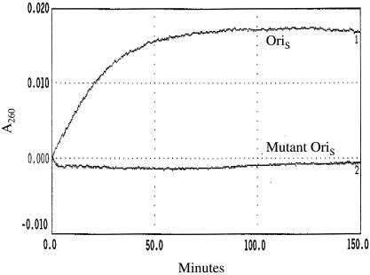
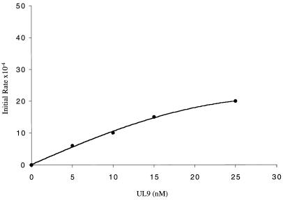
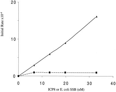
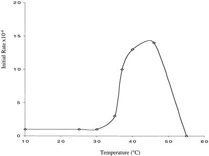
The increase in A260 as a consequence of the unwinding of Oris by the UL9 protein–ICP8 complex is shown in Fig. 1. The reaction proceeded linearly for 40 min. ATP was not required for the reaction, and there was no effect of adding the nonhydrolyzable ATP analogue ATPγS (data not shown). The initial rate was linearly dependent on the concentration of UL9 protein and ICP8 (Figs. 2 and 3). However, the same plateau value was reached over a 5-fold range of UL9 protein–ICP8 concentrations (4–20 pmol; data not shown). ICP8 could not be replaced by the heterologous E. coli single-strand DNA binding protein, SSB (Fig. 3). A mutant Oris with mutations in Boxes I and II that was unable to bind UL9 protein (7) showed no increase in A260 (Fig. 1). The reaction depended on Mg2+ and was optimal at 10–20 mM Mg2+. In agreement with earlier measurements of helicase activity by nondenaturing PAGE, the reaction was optimal at 40–45°C and declined rapidly at higher temperatures, presumably as a result of thermal denaturation of the enzyme (2) (Fig. 4). The Tm for the reaction was 37°C. The inability to observe any reaction at 30°C suggests that some thermally induced destabilization of the A + T-rich sequence is necessary for the reaction (see below).
Figure 1.
Spectrophotometric measurement of unwinding of Oris. Reaction mixtures prepared as described in Materials and Methods contained 50 nM Oris(1), or mutant Oris(2), 4 nM UL9 protein, and 5 nM ICP8. Increase in A260 was measured as described in Materials and Methods.
Figure 2.
Effect of UL9 protein concentration on initial rate of unwinding of Oris. Reaction mixtures were prepared as described in Materials and Methods with 320 nM Oris, 13.2 nM ICP8, and the indicated concentrations of UL9 protein. Initial rate units (ΔA260/min) were determined from the increase in A260 at 15 min.
Figure 3.
Effect of ICP8 concentration on initial rate of unwinding of Oris. Reaction mixtures were prepared as described in Materials and Methods with 320 nM Oris, 5 nM UL9 protein, and the indicated concentrations of ICP8 (▴) or E. coli SSB (■).
Figure 4.
Effect of temperature on initial rate of unwinding of Oris. Reaction mixtures were prepared as described in Materials and Methods with 52 nM Oris, 5.6 nM UL9 protein, and 5.2 nM ICP8. Temperature was varied from 10°C to 55°C as indicated.
Unwinding of A + T-Rich Sequence Is the Initial Step in Unwinding of Oris.
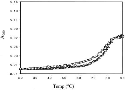
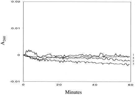
The thermal denaturation profile of Oris is shown in Fig. 5. The Tm of 80°C is consistent with the G + C content of Oris (49%). The maximum increase in A260 that occurred on incubation of Oris with the UL9 protein and ICP8 was 23% of that obtained on complete thermal denaturation (Figs. 1 and 5). Thus, as measured by the spectrophotometric assay, 23% of the Oris duplex was unwound by the UL9 protein–ICP8 complex. This value is in good agreement with the proportion of Oris comprising the A + T-rich sequence (18/63 = 28%), suggesting that it is this sequence that is being unwound in the initial ATP-independent phase of the reaction. This idea was supported by an experiment using an Oris analogue in which the A + T-rich sequence was replaced by two 18-nt oligodeoxythymidylate sequences to create an unpaired “bubble” between Boxes I and II (OrisT18) (4). As shown in Fig. 6, there was no increase in A260 with OrisT18 under conditions in which an increase of 23% of maximum was observed with Oris. Because the oligodeoxythymidylate sequences in OrisT18 were already unpaired, no hyperchromic effect was to be expected.
Figure 5.
Temperature melting curve of Oris. DNA melting was measured with a Cary IE UV-visible spectrophotometer equipped with a temperature control unit. The temperature was increased or decreased at a rate of 1°C per min and held at that temperature for 30 min. The reaction curvette contained 500 nM Oris in 50 mM Tris⋅HCl (pH 8.15). The reference curvette contained the Tris buffer without Oris. The temperature ranges were 20° to 90°C (▵) and 90° to 20°C (□).
Figure 6.
Lack of hyperchromic effect with OrisT18 and Oris(GC)18. Reaction mixtures were prepared as described in Materials and Methods with 340 nM OrisT18 and 4 nM UL9 protein (1), 340 mM OrisT18 and 5 nM ICP8 (2), 340 mM OrisT18 and 4 nM UL9 protein, and 5 nM ICP8 (3) or 53 nM Oris(GC)18, 4 nM UL9 protein, and 5 nM ICP8 (4).
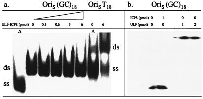
Further evidence that a thermally induced destabilization of the A + T-rich sequence is required for the initial ATP-independent step in unwinding of Oris was provided by an experiment in which the A and T residues of the A + T-rich sequence were replaced by G and C [Oris(GC)18]. With this Oris analogue in which the G + C sequence should remain fully hydrogen bonded at 37°C, there was no increase in A260 (Fig. 6). Furthermore, unlike Oris T18, which could be efficiently unwound by the helicase activity of the UL9 protein–ICP8 complex (4), Oris(GC)18 was essentially inert (Fig. 7). The lack of helicase activity was not a consequence of the failure of UL9 protein to bind Oris(GC)18 as judged by a gel mobility shift analysis (Fig. 7).
Figure 7.
Lack of ATP-dependent unwinding of Oris(GC)18 by UL9 protein-ICP8 complex. (a) Reaction mixtures for helicase measurements were prepared as described in Materials and Methods with 1 pmol OrisT18 or 1 pmol Oris(GC)18, and the indicated amounts of UL9 protein and ICP8. ▵, OrisT18 and Oris(GC)18, heated at 100°C for 5 min. Incubation and PAGE were performed as described in Materials and Methods. (b) Reaction mixtures for gel mobility shift measurements were prepared as described in Materials and Methods with 0.5 pmol Oris(GC)18 and the indicated amounts of UL9 protein and ICP8. Samples were processed as described in Materials and Methods. ds, double stranded; ss, single stranded.
Single-Stranded Character of A + T-Rich Sequence.
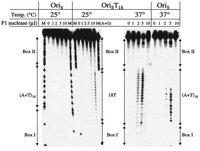
P1 nuclease has a strong preference for single- over double-stranded DNA (8). Treatment of OrisT18 with P1 nuclease at both 25°C and 37°C produced a digestion pattern consistent with the presence of oligo(dT)18. In contrast, the pattern observed on nuclease digestion of the wild-type Oris duplex at 37°C indicated that the A + T sequence was largely destabilized at 37°C but not at 25°C. As expected, Boxes I and II of neither substrate were P1 nuclease-sensitive at either temperature (Fig. 8).
Figure 8.
P1 nuclease sensitivity of Oris and OrisT18. Reaction mixtures prepared as described in Materials and Methods with 1 pmol of 32P-labeled Oris or OrisT18 and the indicated amounts of P1 nuclease and incubated at 25°C or 37°C. The products were analyzed by 10% PAGE in the presence of urea-formamide as described in Materials and Methods. M, markers.
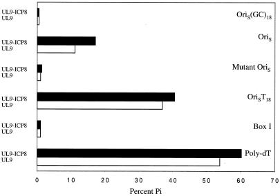
Further support for the single-stranded character of the A + T-rich sequence was provided by measurement of the single-strand DNA-dependent ATPase activity of the UL9 protein (9). As shown in Fig. 9, although the fully duplex Box I supported little ATPase activity at 37°C, Oris showed substantial ATPase activity, similar to that observed with OrisT18 and with poly(dT)300–3000. In contrast, only a low level of DNA-dependent ATPase activity was observed with Oris at 25°C, presumably a manifestation of the DNA-independent ATPase activity of the UL9 protein (9) (data not shown). A mutant Oris that is unable to bind the UL9 protein did not support the ATPase activity of the UL9 protein (Fig. 9). Similarly, no DNA-dependent ATPase activity was observed with Oris(GC)18.
Figure 9.
DNA-dependent ATPase activity of UL9 protein. Reactions were performed as described in Materials and Methods with 1 pmol UL9 protein, 1 pmol ICP8, 3 mM [γ-32P]ATP, and 7.4 pmol poly(dT), 5.7 pmol Box I, 13 pmol Oris, 11 pmol OrisT18, 7.7 pmol mutant Oris or Oris(GC)18.
Discussion
The spectrophotometric assay that we have developed which permits the continuous monitoring of helicase activity has provided some insight into the initial steps in the unwinding of Oris by the UL9 protein–ICP8 complex. The A + T-rich sequence linking boxes I and II of Oris, because of its low Tm relative to the G + C-rich boxes I and II, possesses sufficient single-strand character at 37°C to permit the binding of ICP8. However, complete destabilization of the duplex A + T-rich sequence does not occur under these conditions, as judged by the lack of hyperchromicity on ICP8 binding. The UL9 protein can, however, bind to the high-affinity boxes I and II and form a complex with ICP8 bound to the A + T-rich sequence. The resulting UL9 protein–ICP8 complex, presumably using the energy of binding, can then completely unwind the A + T-rich sequence in an ATP-independent reaction. As observed previously, a specific UL9 protein–ICP8 complex is required (2–4); the E. coli SSB, although capable of binding to the A + T-rich sequence (10), is unable to promote its unwinding, presumably because of its inability to interact with the UL9 protein. In the presence of an energy source, i.e., ATP, the UL9 protein–ICP8 complex can promote the complete unwinding of Oris to permit access of the replication machinery to the origin sequences.
We had earlier suggested that the unwinding of the A + T-rich sequence depended on transcription from the transactivator binding sequences flanking Oris, i.e., transcriptional activation (3). Although transcription may indeed activate the initiation of HSV-1 DNA replication at Oris in vivo, the findings reported here suggest that this is not an obligatory event.
Earlier experiments by Koff et al. (11) had shown that the A + T-rich sequence at the HSV-2 Oris became DNase I sensitive on binding of the UL9 protein. In KMnO4-sensitivity measurements, they also observed the increased reactivity of the thymidylate residues of the A + T sequence on addition of the UL9 protein. Based on these findings, they suggested that binding of UL9 to Boxes I and II resulted in the destabilization of the A + T sequence. Our studies have indicated that the A + T sequence is partially unwound at 37°C, even in the absence of UL9 protein. This reaction might, however, be facilitated by the binding of UL9 protein.
Acknowledgments
We thank Drs. Steven Matson, Per Elias, and Paul Boehmer for their comments and suggestions. This work was supported by National Institutes of Health Research Grant AI26538.
Abbreviations
- HSV-1
herpes simplex virus type 1
- SSB
single-stranded DNA binding protein
- Tm
melting temperature
References
- 1.Lehman I R, Boehmer P E. J Biol Chem. 1999;274:28059–28062. doi: 10.1074/jbc.274.40.28059. [DOI] [PubMed] [Google Scholar]
- 2.Boehmer P E, Dodson M S, Lehman I R. J Biol Chem. 1993;268:1220–1225. [PubMed] [Google Scholar]
- 3.Lee S S-K, Lehman I R. Proc Natl Acad Sci USA. 1997;94:2838–2842. doi: 10.1073/pnas.94.7.2838. [DOI] [PMC free article] [PubMed] [Google Scholar]
- 4.He X, Lehman I R. J Virol. 2000;74:5726–5728. doi: 10.1128/jvi.74.12.5726-5728.2000. [DOI] [PMC free article] [PubMed] [Google Scholar]
- 5.Boehmer P E, Lehman I R. J Virol. 1993;67:711–715. doi: 10.1128/jvi.67.2.711-715.1993. [DOI] [PMC free article] [PubMed] [Google Scholar]
- 6.Lee S S-K, Dong Q, Wang T S-F, Lehman I R. Proc Natl Acad Sci USA. 1995;92:7882–7886. doi: 10.1073/pnas.92.17.7882. [DOI] [PMC free article] [PubMed] [Google Scholar]
- 7.Hernandez T R, Dutch R E, Lehman I R, Gustafsson C, Elias P. J Virol. 1991;65:1649–1652. doi: 10.1128/jvi.65.3.1649-1652.1991. [DOI] [PMC free article] [PubMed] [Google Scholar]
- 8.Chiu S K, Rao B J, Staory R M, Radding C M. Biochemistry. 1993;32:13246–13155. doi: 10.1021/bi00211a025. [DOI] [PubMed] [Google Scholar]
- 9.Dodson M S, Lehman I R. J Biol Chem. 1993;268:1213–1219. [PubMed] [Google Scholar]
- 10.Makhov A M, Boehmer P E, Lehman I R, Griffith J D. EMBO J. 1996;15:1742–1750. [PMC free article] [PubMed] [Google Scholar]
- 11.Koff A, Schwedes J F, Tegtmeyer P. J Virol. 1991;65:3284–3292. doi: 10.1128/jvi.65.6.3284-3292.1991. [DOI] [PMC free article] [PubMed] [Google Scholar]


