Abstract
Leptothrix ochracea is a common inhabitant of freshwater iron seeps and iron-rich wetlands. Its defining characteristic is copious production of extracellular sheaths encrusted with iron oxyhydroxides. Surprisingly, over 90% of these sheaths are empty, hence, what appears to be an abundant population of iron-oxidizing bacteria, consists of relatively few cells. Because L. ochracea has proven difficult to cultivate, its identification is based solely on habitat preference and morphology. We utilized cultivation-independent techniques to resolve this long-standing enigma. By selecting the actively growing edge of a Leptothrix-containing iron mat, a conventional SSU rRNA gene clone library was obtained that had 29 clones (42% of the total library) related to the Leptothrix/Sphaerotilus group (≤96% identical to cultured representatives). A pyrotagged library of the V4 hypervariable region constructed from the bulk mat showed that 7.2% of the total sequences also belonged to the Leptothrix/Sphaerotilus group. Sorting of individual L. ochracea sheaths, followed by whole genome amplification (WGA) and PCR identified a SSU rRNA sequence that clustered closely with the putative Leptothrix clones and pyrotags. Using these data, a fluorescence in-situ hybridization (FISH) probe, Lepto175, was designed that bound to ensheathed cells. Quantitative use of this probe demonstrated that up to 35% of microbial cells in an actively accreting iron mat were L. ochracea. The SSU rRNA gene of L. ochracea shares 96% homology with its closet cultivated relative, L. cholodnii, This establishes that L. ochracea is indeed related to this group of morphologically similar, filamentous, sheathed microorganisms.
Introduction
L. ochracea is a member of the neutrophilic, freshwater iron-oxidizing bacteria (FeOB), a group that accelerates Fe(II) oxidation in chalybeate waters and forms visible ocherous mats. In addition to playing an important role in the iron cycle, these microbial mats accrete rapidly and may decrease water flow, and sorb nutrients [1]–[3]. The FeOB are considered nuisance organisms when they cause biofouling of water distribution pipelines, or influence biocorrosion [4], [5]. In Europe, they have been recognized to have a beneficial role in water filtration systems due to the sorptive capacities of the Fe-oxyhydroxides [6], [7].
L. ochracea is often the most morphologically conspicuous member of FeOB communities due to its copious production of 1–2 micron wide smooth filamentous sheaths that are mostly devoid of cells [1], [8]–[10]. L. ochracea was first described in 1888 by Winogradsky [9], who identified the organism based on its distinctive sheath morphology. In the early days of light microscopy, it was natural to believe that these filamentous sheaths were synonymous with the abundance of L. ochracea cells [11]. Subsequent studies using epifluorescence microscopy coupled with nucleic acid binding dyes revealed the majority of sheaths were empty remains left by L. ochracea as it grew and oxidized Fe(II) [12].
Following the description of L. ochracea, other species of Leptothrix have been described along with a related genus, Sphaerotilus. The classification of these organisms was based on sheath production as well as Fe and/or Mn oxidation that resulted in metal impregnated sheaths [13]. Phylogenetic analysis has confirmed that these morphologically similar organisms are indeed close relatives, clustering together in the order Burkholderiales of the Betaproteobacteria [14]–[16]. Species in this cluster, e.g. L. cholodnii, L. discophora, S. natans, have been isolated and shown to be heterotrophs able to grow on a variety of carbon sources [10]. They may oxidize Fe(II) or Mn(II), and in the case of L. discophora Mn and Fe-oxidizing proteins have been described [17], [18]; however they do not require, beyond trace amounts, Fe or Mn for growth [13], [19]. Because of historical precedence, L. ochracea has remained the type species of the Leptothrix genus [20]. However, its taxonomic affiliation is based solely on morphological traits of cell shape and sheath formation, along with Fe-oxidation. Subsequent attempts to clarify L. ochracea's taxonomic affiliation and determine its physiology have generated more controversy than clarity [21].
We set out to determine the molecular phylogeny of L. ochracea by taking advantage of knowledge about its natural history and combining this with cultivation independent approaches. First, we characterized the microbial composition in an active freshwater Fe-oxidizing mat using a combination of SSU rDNA clone libraries, pyrotags, and DNA sequencing of individual cell filaments. We used this information to design a fluorescent in situ hybridization (FISH) probe putatively targeting L. ochracea. Hybridization of this probe to cells inside L. ochracea sheaths confirmed that the obtained SSU rRNA sequences were indeed of L. ochracea, enabling a detailed phylogenetic analysis.
Methods
Characterization of a freshwater iron seep and measurement of physiochemical parameters
All environmental samples were collected from flocculant iron mats in a soft-water, first-order ephemeral, intermittent stream located near Lakeside Drive (LD) in Boothbay Harbor, ME (43°51.699′N 69°38.929′W) during August 2008 and July 2009. The site was located two miles from our laboratory, and was visited on a weekly and even daily basis. Routine measurements were taken for water temperature, pH (Oakton 110 series meter), and Fe(II) concentrations (determined by the ferrozine method; [22]). Oxygen in mats was measured by ISO2 Oxygen meter and a OXELP probe (World Precision Instruments, Sarasota FL). For the cultivation-independent studies, samples were collected from mats that had formed within the previous 24 hrs. To confirm these mat samples were active, they were examined by phase contrast and epifluorescence microscopy with an Olympus BX60 microscope (Center Valley, PA). Samples retained for analysis were dominated by characteristic smooth microtubular sheaths of L. ochracea, and approximately 10% of the sheaths contained cells.
Several attempts were made to isolate Leptothrix or Sphaerotilus from LD mat samples using either Sphaerotilus medium [23] or agar plates with autoclaved sterilized LD waters and agarose (1.5% final concentration).
SSU rDNA gene V4 region pyrosequencing library
A tagged pyrosequencing library of the hypervariable V4 region was generated from frozen LD bulk mat samples. The frozen mat was thawed, concentrated in an Eppendorf centrifuge (4000 x g) and resuspended in 20 mM phosphate buffer (pH 7.4). This phosphate wash was repeated twice prior to extracting the DNA using the MoBio Soil DNA extraction kit. Extracted DNA was sent to the Michigan State University sequencing facility (http://rtsf.msu.edu/) for pyrosequencing using the FLX System (454 Life Sciences, Branford, CT, USA) with V4 primers ( Table 1 ) to target the V4 hypervariable region of SSU rRNA gene.
Table 1. Probes and primers used in this study.
| Probe/primer | Sequence (5′–3′) | Reference |
| EUB338 | cy3 gct gcc tcc cgt agg agt | 63 |
| NON338 | cy3 act cct acg gga ggc agc | 64 |
| BET 42a | cy3 gcc ttc cca ctt cgt tt | 38 |
| GAM42a | Fl gcc ttc cca cat cgt tt | 38 |
| PS-1 | cy3 gat tgc tcc tct acc gt | 39 |
| Lepto 175 | cy3 atc cac aga tca cat gcg | this study |
| V4-F | ayt ggg ydt aaa gng | 65 |
| V4-R1 | tac nvg ggt atc taa tcc | 65 |
| V4-R2 | tac crg ggt htc taa tcc | 65 |
| V4-R3 | tac cag agt atc taa ttc | 65 |
| V4-R4 | tac dsr ggt mtc taa tcn | 65 |
| 27F | agr gtt yga tym tgg ctc ag | 65 |
| 907R | ccg tca att cmt ttr agt tt | 65 |
| 1492R | tac ggy tac ctt gtt acg act t | 66 |
| gyrB F | kcg caa gcg scc sgg cat gta | 67 |
| gyrB R | ccg tcs acg tcg gcr tcg gtc at | 67 |
SSU rDNA clone library
To select for active L. ochracea, freshly precipitated mat samples (15 mL) were collected from the edges of the actively growing microbial mat with a sterile pipette. To enrich ensheathed cells and decrease the number of planktonic cells, the samples were filtered through a sterile 8 µm mesh filter (Biodesign Inc. of New York) mounted on a 25 mm glass frit support (VWR) using a vacuum pump. The DNA was extracted, amplified, cloned and sequenced using standard methods and is detailed in File S1.
Single cell genomic analysis
Single cell sorting, whole genome amplification (WGA) and PCR-based analyses were performed at the Bigelow Laboratory Single Cell Genomics Center (www.bigelow.org/scgc). Iron mat samples were sheared by vortexing between 10 and 60 seconds, stained with 5 µM SYTO-9 nucleic acid stain (Invitrogen) and passed through a 70 µm mesh-size cell strainer (BD). Using a MoFlo™(Beckman Coulter) flow cytometer equipped with a 70 µm nozzle orifice and using a 488 nm laser excitation, fluorescence activated cell sorting (FACS) from LD iron mat samples was done to identify a sort region that would yield ensheathed L. ochracea cells. The cytometer was triggered on SYTO-9 fluorescence (green), and between 1000–2000 particles were collected from different gated regions for microscopic observation. The presence of ensheathed L. ochracea cells was confirmed from a gated region using phase contrast and epifluorescence microscopy (Figure S1).
For downstream DNA analyses, individual target particles were sorted using side scatter trigger and “purify 0.5 drop” mode and deposited into 384-well PCR microplates containing 600 nL per well of the prepGEM Bacteria (ZyGEM) reaction mix. The microplates were then stored at −80°C until further processing. Of the 384 wells, 311 were dedicated for single sheath particles, 62 were used as negative controls (no droplet deposition), 3 wells received 10 sheath particles each (positive control type 1) and 8 wells received 40 fg human DNA (Promega, positive control type 2). Prior to single cell sorting, the cytometer was cleaned thoroughly with 10% bleach. A 1% NaCl solution (0.2 µm filtered and UV treated) was used as sheath fluid. All tubes, plates and reagents were UV treated to remove DNA contamination. Full details on cleaning methods and instrument preparation are described previously in Stepanauskas and Sieracki [24].
To lyse the cells, microplates with the sorted material were incubated at 37°C and 75°C, 20 min each. This sequentially activated lysozyme and the prepGEM protease. After the enzymatic treatment, cold KOH was used to ensure complete cell lysis and to denature DNA [25]. Genomic DNA from the lysed cells was then amplified using multiple displacement amplification (MDA) [26] in 10 µL final volume, using Repliphi (Epicenter) polymerase and buffers and phosphorylated random hexamers (NNNN*N*N; IDT). The MDA reactions were run at 30°C for 16 h, then inactivated by a 10 min incubation at 65°C. The amplified genomic DNA was stored at −80°C until further processing. We refer to the MDA products originating from individual cells as single amplified genomes (SAGs).
The MDA products were screened for the presence of the bacterial SSU rRNA gene by PCR, using 500 nL of 50-fold diluted WGA products as template in a 5 µL real-time PCR with primers 27F and 907R ( Table 1 ) (Integrated DNA Technologies, IDT). Real-time PCR was performed on a LightCycler 480 using SYBR Green I master mix (Roche). We used the real-time PCR kinetics and melting curve analysis to detect successful reactions, the amplicons of which were sequenced from both ends by Beckman Coulter Genomics facility (http://www.beckmangenomics.com/genomic_services/sample_submission.html) using Sanger technology.
Sequence analysis
The SSU rRNA gene sequences from SAGs and clones were assembled and manually curated using Geneious (Biomatters Ltd. Aukland, New Zealand) or Sequencer (Gene Codes Corporation, Ann Arbor, MI) software. The consensus sequences were aligned using the Silva aligner (www.arb-silva.de, March 23, 2010). The phylogeny was determined with the ARB program and RAxML (Neighbor Joining tree; [27] Maximum Liklihood; [28]).
High-quality pyrosequencing reads were identified using PyroTagger [26]. Using the BLAST 2.2.22+ program (ftp://ftp.ncbi.nlm.nih.gov/blast/), we identified reads that were ≥99% identical to the V4 region in the putative L. ochracea clones and SAGs. These closely related sequences were then aligned using MUSCLE [30] within the Geneious software and evolutionary trees were constructed in ARB. The analysis of SSU rRNA gene sequences is detailed in File S1.
The following sequences were submitted to GenBank: the complete SSU rRNA gene sequence from L. ochracea clones (HQ317218-HQ317231), the complete SSU rDNA gene sequences from single amplified genome AAA018-L12 (HQ290516), the partial gene sequence from the L. ochracea gyrB gene sequence (HQ290424) the non-chimeric partial SSU rRNA gene sequence clone libraries (HQ290425-HQ290476) and the partial SSU rRNA gene sequences from the single amplified genomes (HQ290477-HQ290515). The cleaned pyrosequencing hypervariable V4 region reads were submitted to the NCBI short read archive(http://trace.ncbi.nlm.nih.gov/Traces/sra/sra.cgi?) under the accession number (SRP005891).
FISH analysis
For FISH, mat samples from either LD, or cultivated control strains were fixed at 4°C in 2.5% paraformaldehyde (final conc.; per Fuchs et al., [31]) for 90 min. After fixation samples were rinsed twice with PBS via centrifugation at 2150 x g, resuspended in 1∶1 PBS:Molecular Biology Grade Ethanol, Sigma and stored at −20°C until use. A FISH probe for L. ochracea (Lepto175; Table 1 ) was designed using the SSU rRNA gene sequences from the clone and SAG libraries and the ARB software. The probe development and hybridizations were performed using standard methods [31] and are detailed in File S1. We found that iron oxyhydroxides interfere with FISH. To reduce this interference, the sample was dried on ClearCell slides with 7 mm diameter wells (ER-279W-2; ThermoScientific), not dehydrated with an ethanol series, and prehybridized with the blocking reagents typically used in CARD-FISH [32].
Cell counts and Microscopy
Phase contrast and fluorescence microscopy were done using an epifluoresence Olympus BX60 microscope equipped with a QICAM Fast CTD camera (Qimaging, Surrey BC).
The percentage of L. ochracea cells in freshly formed LD mats was determined by FISH using six samples collected in 2008 or 2009 and are detailed in File S1. To determine the cell width and length, and sheath width of L. ochracea cells, and the total percent of sheaths filled with cells, photomicrographs were obtained from gluteraldehyde preserved samples, and analyzed using the ImageJ (http://rsbweb.nih.gov/ij/ [33]) plot profile tool.
Scanning Electron Microscopy of L. ochracea sheaths was done on freshly precipitated L. ochracea-rich Fe-mats using a Hitachi S-4700 FESEM at DBI Bioimaging (http://www.dbi.udel.edu/bioimaging/fesem.html) at the University of Delaware.
Results
Physicochemical parameters of LD Fe-mats and identification of L. ochracea
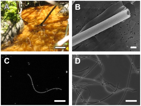
The freshly precipitated iron mats in the stream on LD were used as the source material for identification and characterization of L. ochracea. The mats at LD consisted of a loose floc of Fe-oxyhydroxides that at times filled the entire depth of the 15 cm deep water column ( Figure 1A ), and were most abundant during June, July, and August. The presence of Fe-mats was sensitive to large rainfall events (>0.5 cm day−1) which flushed the accumulated iron mat downstream; however once normal flow rates (<3 cm s−1) resumed, and so long as Fe(II) concentrations were high (> 70 µM) the mats reestablished rapidly, typically in less than a week. During August 2008 and July 2009, when samples were taken for the analyses reported here, the water was consistently 15°C–17°C, the pH 6.0–6.5 and the Fe(II) 70 µM–160 µM. The stream was approximately half-saturated with O2 (approximately 150 µM at 15°C) and profiles within the mats revealed a gradual decrease in O2 concentration with depth. This confirmed previous reports of low O2 demand in this type of iron mats [34], [35]. Smooth tubular iron oxide encrusted sheaths formed the primary mat matrix. Based on observations by light microscopy, it was noted that samples collected at the edges of the mat contained more sheathed cells than deeper layers, and that the sheaths appeared less mineralized.
Figure 1. Photo-montage of L. ochracea sheaths and cells from Lakeside Drive.
Summer time bloom of typical LD iron mats (A), Scanning electron micrograph of a typical L. ochracea sheath (B). Epi-fluorescence of Syto 13 stained cells (C) and phase contrast image (D) of filamentous cells and sheaths from LD iron mats. Scale bars are 10 cm, 10 µm, 10 µm and 10 µm, respectively.
The putative L. ochracea sheathed cells ( Figure 1B & 1C ) were 1.0 µm ± 0.14 (σ, n = 32) wide and 3.04 µM ± 0.92 (σ, n = 23) long. The microtubular sheaths that encased the cells were free of unstructured particulate oxides ( Figure 1D ) and measured 1.1 µM ± 0.95 (σ, n = 15) wide. We estimated that 9.4% ± 3.32 (σ, n = 9 fields) of the sheaths in actively growing mat were filled with cells. Attempts to enrich and isolate heterotrophic Leptothrix spp. or Sphaerotilus spp. from LD mats did not indicate they were abundant. Plating mat samples on Sphaerotilus agar and LD waters yielded very few filamentous bacterial colonies that matched the morphological description of Leptothrix or Sphaerotilus (only 2 colonies present at the 100x dilution), indicating these heterotrophs were not abundant.
Microbial diversity in the iron mats
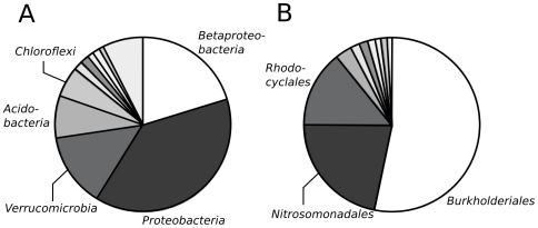
After processing and filtering the pyrosequencing library, a total of 9864 reads out of 16786 (59%) were deemed to be of suitable quality for phylogenetic analysis. In total, 8607 sequences that could be assigned to an OTU (an OTU is defined as sequences that have ≥97% identity) that contained ≥ 2 members, the remaining 1257 sequences were singletons. Overall, the largest number of OTUs (222) and greatest proportion of sequences (19.8%) were affiliated with the Betaproteobacteria ( Figure 2 ). The Burkholderiales order and the Comamonadaceae family which include the cultivated Leptothrix and Sphaerotilus species and contained 10.3% and 9.6% of the total sequences, respectively. The OTU that was most closely affiliated with Leptothrix spp. contained 667 sequences. Representatives from Gallionellaceae (including Sideroxydans, Gallionella), that include freshwater, lithotrophic iron-oxidizing bacteria, were 2.5% of the sequences, and bacterial groups known to include Fe-reducers, e.g. Geobacter, Rhodoferax, Ferribacterium, and Pelobacter, were 6.1% of the sequences. Several other abundant OTUs (containing >100 sequences each) were affiliated with the Gammaproteobacteria the Verrucomicrobia, and the Alphaproteobacteria.
Figure 2. Bacterial composition in the bulk samples of Fe-oxidizing mats as determined by pyrosequencing of the SSU rDNA V4 region.
Fraction of the various bacterial phyla (A), as determined by the RDP Classifier with ≥80% confidence; the unlabeled sections are (clockwise, starting after Chloroflexi) Gemmatimonadetes, unclassified, WS3, ZB2 and Chlamydiae and phyla that form less than 1% of the total sequences. A subset of the data that breaks out the different orders of Betaproteobacteria (B), as determined by alignment and parsimony analysis with the Silva database (release 102); the respective unlabeled orders are (clockwise), staring after Rhodocyclales unassigned, SCI-84, Neisseriales, UCT N117, Hydrogenophilales, 070125-BRIC7-5 and TRA3-20.
The conventional clone library contained a total of 69 sequences after quality control processing, and 41% of the total sequences, (Figure S2) that were ≥ 99% similar to one another and were affiliated with the Betaproteobacteria. When the phylogeny of this group was analyzed by distance algorithms it was clearly affiliated with the Burkholderiales family with its closest relatives being in the Leptothrix/Sphaerotilus group; but still distinct (≤96% identity) from the cultivated members of the Leptothrix and Sphaerotilus genera. Other sequence groupings from the SSU rRNA gene clone library were far less abundant. Therefore, it was concluded that the most abundant sequence cluster represented putative clones of L. ochracea.
Single cell analysis
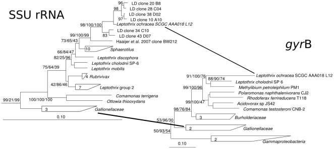
The phylogenetic diversity of SAGs was similar to the diversity of the SSU rRNA gene clone library and the pyrosequencing data set (Figure S2), and contained representatives of genera known to oxidize iron or reduce iron. One SAG (SCGC AAA018-L12) was ≥99% identical to the putative L. ochracea SSU rRNA clones and >97% identical to only 2 sequences in GenBank (>99% for AY947983; [36], and 97% for EF079081; [37]). Furthermore it was only 95% similar to Leptothrix cholodnii SP-6, its closest relative among the cultured Leptothrix/Sphaerotilus group. A Maximum Likelihood phylogeny with other taxa in the Betaproteobacteria grouped the full-length sequence from the putative L. ochracea SAG and several clones from LD with the members of the Leptothrix/Sphaerotilus group ( Figure 3 ). Phylogenic analysis of the gyrB gene sequence from the L. ochracea SAG also clustered with the gyrB gene sequence of L. cholodnii SP-6. A similarity matrix analysis indicated that the putative L. ochracea SAG SSU rRNA gene was distinctly different from all cultivated Leptothrix/Sphaerotilus species, with its closest relative being L. cholodnii strain SP-6 (96.3% identical). For all downstream in silico analysis and development of FISH probes, this SAG sequence was used as the putative L. ochracea SSU rRNA gene sequence.
Figure 3. Phylogenetic tree of the SSU rRNA and gyrB genes from Leptothrix and several type species from the Betaproteobacteria as determined by Neighbor-Joining, with Jukes-Canter correction.
Node percentages represent the bootstrap values from Neighbor Joining, Maximum Liklihood (RAxML with a Gamma distribution mode) and Parsimony (with phylip) methods. The tree shown is the most-likely tree from 1000 replicate trees with Sulfitobacter guttiformis (Y16427) as the outgroup.
Enumeration of L. ochracea with fluorescence in situ hybridization (FISH)
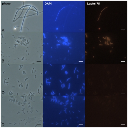
To confirm the phylogenetic affiliation of L. ochracea and link it to its recognized morphology, a FISH probe for the SSU rRNA gene was developed based on the sequence information gathered above. Initial results showed that the general Betaproteobacteria probe BET 42a bound to L. ochracea cells when used with GAM 42a as a competitor probe [38]. Furthermore, a Leptothrix specific probe (PS-1), designed to target L. cholodnii SP-6 [39] bound to L. ochracea, but only at low stringency (<5% formamide). Sequence comparison of the PS-1 probe and the putative L. ochracea SSU rRNA gene sequence showed a 2 bp mismatch ( Table S1 ). Therefore, the Lepto175 probe was designed to specifically target the putative L. ochracea sequence. The probe sequence exactly matched 3 environmental sequences (AY947983, AM778004, and EF079081; [36], [37]) and the isolate Ottowia thiooxydans (AJ537466;[40]), a nonsheath-forming unicellular bacterium that is not known to oxidize Fe(II) match or hybridize with the PS-1 probe (Table S1). Under stringent conditions (up to 20% formamide; Figure S3) the probe bound well to ensheathed Leptothrix ochracea cells, ( Figure 4 ). This probe did not bind to S. natans (5 bp mismatch), or to L. cholodnii (4 bp mismatch) under stringent conditions (15% formamide) ( Figure 4 ).
Figure 4. Micrographs of LD iron-mats (row A), O. thiooyxgens, (row B), S. natans (row C) and L. cholodnii SP-6 (row D) viewed with phase contrast, epi-florescence stained with DAPI, and epi-florescence with the Lepto175 probe applied.
Scale bar is 5 µm. Cells were hybridized with the Lepto175 probe (hybridized at a 15% formamide concentration).
To assess natural populations of L. ochracea mats, were collected from the periphery of the LD mat from different days and years. Ensheathed cells that bound to the Lepto175 probe composed 35–85% (n = 7) of the total bacterial population able to hybridize with universal bacterial probe EUB 338, and up to 32% (n = 6) of the total microbial population stained by the DNA dye Syto13. Cells that hybridized to the Lepto175 probe were all ensheathed and only filled approximately 10% of the sheaths present. These observations showed that the cell morphology was consistent with the historical description of L. ochracea.
Microdiversity analysis
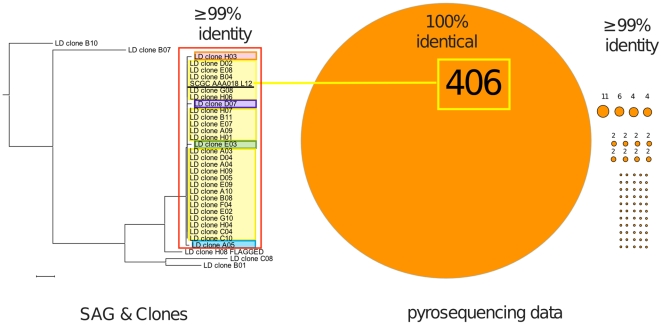
To further understand the population structure of L. ochracea within these iron mats, we analyzed the microdiversity of sequences obtained both through clone libraries and pyrosequencing. To do this, a custom database was built from the tagged pyrosequences and then subjected to BLASTn similarity searches using the putative L. ochracea sequences from SAG and clone libraries (see details in Material and Methods). From these, 501 related sequences (5% of the total reads) that were ≥ 99% similar to the putative L. ochracea clones and the SAG were used to define the microdiversity of L. ochracea ribotypes. Clustering analysis of highly similar pyrosequencing reads suggested that 67 unique ribotypes were present ( Figure 5 ). Remarkably, the dominant ribotype found in the pyrosequencing data set (406 reads) was identical to the hypervariable region V4 of the several clones and the SAG SCGC AAA018-L12. This suggests that the cell or filament of cells isolated by FACS from the Fe-mats was probably the most abundant and dominant L. ochracea ribotype in the environment. Other putative L. ochracea ribotypes (those with ≥99% to the SAG) represented only 18% of the L. ochracea pyrosequencing reads and were not identical to any of the sequences in the clone library.
Figure 5. Microdiversity of L. ochracea clones and pyrosequencing reads.
The Neighbor-Joining tree was constructed using the Jukes-Cantor substitution model with sequences of the hypervariable region V4 of the SSU rRNA gene from SAG and clones that matched the Lepto175 probe. Ribotypes boxed in red had ≥ 99% identity to one another and each highlighted clone(s) represent a unique ribotype in the clone library. Each circle or dot represents a unique ribotype of L. ochracea found in the pyrosequencing library that was either ≥100 or 99% identical to L. ochracea SAG and clones boxed in yellow.
Discussion
Through the use of SSU rRNA gene analysis, WGA of single cells, and specific identification of cells in environmental samples using FISH we have confirmed that L. ochracea is phylogenetically related to other species of Leptothrix and Sphaerotilus. The results presented here settle a 120 year old mystery concerning the provenance of L. ochracea [20], [21]. In this case, the common traits of the ensheathed filamentous cells, the capacity to oxidize Fe(II), and the phylogeny are linked in the Leptothrix/Sphaerotilus group. The phylogenetic analyses, based on the SSU rRNA gene sequences of one L. ochracea SAG, and several clones, show that L. ochracea forms a unique cluster within this group ( Figure 3 ). Among the known species of Leptothrix and Sphaerotilus, its closest relative is L. chlodonii. However, the different treeing methods used to generate both the SSU rRNA gene phylogeny and the gyrB gene phylogeny (Maximum Liklihood, Neighbor Joining, Parsimony) indicate that L. ochracea is related equi-distant from known Leptothrix and Sphaerotilus species. Based on this phylogenetic analysis, it is reasonable to conclude that L ochracea is a relative of Leptothrix and Sphaerotilus spp., but that it represents a unique lineage, which is consistent with its apparent differences in morphology and habitat.
Ecological implications
Given the apparent differences in physiology among the cultivated members of the Leptothrix/Sphaerotilus cluster, it was not obvious that L. ochracea would share a close phylogenetic relationship with other Leptothrix and Sphaerotilus spp. Both sheath formation and Fe-oxidation are properties found throughout the bacterial tree of life [15], [41]–[45]. Our cultivation-based attempts at enriching heterotrophic Leptothrix and Sphaerotilus spp. from LD iron mats also yielded few results, confirming other studies that the heterotrophic species of Leptothrix or Sphaerotilus are not common members of microbial iron mat communities [1], [12]. Our working hypothesis had been that L. ochracea might be more closely related to another family of Betaproteobacteria, the Gallionellaceae, which consist of known chemolithotrophic FeOB that are found in freshwater Fe-oxidizing communities. The genera, from this family (Sideroxydans and Gallionella) have been shown to be obligate, lithotrophic FeOB [1], [46], [47]; furthermore, G. ferruginea produces an Fe-oxide encrusted extracellular stalk. Recent community diversity studies [37], [48]–[50] of microbial iron mat communities similar to the one at LD have found that significant fractions (10–25%) of SSU rRNA clones clustered within the Gallionellaceae. Most of these mats were reported to have abundant L. ochracea type sheaths associated with them, as opposed to the stalks of G. ferruginea; however only a small fraction (0–3%) of the clones were related to Leptothrix spp. This contrasts to the results reported here, where 5% of pyrotagged community from the bulk mat and 40% of the clones from the actively growing periphery of the mat belonged to the Leptothrix clade. While members of the Gallionellaceae were present in LD iron mats, they only made up 2.5% of the pyrotagged library, and 4.3% of the clone library (Figure S2).
A previous model suggested that L. ochracea was most active at the periphery of iron mats where it contributed to the accretion of new mat [1]. The evidence obtained with the use of the Lepto175 FISH probe supports this model. Analysis of the actively accreting portion of the LD mat indicated that up to 32% of the cells belonged to L. ochracea but only 9% of the sheaths actually contain cells. These observations suggests that despite sheathed cells being relatively rare in the bulk mat, they still make up a significant percentage of the total population in the mat periphery, where they contribute substantially to biotic iron oxidation and to new mat formation. It is likely that other FeOB, e.g. Sideroxydans spp., colonize the matrix of sheaths left behind and continue to oxidize Fe, and that older mat becomes host to other bacterial populations as well, including Fe-reducing bacteria (insert Emerson, field studies AEM reference here). The complexities of these interactions remain to be worked out; however a growing array of tools should assist in developing a more detailed understanding of how different microbial populations interact and control the processes occurring in these Fe-dominated ecosystems.
Detecting rarely observed sequences
Single cell-based techniques have proven effective at both identifying dominant members of microbial ecosystems that have resisted cultivation, and demonstrating that numerically minor members of a community may have a significant impact on ecosystem function [51]. In this case, we have taken advantage of unique morphological characteristics of L. ochracea to use FACS to isolate single cells or filaments of cells. In general, isolation of cells using FACS has focused on planktonic cells that grow predominantly in the liquid phase, rather than in consolidated microbial flocs or mats.
Because L. ochracea grows as filaments of cells, it provides a good signature for FACS sorting. For example, L. ochracea, L. cholodnii and S. natans filaments have a distinctively higher relative florescence and a higher relative side scatter than single cells (Figure S1; [52], [53]). The advent of MDA technology for amplification of single cells makes it possible to acquire enough genomic material for sequencing of specific genes, in this case the SSU rRNA and the gyrB gene, and ultimately sequencing the entire genome. Fortuitously, the presence of the sheath, or the Fe-oxyhydroxides that encrust it, do not appear to preclude either cell sorting or WGA, although it is possible that the presence of the sheath may have reduced the efficiency of the process, which could explain the low overall recovery of L. ochracea-type cells. It is also interesting to note that one of the SAGs we acquired was closely related to the filamentous, methane-oxidizing bacterium, (Crenothrix polyspora [44]; SCGC AAA018-I2), suggesting this approach could be used to target other filamentous bacteria.
Microdiversity of L. ochracea
Analysis of unique SSU rRNA gene sequences that cluster together with a >99% SSU rRNA gene sequence similarity can reveal the presence of microdiversity within a species and help delineate fine-scale patterns of community composition [54]. The large amount of information generated by pyrosequencing makes it possible to begin to assess population diversity of known ribotypes in complex communities. Analysis of the pyrosequencing data set and the clone library revealed up to 67 different individual L. ochracea ribotypes and 5 different ribotypes, respectively in the LD Fe-mats. Caution must be taken in interpreting these results due to pyrosequencing artifacts [55]; however, the sheer number of identical, or closely related clones, provides evidence for substantial populations of very closely related, and possibly, clonal ribotypes. It is also possible that minor sequence variation between multiple copies of the SSU rRNA gene within an individual organism could account for some of the observed microdiversity (see, for example, Acinas et al. [54]), the genome of the phylogenetically related L. cholodnii has only 2 SSU rRNA copies that are 100% identical to one another. Most notably the pyrosequencing data set contained a dominant L. ochracea ribotype that was identical to the L. ochracea SAG, as well as being abundant in the clone library ( Figure 4 ). This is corroborative evidence that this ribotype was one of the most abundant individual ribotypes in the Fe-mat and indicates it may play a significant role in Fe-oxidation. Other studies have shown that individual ribotypes are often thought to adapt to specific resources or environmental conditions [56], [57], and it also known that spatial and temporal resource partitioning can select for specific ribotypes [58]. It is unknown why one particular ribotype of L. ochracea appears dominant in the LD iron mat, but it will be interesting to determine the temporal and spatial dynamics of L. ochracea ribotypes in other iron mats.
Physiological implications
The findings that L. ochracea belongs to the Leptothrix/Sphaerotilus group leaves open the question of its physiology. L. ochracea cells must excrete a large amount of Fe-oxyhydroxide coated sheath, implying this organism oxidizes large amounts of Fe(II) relative to the cell biomass that is produced. This is consistent with the requirements of a cell that uses Fe(II) as an energy source, since the thermodynamics of Fe(II) oxidation are poor [59], [60]. This contrasts with the known members of the Leptothrix/Spaerotilus group that are heterotrophic, and whose sheaths are normally filled with cells. The benefit they get from either Fe(II) or Mn(II) oxidation remains unknown. S. natans is typically thought of as an organism that grows in organic rich environments, e.g. sewage sludge [4], [23]; while other Leptothrix spp are often found in freshwater wetlands [45], [61], but not necessarily associated with chalybeate waters. While L. ochracea has yet to be obtained in pure culture, enrichment studies using flow-through laboratory microcosms have shown that it requires significant amounts of Fe(II) to grow, and may also require natural waters from its habitat [9], [10], [12] Fleming unpublished results). Nonetheless, attempts to enrich or isolate L. ochracea using static gradient methods or fed batch cultures typically used to isolate Gallionella spp. or Sideroxydans spp. [62] have not shown success (Emerson, unpublished results). From the single cell analysis done here, we have identified a SAG from L. ochracea, and this has been submitted for whole genome sequencing. The genome sequence coupled with further ecological and physiological investigations will hopefully establish the true metabolism of this enigmatic microbe.
Supporting Information
Flow cytometry dot plot of the Fe-oxidizing mat sample stained with SYTO-9 (A) and excited with a 488 nm laser. Displayed are side scatter (SSC) and green fluorescence (FL1) signals. Region R3 was used as the sorting gate. Phase contrast (B) and epifluorescence micrographs of the sorted material (C). Mat samples for single cell genomics were also collected from the margins of the growing Fe-mats and filtered to enrich for filamentous sheaths. Prior to single cell sorting, samples were confirmed to contain between 6–12% ensheathed cells. The high relative SYTO-9 (nucleic acid) fluorescence and high relative side scatter settings on the cell sorter were used to select for a size range consistent with filaments of cells with the approximate size range of L. ochracea. These cytometer settings were tested previously with ensheathed L. cholodonii SP-6 cells and observed to select for ensheathed cells. With Fe-mat samples the empty Fe-oxide encrusted sheaths did not fluoresce, nor did they appear to interfere with the operation of the cell sorter. Scale bar on the micrographs is 10 µm.
(TIF)
Neighbor-joining tree of a partial SSU rRNA gene sequence from non-chimeric clones and single amplified genomes of bacteria. The tree was generated using the ARB neighbor joining package with Jukes-Cantor correction. Percentages represent the bootstrap values at each node for 1000 replicate trees and percentages less than 50% are not shown. Originally, sequences were inserted by parsimony into the Silva SSURef NR 99% release 102, (Feb 13, 2010) database and used to assign phyla, orders, classes, families or genus. The number of clones and SAGs (respectively) are listed for each grouping and 4 cultivated Leptothrix/Sphaerotilus sequences (*) were included as a reference (D16214, L33975, CP001013, Z18534).
(TIF)
Stringency of L. ochracea specific FISH probe, Lepto175 to L. ochracea cells present in LD mat samples collected August 2008 (closed symbols) and O. thiooxydans cells grown in R2A (open circles (check a)) or caso- agar (open squares (check b)). The relative fluorescence was normalized to hybridizations performed at 5% or 0% formamide. Hybridizations on LD mats were performed on 3 separate occasions using the same conditions and O. thiooxygens cells were both tested with the Lepto175 probe on the same day as the LD mat with closed triangles. Background was ≤ than 20% of relative fluorescence.
(TIF)
The ability of FISH probes to bind to select species of the Betaproteobacteria.
(DOC)
Information regarding additional materials and methods used to generate SSU rRNA gene sequence clone libraries, to analyze the sequence reads, develop the Lepto175 and to perform FISH on Lakeside Drive iron-mat samples.
(DOC)
Acknowledgments
The authors would like to thank Dr. Woo Jun Sul and Dr. James Tiedje for help and advice with the pyrosequencing, and the MSU sequencing facility for running the pyrosequencing samples. We are indebted to Dr. Clara Chan and Debbie Powell at the University of Delaware for the SEM imaging. At Bigelow, we would also like to thank Dr. Micheal Sieracki for access to flow cytometry, Dr. David McClellan for aiding in the phylogenetic determination of L. ochracea, and Dr. Joyce McBeth for helpful discussions. David Gaecklan and Steve Bryer kindly provided access to the LD iron-seep.
Dedication
We dedicate this paper to the memory of Lee Adams III who passed away in a mountaineering accident in July, 2010. Lee did seminal work on the physiology of Leptothrix and elucidating the mechanism of Mn oxidation.
Footnotes
Competing Interests: The authors have declared that no competing interests exist.
Funding: EJF and DE were funded in part by NSF grant IOS 0951077 (http://www.nsf.gov/) and the NASA Astrobiology Institute (http://astrobiology.nasa.gov/nai/). AEL was funded by Bigelow Laboratory NSF REU program and a Swarthmore College summer HHMI grant (http://www.swarthmore.edu/hhmi.xml). Instrumentation and infrastructure for the Bigelow Laboratory Single Cell Genomics Center were sponsored by the NSF MRI grants OCE-821374 and OCE-1019242 to RS and by the Maine Technology Institute (http://www.mainetechnology.org/). The development of single cell genomics methods was supported by NSF SGER grants EF-0633142 and MCB-738232 to RS. The funders had no role in study design, data collection and analysis, decision to publish, or preparation of the manuscript.
References
- 1.Emerson D, Weiss JV. Bacterial iron oxidation in circumneutral freshwater habitats: findings from the field and the laboratory. Geomicrobiol J. 2004;21:405–414. [Google Scholar]
- 2.Langley S, Gault AG, Ibrahim A, Takahashi Y, Renaud R, et al. Strontium desorption from bacteriogenic iron oxides (BIOS) subjected to microbial Fe(III) reduction. Chem Geol. 2009;262:217–228. [Google Scholar]
- 3.Rentz JA, Turner IP, Ullman JL. Removal of phosphorus from solution using biogenic iron oxides. Water Res. 2009;43:2029–2035. doi: 10.1016/j.watres.2009.02.021. [DOI] [PubMed] [Google Scholar]
- 4.Mulder EG. Iron Bacteria, particularly those of the Sphaerotilus-Leptothrix Group, and Industrial Problems. J Appl Microbiol. 1964;27:151–173. [Google Scholar]
- 5.Little BJ, Lee JS. Hoboken, NJ: Wiley-Inverscience; 2007. Microbiologically influenced corrosion.279 [Google Scholar]
- 6.Mouchet P. From conventional to biological removal of iron and manganese in France. J Am Water Works Assoc. 1992;84:158–166. [Google Scholar]
- 7.Katsoyiannis I, Zouboulis A. Use of iron- and manganese-oxidizing bacteria for the combined removal of iron, manganese and arsenic from contaminated groundwater. Water Qual Res J Can. 2006;41:117–129. [Google Scholar]
- 8.Kützing FT. Leipzig: F.A. Brockhaus; 1843. Phycologia Generalis oder Anatomie, Physiologie und Systemkunde der Tange.538 [Google Scholar]
- 9.Winogradsky S. Über Eisenbakterien. Botanishe Zeitung. 1888;46:261–271. [Google Scholar]
- 10.Mulder E, van Veen W. Investigations on the Sphaerotilus-Leptothrix group. A Van Leeuw. 1963;29:121–153. doi: 10.1007/BF02046045. [DOI] [PubMed] [Google Scholar]
- 11.Harder EC. Iron Bacteria. Science. 1915;42:310–311. doi: 10.1126/science.42.1079.310. [DOI] [PubMed] [Google Scholar]
- 12.Emerson D, Revsbech NP. Investigation of an iron-oxidizing microbial mat community located near Aarhus, Denmark: laboratory studies. Appl Environ Microbiol. 1994;4032-4038 doi: 10.1128/aem.60.11.4032-4038.1994. [DOI] [PMC free article] [PubMed] [Google Scholar]
- 13.van Veen W, Mulder E, Deinema M. The Sphaerotilus-Leptothrix group of bacteria. Microbiol Rev. 1978;42:329–356. doi: 10.1128/mr.42.2.329-356.1978. [DOI] [PMC free article] [PubMed] [Google Scholar]
- 14.Willems A, Gillis M, De L. Transfer of Rhodocyclus gelatinosus to Rubrivivax gelatinosus gen nov, comb nov, and phylogenetic relationships with Leptothrix, Sphaerotilus natans, Pseudomonas saccharophila, and Alcaligenes latus. Int J Syst Evol Micr. 1991;41:65–73. [Google Scholar]
- 15.Corstjens PL, Muyzer G. Phylogenetic Analysis of the Metal-oxidizing Bacteria Leptothrix discophera and Sphaerotilus natans using 16S rDNA sequencing data. System Appl Microbiol. 1993;16:219–223. [Google Scholar]
- 16.Siering P, Ghiorse WC. Phylogeny of the Sphaerotilus-Leptothrix group inferred from morphological comparisons, genomic fingerprinting, and 16S ribosomal DNA sequence analyses. Int J Syst Evol Micr. 1996;46:173–182. doi: 10.1099/00207713-46-1-173. [DOI] [PubMed] [Google Scholar]
- 17.Adams LF, Ghiorse WC. Characterization of extracellular Mn(II) oxidizing activity and isolation of a Mn(II) oxidizing protein from Leptothrix discophora SS1. J Bacteriol. 1987;169:1279–1285. doi: 10.1128/jb.169.3.1279-1285.1987. [DOI] [PMC free article] [PubMed] [Google Scholar]
- 18.Corstjens PLAM, de Vrind JPM, Westbroek P, de Vrind EM, de Jong Enzymatic iron oxidation by Leptothrix discophora: Identification of an iron-oxidizing protein. Appl Environ Microbiol. 1992;58:450–454. doi: 10.1128/aem.58.2.450-454.1992. [DOI] [PMC free article] [PubMed] [Google Scholar]
- 19.Adams, LF, Ghiorse WC. Influence of manganese on growth of a sheathless strain of Leptothrix discophora SS-1. Appl Environ Microbiol. 1985;49:556–562. doi: 10.1128/aem.49.3.556-562.1985. [DOI] [PMC free article] [PubMed] [Google Scholar]
- 20.Spring S, Kämpfer P. Genus Incertae Sedis XIII. Leptothrix Kützing 1843, 184. Bergey's Manual of Determinative Bacteriology. 2006;740-745 [Google Scholar]
- 21.Ghiorse WC. Biology of iron-and manganese-depositing bacteria. Annu Rev Microbiol. 1984;38:515–550. doi: 10.1146/annurev.mi.38.100184.002503. [DOI] [PubMed] [Google Scholar]
- 22.Stookey LL. Ferrozine- a new spectrophotometric reagent for iron. Anal Chem. 1970;42:779–781. [Google Scholar]
- 23.Stokes JL. Studies on the filamentous sheathed iron bacterium Sphaerotilus natans. J Bacteriol. 1954;67:278–291. doi: 10.1128/jb.67.3.278-291.1954. [DOI] [PMC free article] [PubMed] [Google Scholar]
- 24.Stepanauskas R, Sieracki ME. Matching phylogeny and metabolism in the uncultured marine bacteria, one cell at a time. Proc Natl Acad Sci USA. 2007;104:9052–9057. doi: 10.1073/pnas.0700496104. [DOI] [PMC free article] [PubMed] [Google Scholar]
- 25.Raghunathan A, Ferguson HRJ, Bornarth CJ, Song W, Driscoll M, et al. Genomic DNA amplification from a single bacterium. Appl Environ Microbiol. 2005;71:3342–3347. doi: 10.1128/AEM.71.6.3342-3347.2005. [DOI] [PMC free article] [PubMed] [Google Scholar]
- 26.Dean FB, Hosono S, Fang L, Wu X, Faruqi AF, et al. Comprehensive human genome amplification using multiple displacement amplification. P Natl Acd Sci USA. 2002;99:5261–5266. doi: 10.1073/pnas.082089499. [DOI] [PMC free article] [PubMed] [Google Scholar]
- 27.Ludwig W, Strunk O, Westram R, Richter L, Meier H, et al. ARB: a software environment for sequence data. Nucleic Acids Res. 2004;32:1363–1371. doi: 10.1093/nar/gkh293. [DOI] [PMC free article] [PubMed] [Google Scholar]
- 28.Stamatakis A. RAxML-VI-HPC: maximum likelihood-based phylogenetic analyses with thousands of taxa and mixed models. Bioinformatics. 2006;22:2688–2690. doi: 10.1093/bioinformatics/btl446. [DOI] [PubMed] [Google Scholar]
- 29.Kunin V, Hugenholtz P. PyroTagger: a fast, accurate pipeline for analysis for analysis of rRNA amplicon pyrosequencing data. The Open J. 2010;1:1–8. [Google Scholar]
- 30.Edgar RC. MUSCLE: multiple sequence alignment with high accuracy and high throughput. Nucleic Acids Res. 2004;32:1792–1797. doi: 10.1093/nar/gkh340. [DOI] [PMC free article] [PubMed] [Google Scholar]
- 31.Fuchs B, Pernthaler J, Amann R. Single cell identification by fluorescence in situ hybridization. In: Reddy CA, Beveridge TJ, Breznak JA, Marzluf G, Schmidt TM, Snyder LR, editors. Methods for General and Molecular Microbiology, 3rd Ed. Washington DC: ASM Press; 2007. pp. 886–896. [Google Scholar]
- 32.Pernthaler A, Pernthaler J, Amann R. Fluorescence in situ Hybridization and catalyzed reporter deposition for the identification of marine bacteria. Appl Environ Microbiol. 2002;68:3094–3101. doi: 10.1128/AEM.68.6.3094-3101.2002. [DOI] [PMC free article] [PubMed] [Google Scholar]
- 33.Collins TJ. ImageJ for microscopy. Biotech. 2007;43:S25–S30. doi: 10.2144/000112517. [DOI] [PubMed] [Google Scholar]
- 34.Emerson D, Revsbech NP. Investigation of an iron-oxidizing microbial mat community located near Aarhus, Denmark: Field Studies. Appl Environ Microbiol. 1994;60:4022–4031. doi: 10.1128/aem.60.11.4022-4031.1994. [DOI] [PMC free article] [PubMed] [Google Scholar]
- 35.Druschel G, Emerson D, Sutka R, Suchecki P, Luther G., III Low-oxygen and chemical kinetic constraints on the geochemical niche of neutrophilic iron(II) oxidizing microorganisms. Geochim Cosmochim Acta. 2008;72:3358–3370. [Google Scholar]
- 36.Crump BC, Hobbie JE. Synchrony and seasonality in bacterioplankton communities of two temperate rivers. Limnol Oceanogr. 2005;50:1718–1729. [Google Scholar]
- 37.Haaijer S, Harhangi H, Meijerink B, Strous M, Pol A, et al. Bacteria associated with iron seeps in a sulfur-rich, neutral pH, freshwater ecosystem. ISME J. 2008;2:1231–1242. doi: 10.1038/ismej.2008.75. [DOI] [PubMed] [Google Scholar]
- 38.Manz W, Amann R, Wagner M, Schleifer K. Phylogenetic oligodeoxynucleotide probes for the major subclasses of Proteobacteria: problems and solutions. System Appl Microbiol. 1992;15:593–600. [Google Scholar]
- 39.Siering P, Ghiorse WC. Development and application of 16S rRNA-targeted probes for detection of iron-and manganese-oxidizing sheathed bacteria in environmental samples. Appl Environ Microbiol. 1997;63:644–651. doi: 10.1128/aem.63.2.644-651.1997. [DOI] [PMC free article] [PubMed] [Google Scholar]
- 40.Spring S, Jackel U, Wagner M, Kampfer P. Ottowia thiooxydans gen. nov, sp nov, a novel facultatively anaerobic, N2O-producing bacterium isolated from activated sludge, and transfer of Aquaspirillum gracile to Hylemonella gracilis gen nov, comb nov. Int J Syst Evol Micr. 2004;54:99–106. doi: 10.1099/ijs.0.02727-0. [DOI] [PubMed] [Google Scholar]
- 41.Lane D, Harrison A, Jr, Stahl D, Pace B, Giovannoni S, et al. Evolutionary relationships among sulfur-and iron-oxidizing eubacteria. J Bacteriol. 1992;174:269. doi: 10.1128/jb.174.1.269-278.1992. [DOI] [PMC free article] [PubMed] [Google Scholar]
- 42.Thomsen TR, Kjellerup BV, Nielsen JL, Hugenholtz P, Nielsen PH. In situ studies of the phylogeny and physiology of filamentous bacteria with attached growth. Environ Microbiol. 2002;4:383–391. doi: 10.1046/j.1462-2920.2002.00316.x. [DOI] [PubMed] [Google Scholar]
- 43.Schauer M, Hahn M. Diversity and phylogenetic affiliations of morphologically conspicuous large filamentous bacteria occurring in the pelagic zones of a broad spectrum of freshwater habitats. Appl Environ Microbiol. 2005;71:1931–1940. doi: 10.1128/AEM.71.4.1931-1940.2005. [DOI] [PMC free article] [PubMed] [Google Scholar]
- 44.Stoecker K, Bendinger B, Schöning B, Nielsen PH. Cohn's Crenothrix is a filamentous methane oxidizer with an unusual methane monooxygenase. P Natl Acd Sci USA. 2006;2363-2367 doi: 10.1073/pnas.0506361103. [DOI] [PMC free article] [PubMed] [Google Scholar]
- 45.Emerson D, Fleming EJ, McBeth JM. Iron-oxidizing bacteria: an environmental and genomic perspective. Annu Rev Microbiol. 2010;64:561–583. doi: 10.1146/annurev.micro.112408.134208. [DOI] [PubMed] [Google Scholar]
- 46.Hallbeck L, Pedersen K. Autotrophic and mixotrophic growth of Gallionella ferruginea. Microbiol. 1991;137:2657–2661. [Google Scholar]
- 47.Emerson D, Moyer C. Isolation and characterization of novel iron-oxidizing bacteria that grow at circumneutral pH. Appl Environ Microbiol. 1997;68:3085–3093. doi: 10.1128/aem.63.12.4784-4792.1997. [DOI] [PMC free article] [PubMed] [Google Scholar]
- 48.Duckworth O, Holmstrom S, Pena J, Sposito G. Biogeochemistry of iron oxidation in a circumneutral freshwater habitat. Chem Geol. 2009;260:149–158. [Google Scholar]
- 49.Blöthe M, Roden E. Microbial iron redox cycling in a circumneutral pH groundwater seep. Appl Environ Microbiol. 2009;75:468–473. doi: 10.1128/AEM.01817-08. [DOI] [PMC free article] [PubMed] [Google Scholar]
- 50.Bruun A, Finster K, Gunnlaugsson H, Nørnberg P, Friedrich MW. A comprehensive investigation of iron cycling in a freshwater seep including microscopy, cultivation and molecular community analysis. Geomicrobiol J. 2010;27:15–34. [Google Scholar]
- 51.Musat N, Halm H, Winterholler B, Hoppe P, Peduzzi S, et al. A single-cell view on the ecophysiology of anaerobic phototrophic bacteria. Proc Natl Acad Sci USA. 2008;105:17861–17866. doi: 10.1073/pnas.0809329105. [DOI] [PMC free article] [PubMed] [Google Scholar]
- 52.Wallner G, Erhart R, Amann R. Flow cytometric analysis of activated sludge with rRNA-targeted probes. Appl Environ Microbiol. 1995;61:1859–1866. doi: 10.1128/aem.61.5.1859-1866.1995. [DOI] [PMC free article] [PubMed] [Google Scholar]
- 53.Wallner G, Fuchs B, Spring S, Beisker W, Amann R. Flow sorting of microorganisms for molecular analysis. Appl Environ Microbiol. 1997;63:4223–4231. doi: 10.1128/aem.63.11.4223-4231.1997. [DOI] [PMC free article] [PubMed] [Google Scholar]
- 54.Acinas SG, Marcelino LA, Klepac-Ceraj V, Polz MF. Divergence and redundancy of 16S rRNA sequences in genomes with multiple rrn operons. J Bacteriol. 2004;186:2629–2635. doi: 10.1128/JB.186.9.2629-2635.2004. [DOI] [PMC free article] [PubMed] [Google Scholar]
- 55.Kunin V, Engelbrektson A, Ochman H, Hugenholtz P. Wrinkles in the rare biosphere: pyrosequencing errors can lead to artificial inflation of diversity estimates. Environ Microbiol. 2010;12:118–123. doi: 10.1111/j.1462-2920.2009.02051.x. [DOI] [PubMed] [Google Scholar]
- 56.Hunt DE, David LA, Gevers D, Preheim SP, Alm EJ, et al. Resource partitioning and sympatric differentiation among closely related bacterioplankton. Science. 2008;320:1081–1085. doi: 10.1126/science.1157890. [DOI] [PubMed] [Google Scholar]
- 57.Denef V, Kalnejais L, Mueller R, Wilmes P, Baker B, et al. Inaugural Article: Proteogenomic basis for ecological divergence of closely related bacteria in natural acidophilic microbial communities. P Natl Acd Sci USA. 2010;107:2383–2390. doi: 10.1073/pnas.0907041107. [DOI] [PMC free article] [PubMed] [Google Scholar]
- 58.Polz MF, Bertilsson S, Acinas SG, Hunt D. A(r)ray of hope in analysis of the function and diversity of microbial communities. Biol Bull. 2003;204:196–199. doi: 10.2307/1543558. [DOI] [PubMed] [Google Scholar]
- 59.Neubauer SC, Emerson D, Megonigal JP. Life at the energetic edge: kinetics of circumnueutral iron oxidation by lithotrophic iron-oxidizing bacteria isolated from the wetland-plant rhizosphere. Appl Environ Microbiol. 2002;68:3988–3995. doi: 10.1128/AEM.68.8.3988-3995.2002. [DOI] [PMC free article] [PubMed] [Google Scholar]
- 60.Roden EE, Sobolev D, Glazer B, Luther GW. Potential for microscale bacterial Fe redox cycling at the aerobic-anaerobic interface. Geomicrobiol J. 2004;21:379–391. [Google Scholar]
- 61.Ghiorse WC, Ehrlich HL. Microbial biomineralization of iron and manganese. Catena Supplement 21. 1993;21:xx–yy. [Google Scholar]
- 62.Emerson D, Merrill-Floyd M. Enrichment and isolation of iron-oxidizing bacteria at neutral pH. Method Enzymol. 2005;397:112–123. doi: 10.1016/S0076-6879(05)97006-7. [DOI] [PubMed] [Google Scholar]
- 63.Amann R, Krumholz L, Stahl D. Fluorescent-oligonucleotide probing of whole cells for determinative, phylogenetic, and environmental studies in microbiology. J Bacteriol. 1990;172:762. doi: 10.1128/jb.172.2.762-770.1990. [DOI] [PMC free article] [PubMed] [Google Scholar]
- 64.Wallner G, Amann R, Beisker W. Optimizing fluorescent in situ hybridization with rRNA-targeted oligonucleotide probes for flow cytometric identification of microorganisms. Cytometry. 1993;14:136–143. doi: 10.1002/cyto.990140205. [DOI] [PubMed] [Google Scholar]
- 65.Cole J, Wang Q, Cardenas E, Fish J, Chai B, et al. The Ribosomal Database Project: improved alignments and new tools for rRNA analysis. Nucleic Acids Res. 2009;37:D141–D145. doi: 10.1093/nar/gkn879. [DOI] [PMC free article] [PubMed] [Google Scholar]
- 66.Lane DJ. 16S/23S rRNA Sequencing. In: Steckebrandt E, Goodfellow M, editors. Nucleic Acid Techniques in Bacterial Systematics. New York, NY: John Wiley & SonsLtd; 1991. pp. 115–176. [Google Scholar]
- 67.Yamamoto S, Harayama S. PCR amplification and direct sequencing of gyrB genes with universal primers and their application to the detection and taxonomic analysis of Pseudomonas putida strains. Appl Environ Microbiol. 1995;61:1104–1109. doi: 10.1128/aem.61.3.1104-1109.1995. [DOI] [PMC free article] [PubMed] [Google Scholar]
Associated Data
This section collects any data citations, data availability statements, or supplementary materials included in this article.
Supplementary Materials
Flow cytometry dot plot of the Fe-oxidizing mat sample stained with SYTO-9 (A) and excited with a 488 nm laser. Displayed are side scatter (SSC) and green fluorescence (FL1) signals. Region R3 was used as the sorting gate. Phase contrast (B) and epifluorescence micrographs of the sorted material (C). Mat samples for single cell genomics were also collected from the margins of the growing Fe-mats and filtered to enrich for filamentous sheaths. Prior to single cell sorting, samples were confirmed to contain between 6–12% ensheathed cells. The high relative SYTO-9 (nucleic acid) fluorescence and high relative side scatter settings on the cell sorter were used to select for a size range consistent with filaments of cells with the approximate size range of L. ochracea. These cytometer settings were tested previously with ensheathed L. cholodonii SP-6 cells and observed to select for ensheathed cells. With Fe-mat samples the empty Fe-oxide encrusted sheaths did not fluoresce, nor did they appear to interfere with the operation of the cell sorter. Scale bar on the micrographs is 10 µm.
(TIF)
Neighbor-joining tree of a partial SSU rRNA gene sequence from non-chimeric clones and single amplified genomes of bacteria. The tree was generated using the ARB neighbor joining package with Jukes-Cantor correction. Percentages represent the bootstrap values at each node for 1000 replicate trees and percentages less than 50% are not shown. Originally, sequences were inserted by parsimony into the Silva SSURef NR 99% release 102, (Feb 13, 2010) database and used to assign phyla, orders, classes, families or genus. The number of clones and SAGs (respectively) are listed for each grouping and 4 cultivated Leptothrix/Sphaerotilus sequences (*) were included as a reference (D16214, L33975, CP001013, Z18534).
(TIF)
Stringency of L. ochracea specific FISH probe, Lepto175 to L. ochracea cells present in LD mat samples collected August 2008 (closed symbols) and O. thiooxydans cells grown in R2A (open circles (check a)) or caso- agar (open squares (check b)). The relative fluorescence was normalized to hybridizations performed at 5% or 0% formamide. Hybridizations on LD mats were performed on 3 separate occasions using the same conditions and O. thiooxygens cells were both tested with the Lepto175 probe on the same day as the LD mat with closed triangles. Background was ≤ than 20% of relative fluorescence.
(TIF)
The ability of FISH probes to bind to select species of the Betaproteobacteria.
(DOC)
Information regarding additional materials and methods used to generate SSU rRNA gene sequence clone libraries, to analyze the sequence reads, develop the Lepto175 and to perform FISH on Lakeside Drive iron-mat samples.
(DOC)


