G&H What are the advantages and disadvantages of bowel ultrasound for diagnosing and monitoring Crohn's disease?
EC Most published studies have found bowel ultrasound to be a useful tool in the management of Crohn's disease (CD). Indeed, it is frequently the imaging technique of choice for screening patients with clinically suspected CD. In these patients, bowel ultrasound—which is well accepted by patients, noninvasive, and low cost—may be the first diagnostic tool employed, especially in young patients, as it can be used for preliminary diagnostic work-up prior to invasive tests. However, the most important application of bowel ultrasound is in the follow-up of patients who have already been diagnosed with CD, in whom it may be used to assess the site and extent of the lesions and to ensure the early detection of intra-abdominal complications, in particular abscesses and stenoses.
The disadvantages of bowel ultrasound include the fact that it is an operator-dependent technique; it can result in occasional false-negatives, especially when lesions in the small bowel are superficial and rare; and it often cannot be performed on obese patients.
G&H How does the information provided by bowel ultrasound differ from the information provided by endoscopy?
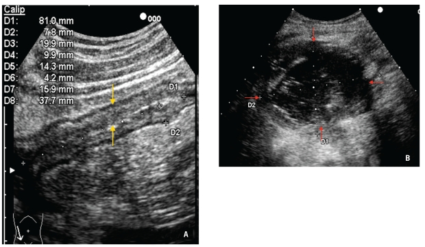
EC Bowel ultrasound provides information regarding intestinal and extraintestinal lesions, including bowel wall thickening, stenosis (with or without dilation), and the presence of abscesses and/or fistulae (Figure 1). In contrast, colonoscopy assesses the mucosal surface and provides a direct view of CD lesions in the colon and terminal ileum.
G&H How does the addition of an oral contrast agent improve bowel ultrasound?
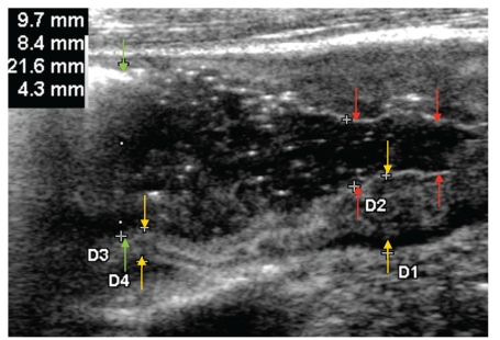
EC The use of oral contrast agents, such as polyethylene glycol solution, has been proposed as a way to improve the detection of CD lesions during bowel ultrasound. The use of polyethylene glycol solution appears to reduce interobserver variability among sonographers and increase ultrasonography's sensitivity for determining the extent of CD lesions, measuring the small bowel lumen, and detecting bowel complications. This technique therefore has value for facilitating early diagnosis and monitoring of CD, and bowel ultrasound may be used as an alternative to invasive procedures for assessing ileal lesions and monitoring their progression over time (Figure 2). From a practical standpoint, use of an oral contrast agent does not alter the procedure greatly; the same equipment is used—with the addition of 375–800 mL of oral contrast fluid—and the procedure duration ranges from 25 minutes to 1 hour.
G&H Why is it important to detect CD recurrences promptly following ileocolonic resection?
EC Surgical resection of the affected bowel is required in almost two thirds of CD patients. Ileocolonoscopy represents the gold standard for detecting postoperative recurrence and is usually performed 6–12 months after ileocolonic resection. In order to prevent possible clinical relapse, the current management of patients following curative resection involves more aggressive medical treatment if patients have severe endoscopic postoperative recurrence. For this reason, prompt detection of postoperative recurrence is mandatory.
G&H In your experience, how well does bowel ultrasound correlate with endoscopy for the detection of postsurgical recurrence of CD?
EC In a study I coauthored, a significant correlation was observed between perianastomotic bowel wall thickness and the Rutgeerts score (P=.0001; r=0.67). A significant correlation was also observed between perianastomotic bowel wall thickness and the degree of recurrence detected by endoscopy when patients were subgrouped according to the amount of time that had elapsed since surgery; this analysis found a stronger correlation among patients who had had surgery more than 3 years previously.
G&H What are the sensitivity and specificity of bowel ultrasound for detecting postsurgical recurrence in patients with CD?
EC In previous studies, bowel ultrasound without oral contrast solution was found to have a sensitivity of approximately 80% for identifying CD recurrence. However, the use of oral contrast solution increases the sensitivity of ultrasound in patients who are being monitored following ileocolonic resection; in our series, bowel ultrasound with oral contrast solution showed a sensitivity of 92.5%, a positive predictive value of 94%, and an accuracy of 87.5% for detecting CD recurrence. The specificity was very low (20%) because only a small number of patients in our study were negative for recurrence.
G&H What are the advantages and disadvantages of other noninvasive imaging modalities?
EC Like ultrasound, magnetic resonance (MR) imaging and MR enterography avoid the need for exposure to ionizing radiation. The main advantage of MR enterography over ultrasound is that MR enterography provides a panoramic view of the whole abdominal cavity, thus allowing clinicians to detect disease involvement in the small bowel and extraintestinal complications in other abdominal organs.
Capsule endoscopy is another promising technology, as it provides direct visual access to the inside of the small intestine without being invasive. The main drawbacks of capsule endoscopy include possible retention of the capsule, an inability to take biopsy samples, and the high cost of this single-use device.
G&H Do you think bowel ultrasound should be routinely used to assess patients with CD?
EC Yes. Over the past few years, the technical evolution of ultrasound equipment, the use of oral and intravenous contrast agents, and an increase in the expertise of sonographers has enhanced the role ultrasonography plays in the assessment of the gastrointestinal tract. In chronic inflammatory conditions, primarily CD, this technology can be used not only for diagnostic purposes, but it has also been suggested that it could play a role in the management of the disease.
G&H What training is necessary to perform bowel ultrasound?
EC Bowel ultrasound has been largely promoted in continental Europe, where ultrasonography is carried out by physicians; in these countries, it is an integral part of the training curriculum for both internal medicine and gastroenterology, and training is mandatory for every physician who wants to learn this technique. There are no published learning curve studies that define expertise in this technique, but I estimate that approximately 6 months and 100 examinations are needed to gain proficiency. The use of bowel ultrasound is less widespread in the United States; because it is performed by radiologists or technicians, rather than gastroenterologists, it is not a tool that US gastroenterologists can readily use in their practices.
Figure 1.
Yellow arrows indicate bowel wall thickening with stenosis of the terminal ileum in a 30-year-old Crohn's disease patient (A). Ultrasonography of a superficial abscess (red arrows), which appears as a round hypoanechoic lesion with internal echoes and an irregular wall, in a 16-year-old female Crohn's disease patient (B).
Figure 2.
Perianastomotic area in a Crohn's disease patient following ileocolonic resection as assessed by bowel ultrasound with ingestion of an oral contrast agent. Yellow arrows show bowel wall thickening with stenosis (red arrows) and loop dilation above the lesion (green arrows).
Suggested Reading
- Onali S, Calabrese E, Petruzziello C, et al. Endoscopic vs ultrasonographic findings related to Crohn's disease recurrence: a prospective longitudinal study at 3 years. . J Crohns Colitis. 2010;4:319–328. doi: 10.1016/j.crohns.2009.12.010. [DOI] [PubMed] [Google Scholar]
- Calabrese E, Petruzziello C, Onali S, et al. Severity of postoperative recurrence in Crohn's disease: correlation between endoscopic and sonographic findings. Inflamm Bowel Dis. 2009;15:1635–1642. doi: 10.1002/ibd.20948. [DOI] [PubMed] [Google Scholar]
- Maconi G, Radice E, Bareggi E, Bianchi Porro G. Hydrosonography of the gastrointestinal tract. AJR Am J Roentgenol. 2009;193:700–708. doi: 10.2214/AJR.08.1979. [DOI] [PubMed] [Google Scholar]
- Castiglione F, Bucci L, Pesce G, et al. Oral contrast-enhanced sonography for the diagnosis and grading of postsurgical recurrence of Crohn's disease. Inflamm Bowel Dis. 2008;14:1240–1245. doi: 10.1002/ibd.20469. [DOI] [PubMed] [Google Scholar]
- Biancone L, Calabrese E, Petruzziello C, et al. Wireless capsule endoscopy and small intestine contrast ultrasonography in recurrence of Crohn's disease. Inflamm Bowel Dis. 2007;13:1256–1265. doi: 10.1002/ibd.20199. [DOI] [PubMed] [Google Scholar]


