Figure 1.
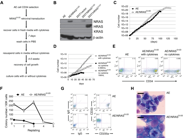
AE/N-RasG12D cells exhibit various features of oncogenic transformation. (A) The “cytokine depletion protocol” for establishing AE/N-RasG12D cells. (B) Western blotting for the indicated proteins. (C) AE and AE/N-RasG12D cells were plated at 0.5 × 10⁶/mL. Cells were counted twice a week, followed by replating at the initial density. (D) AE/N-RasG12D cells grown with cytokine supplementation were plated at 0.5 × 10⁶/mL. AE/N-RasG12D cells grown in the absence of cytokines were plated at 0.5 × 10⁶/mL or 1 × 10⁶/mL. Cells were counted twice a week, followed by replating at the initial density. (E) AE and AE/N-RasG12D cells were stained with allophycocyanin-conjugated anti-CD34 antibody and analyzed by flow cytometry. (F) One representative experiment showing enhanced colony-forming and replating abilities of AE/N-RasG12D cells. CD34+ AE and AE/N-RasG12D cells were plated in methylcellulose media at 5 × 10⁴ cells/mL per plate in triplicate. The number of colonies was scored after 14 days. Cells were collected, washed with phosphate-buffered saline, and replated at 5 × 10⁴ cells/mL per plate in triplicate. (G) Cells collected from methylcellulose media were immunostained with antibodies against the indicated surface markers, followed by flow cytometric analysis. (H) A total of 1 × 10⁵ cells were cytocentrifuged onto glass slides and Wright-Giemsa stained. Pictures were photographed using Leica LAS software with LeicaDMI6000B Inverted Research Microscpoe, 40× dry objective and Leica DFC290HD camera. Bars represent 10 μm.

