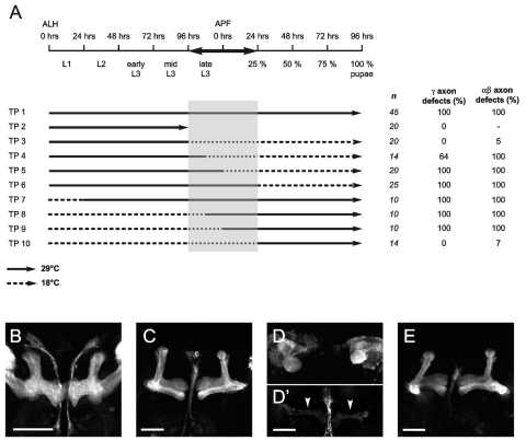
Fig. 3.
Neuroglian function is necessary during late larval and early pupal stages. (A) Diagram of different temperature shifts. The timeline at the top refers to developmental time at 25°C. Wild-type (WT) and Nrg3 embryos were collected at 18°C. First instar larvae were collected within 3 hours of larval hatching (ALH), and then exposed to different temperature programs (TPs 1-10). Broken lines indicate incubation at 18°C (permissive temperature). Black lines represent incubation at 29°C (restrictive temperature). The arrows indicate the time of dissection. αβ and γ lobe phenotypes were scored in anti-FAS2 stained brains. Penetrant neuroanatomical MB defects were found in flies raised at 29°C between late third instar larval (L3) stage and 24 hours after puparium formation (APF). n is the number of brain hemispheres analyzed. (B-E) MBs from Nrg3/−; UAS-GFP::CD8/+;; OK107/+ flies, subjected to one of the TPs depicted in A. (B) MBs from TP 2 larvae are normal. (C) MBs from TP 3 flies do not display mutant phenotypes. (D-D′) Strong MB phenotypes are observed in TP 5 flies. D shows αβ axon stalling phenotypes. D′ displays thin γ neurons (arrowheads; same brain as in D, but image taken at higher high voltage confocal settings). (E) MBs from TP 10 flies do not show mutant phenotypes.

