

Abstract
We screened 1,397 feral horses (Equus caballus) on Sheldon National Wildlife Refuge, Nevada, United States, for IgM and IgG against flavivirus during 2004–2006, 2008, and 2009. Positive serum samples were tested for neutralizing antibodies to West Nile virus (WNV) and St. Louis encephalitis virus (SLEV). One animal was positive for antibody against WNV in 2004, but all others tested in 2004–2006 were negative. In 2008 and 2009, we found evidence of increasing seropositive horses with age, whereas seroprevalence of WNV decreased from 19% in 2008 to 7.2% in 2009. No horses were positive for antibody against SLEV. Being unvaccinated, feral horses can be useful for WNV surveillance.
Little is known about West Nile virus (WNV) in non-domestic (i.e., wild or feral) horses (Equus caballus) beyond serologic surveys that have reported WNV antibody prevalence from < 1% to 63% of animals tested.1–4 The Bureau of Land Management currently manages an estimated 33,700 wild horses in 10 of the western United States.5 As part of the management of these herds, non-domestic horses are routinely rounded up or “gathered” by natural resource management agencies to reduce populations. We report five non-consecutive years of study to determine seroprevalence against WNV in feral horses on Sheldon National Wildlife Refuge in Nevada, United States, by using serum samples obtained during horse gathers.
Sheldon National Wildlife Refuge (Refuge) (Figure 1), managed by the U.S. Fish and Wildlife Service, consists of approximately 575,000 acres in northwest Nevada (41°48′N, 119°14′W). We obtained portions of 1,397 serum samples from horses gathered on the Refuge, originally collected to test for equine infectious anemia, for flavivirus screening. These samples represented 15–40% of the minimum population on the Refuge each year, during 2004–2006 and 2008–2009 (Table 1). Horses were captured primarily by using a helicopter to herd them into a trap corral. Age of captured horses was determined by teeth characteristics and sex was determined.6
Figure 1.
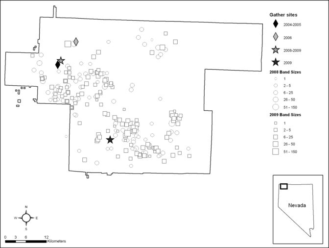
Sheldon National Wildlife Refuge, Nevada, United States. Locations where feral horses were trapped (gather sites) during five years (2004–2006, 2008, and 2009) and numbers of horses per group (band sizes) that were counted during aerial surveys in 2008 and 2009.
Table 1.
Minimum feral horse population and results of testing for antibodies against West Nile virus on Sheldon National Wildlife Refuge, 2004–2006, 2008, and 2009*
| Year | Minimum population | Trap location | Sampling period | No. tested | % Positive (95% confidence interval) |
|---|---|---|---|---|---|
| 2004 | 993 | Swan Creek | Sep–Oct | 403 | 0.2 (0–0.7) |
| 2005 | NC* | Swan Creek | Aug–Sep | 324 | 0 (0–0.9) |
| 2006 | 1,065 | Butler Crossing | Jun | 267 | 0 (0–1.1) |
| 2008 | 1,188 | Little Sheldon | Sep–Oct | 196 | 19 (13–24) |
| 2009 | 1,360 | Little Sheldon | Sept–Oct | 193 | 6.7 (3.2–10) |
| Badger Mountain | Aug | 14 | 14 (0–33) | ||
| 2009 Total | 207 | 7.2† (3.7–11) | |||
| Total | 1,397 | 3.8 (2.8–4.8) |
Horses were not gathered in 2007; NC = not counted.
Significantly less than frequency of seropositive horses in 2008 (P = 0.007, by logistic regression).
Serum samples were heat-inactivated for 30 minutes at 56°C and screened for flavivirus-specific IgM by using a WNV IgM capture enzyme-linked immunosorbent assay (MAC-ELISA) and for flavivirus-specific IgG by using an indirect IgG ELISA.7,8 The assays were modified slightly by the use of recombinant WNV envelope antigen (Henessey Research Associates, Shawnee, KS) diluted 1:100 in phosphate-buffered saline–Tween 20 and the corresponding negative antigen (Henessey Research Associates). The peroxidase substrate system (2,2¢-azino-di [3-ethylbenzthiazoline-6-sulfonate]; Kirkegaard and Perry Laboratories, Gaithersburg, MD) was used as substrate for the anti-flavivirus conjugate diluted 1:6,000 in blocking buffer for the MAC-ELISA and for the horseradish peroxidase–conjugated goat anti-horse IgG di-luted 1:1,500 in phosphate-buffered saline–Tween 20 for the IgG ELISA. Reagent concentrations were determined by checker-board titration. If the optical density of a test sample divided by the optical density of the negative control was ≥ 2.0, the sample was considered provisionally positive for IgM or IgG against WNV. Positive and negative control equine serum samples were used on each ELISA plate.
Serum samples determined to be provisionally positive for IgM or IgG against flavivirus were tested by using a two-fold dilution series and a plaque reduction neutralization test (PRNT) for reactivity to WNV (National Wildlife Health Center American crow [Corvus brachyrhynchos] isolate 16399-3) and St. Louis encephalitis virus (SLEV) (CDC TBH-28).9 Positive and negative control equine (WNV) or avian (SLEV) serum samples, a negative tissue culture control (no virus), and a positive virus control (virus test dose), were included in each PRNT. Sera that exhibited a 90% inhibition of the test dose of the virus were considered positive for the corresponding virus (PRNT90). To differentiate between reactivity to WNV or SLEV, a four-fold difference in titer was required.
Horses sampled in 2008 and 2009 were assigned to four age groups (< 1 year, 1–4 years, 5–9 years, and ≥ 10 years). Horses sampled in 2004–2006 were excluded from statistical analysis because only one animal was seropositive for WNV during that time. We used logistic regression to determine if age or year affected the frequency of seropositive samples and tested for an age effect on seroprevalence within years by using chi-square analysis. We then performed pairwise comparisons of seroprevalence between age groups in 2008 and 2009 by using chi-square analysis with the Bonferroni correction.
In 2004, 1 (0.2%) of 403 horses was seropositive for WNV, but all horses sampled in 2005 and 2006 were negative (Table 1). In 2008, 19% of the horses were seropositive for WNV, which then decreased to 7.2% in 2009 (Table 1). All horses seropositive for WNV were positive by the IgG ELISA and one horse in 2009 was positive for WNV by the MAC-ELISA and the IgG ELISA. The positive horse from 2004 had a PRNT90 titer of 1:160, and titers ranged between 1:20 and 1:640 in horses sampled in 2008 and 2009 (Table 2). Logistic regression on 2008 and 2009 data combined indicated that the frequency of seropositive samples differed by age group (P = 0.001) and year (P = 0.007). In 2009, there was a statistically significant trend of increasing frequency of seropositive samples with age, and the percentage of seropositive samples from horses 5–9 years of age was significantly greater than the percentage in foals and horses 1–4 years of age (Table 3). In 2008, the trend of increasing seropositive samples with age approached significance (Mantel-Haenszel χ2 = 3.476, P = 0.062), and the percentage of seropositive samples from horses 5–9 years of age was significantly greater than the percentage in those 1–4 years of age (Table 3). No horses were positive for antibody against SLEV.
Table 2.
Serum antibody titers against West Nile virus determined by the plaque reduction neutralization test, in feral horses sampled on Sheldon National Wildlife Refuge in 2008 and 2009
| Year | No. tested | No. horses with specific antibody titer for West Nile virus | % Positive (95% confidence interval) | |||||
|---|---|---|---|---|---|---|---|---|
| 1:20 | 1:40 | 1:80 | 1:160 | 1:320 | 1:640 | |||
| 2008 | 196 | 2 | 6 | 8 | 10 | 6 | 5 | 19 (13, 24) |
| 2009 | 207 | 3 | 2 | 1 | 2 | 6 | 1 | 7.2 (3.7, 11) |
Table 3.
Number tested and frequency of feral horses seropositive for West Nile virus on Sheldon National Wildlife Refuge by age group, 2004–2009
| Year | Age group, years | ||||
|---|---|---|---|---|---|
| < 1 | 1–4 | 5–9 | ≥ 10 | Unknown | |
| 2004 | |||||
| No. tested | 89 | 183 | 100 | 31 | 0 |
| % Positive (95% CI*) | 0 (0–3.3) | 0 (0–1.6) | 1 (0–3.0) | 0 (0–9.2) | |
| 2005 | |||||
| No. tested | 70 | 140 | 93 | 19 | 2 |
| % Positive (95% CI) | 0 (0–4.2) | 0 (0–2.1) | 0 (0–3.2) | 0 (0–15) | 0 (0–78) |
| 2006 | |||||
| No. tested | 0 | 146 | 76 | 45 | 0 |
| % Positive (95% CI) | 0 (0–2.0) | 0 (0–3.9) | 0 (0–6.4) | ||
| 2008 | |||||
| No. tested | 0 | 138 | 47 | 11 | 0 |
| % Positive (95% CI) | 14 (8.6–20) | 32† (19–45) | 18 (0–41) | ||
| 2009 | |||||
| No. tested (%) | 57 | 103 | 41 | 2 | 4 |
| % Positive‡ (95% CI) | 1.8 (0–5.2) | 4.8 (0.7–9.0) | 20§ (7.4–32) | 0 (0–78) | 25 (0–67) |
CI = confidence interval.
Significantly greater frequency than 1–4 year age group (χ2 = 6.937, P = 0.008).
Significant trend of increasing frequency of seropositive horses with age in 2009 (Mantel-Haenszel χ2 = 9.018, P = 0.003).
Significantly greater than < 1 year age group (χ2 = 9.016, P = 0.003) and 1–4 year age group (χ2 = 7.672, P = 006).
Our finding of one feral horse seropositive for antibodies against WNV in 2004 is consistent with the fact that the virus was detected for the first time in wild birds and in non-domestic and domestic horses elsewhere in Nevada in 2004.1 It is unclear why none of the horses we sampled in 2005 showed evidence of WNV exposure because WNV was found again in 2005 in wild birds and domestic horses in other areas of Nevada and surrounding states.10 However, we sampled feral horses from relatively small areas distant from the broader statewide surveillance efforts, and conditions within these localized areas may not have been conducive for virus transmission during 2005. In addition, no evidence of WNV exposure was found among 318 passerines of several species that were sampled on the refuge in 2005, which supported the conclusion that WNV activity there was low that year (National Wildlife Health Center, unpublished data). In 2006, feral horses were sampled in June, which was perhaps too early in the WNV transmission season for these horses to have become infected, accounting for the negative results that year.
In all positive horses but one, antibodies to WNV were detected only with the WNV IgG ELISA. The exception was one animal in which antibodies to WNV were detected by the IgG ELISA and the MAC-ELISA. A previous report, citing unpublished data, suggested that IgM to WNV may be detectable in horses for less than three months after infection.11 Most seropositive feral horses were sampled in September and October. Thus, if they had become infected early in the transmission season, IgM to WNV may have decreased to below detectable levels by the time blood was obtained. An experimental study has shown that horses develop low WNV virus titers and that the associated IgM response is weak in some horses, possibly also contributing to our infrequent detection of IgM.7
The evidence for increasing overall WNV seroprevalence with age that we found in feral horses on the Refuge in 2009 and the significantly greater seroprevalence in horses 5–9 years of age than in younger animals in 2008 and 2009 is consistent with increased exposure over time. Similarly, because an earlier report cited unpublished data indicating that antibodies to WNV persist for at least 15 months in horses, we expected to see a greater frequency of seropositive samples from feral horses in 2009 than in 2008, rather than the observed decrease.12 We attribute this primarily to lower WNV activity on the Refuge in 2009. The fact that we did not find a greater prevalence of WNV seropositive samples in horses ≥ 10 years of age than in the other age groups on the Refuge in 2008 and 2009 is inconsistent with the overall age-related trend in animals in 2009, and may have been a result of the small number of horses ≥ 10 years of age that were tested.
Horses are considered dead end hosts of WNV, but wild and feral horses, being unvaccinated, can be useful in WNV surveillance. Although they occupy remote habitats that are generally far removed from human populations, blood samples are routinely obtained from gathered wild and feral horses. Feral horses on the Refuge were negative for antibody to WNV in 2005 and 2006, when WNV was commonly reported in wild bird and veterinary cases elsewhere in Nevada, and the frequency of seropositive samples decreased from 19% in 2008 to 7.2% in 2009. Thus, it remains to be seen if virus activity will persist in horses on the Refuge or if it will occur only sporadically in the future.
ACKNOWLEDGMENTS
We thank the staff of Sheldon-Hart Mountain National Wildlife Refuge Complex for providing logistical support and assistance in the field; Cattoor Livestock Roundup and M. O'Sullivan for gathering horses; L. Pielstick for collecting blood samples; M. Lund, L. Karwal, and C. Carney for providing laboratory assistance; S. Goyal for providing control equine serum samples; M. Samuel for providing consultations on statistics; and T. Rocke and P. Steblein for providing comments on earlier drafts of the manuscript.
Disclaimer: Use of trade, product, or firm names does not imply endorsement by the U.S. Government.
Footnotes
Authors' addresses: J. Christian Franson, Erik K. Hofmeister, and Robert J. Dusek, U.S. Geological Survey, National Wildlife Health Center, Madison, WI, E-mails: jfranson@usgs.gov, ehofmeister@usgs.gov, and rdusek@usgs.gov. Gail H. Collins, U.S. Fish and Wildlife Service, Sheldon-Hart Mountain National Wildlife Refuge Complex, Lakeview, OR, E-mail: gail_collins@fws.gov.
References
- 1.Nevada Department of Agriculture . Nevada Department of Agriculture 2004 West Nile Virus (WNV) Report. Reno, NV: Nevada Department of Agriculture; 2004. [Google Scholar]
- 2.Nevada Department of Agriculture . Nevada Department of Agriculture West Nile Virus (WNV) Report–2005. Reno, NV: Nevada Department of Agriculture; 2005. [Google Scholar]
- 3.Jiménez-Clavero MA, Tejedor CG, Rojo G, Soriguer R, Figuerola J. Serosurvey of West Nile virus in equids and bovids in Spain. Vet Rec. 2007;161:212. doi: 10.1136/vr.161.6.212. [DOI] [PubMed] [Google Scholar]
- 4.Wilkins PA, Glaser AL, McDonnell SM. Passive transfer of naturally acquired specific immunity against West Nile virus to foals in a semi-feral pony herd. J Vet Intern Med. 2006;20:1045–1047. doi: 10.1892/0891-6640(2006)20[1045:ptonas]2.0.co;2. [DOI] [PubMed] [Google Scholar]
- 5.Bureau of Land Management U.S. Department of the Interior, Bureau of Land Management. National Wild Horse and Burro Program. 2010. http://www.blm.gov/wo/st/en/prog/wild_horse_and_burro/wh_b_information_center/Fact_Sheet.html Available at. Accessed July 22, 2010.
- 6.McMullan WC. Dental criteria for estimating age in the horse. Equine Pract. 1983;5:36–43. [Google Scholar]
- 7.Bunning ML, Bowen RA, Cropp CB, Sullivan KG, Davis BS, Komar N, Godsey MS, Baker D, Hettler DL, Holmes DA, Biggerstaff BJ, Mitchell CJ. Experimental infection of horses with West Nile virus. Emerg Infect Dis. 2002;8:380–386. doi: 10.3201/eid0804.010239. [DOI] [PMC free article] [PubMed] [Google Scholar]
- 8.Johnson AJ, Martin DA, Karabatsos N, Roehrig JT. Detection of anti-arboviral immunoglobin G by using a monoclonal antibody-based capture enzyme-linked immunosorbent assay. J Clin Microbiol. 2000;38:1827–1831. doi: 10.1128/jcm.38.5.1827-1831.2000. [DOI] [PMC free article] [PubMed] [Google Scholar]
- 9.Beaty BJ, Calisher CH, Shope RE. Arboviruses. Schmidt NJ, Emmons RW, eds. Diagnostic Procedures for Viral, Rickettsial and Chlamydial Infections. Sixth edition. Washington, DC: American Public Health Association; 1989. pp. 797–855. [Google Scholar]
- 10.U.S. Geological Survey U.S. Geological Survey. West Nile Virus Maps Historical. 2010. http://diseasemaps.usgs.gov/2007/wnv_historical.html Available at. Accessed July 22, 2010.
- 11.Ostlund EN, Andresen JE, Andresen M. West Nile encephalitis. Vet Clin North Am Equine Pract. 2000;16:427–441. doi: 10.1016/s0749-0739(17)30087-1. [DOI] [PubMed] [Google Scholar]
- 12.Ostlund EN, Crom RL, Pedersen DD, Johnson DJ, Williams WO, Schmitt BJ. Equine West Nile encephalitis, United States. Emerg Infect Dis. 2001;7:665–669. doi: 10.3201/eid0704.010412. [DOI] [PMC free article] [PubMed] [Google Scholar]