Abstract
Background:
Aneurysm rupture results in subarachnoid hemorrhage (SAH) with subsequent vasospasm in the cerebral and cerebellar major arteries. In recent years, there has been increasing evidence that hypercholesterolemia plays a role in the pathology of SAH. It is known that hypercholesterolemia is one of the major risk factors for the development of atherosclerosis. Among the factors that have been found to retard the development of atherosclerosis is the intake of a sufficient amount of Vitamin E. An inverse association between serum Vitamin E and coronary heart disease mortality has been demonstrated in epidemiologic studies. Therefore, we tested, in an established model of enhanced cholesterol feed in rabbits, the effects of hypercholesterolemia on vasospasm after SAH by using computed tomography (CT) angiograms of the rabbit basilar artery; in addition, we tested the effects of Vitamin E on these conditions, which have not been studied up to now.
Methods:
In this study rabbits were divided into 3 major groups: control, cholesterol fed, and cholesterol + Vitamin E fed. Hypercholesterolemia was induced by a 2% cholesterol-containing diet. Three rabbit groups were fed rabbit diet; one group was fed a diet that also contained 2% cholesterol and another group was fed a diet containing 2% cholesterol and they received i.m. injections of 50 mg/kg of Vitamin E. After 8 weeks, SAH was induced by the double-hemorrhage method and distilled water was injected into cisterna magna. Blood was taken to measure serum cholesterol and Vitamin E levels. Basilar artery samples were taken for microscopic examination. CT angiography and measurement of basilar artery diameter were performed at days 0 and 3 after SAH.
Results:
Two percent cholesterol diet supplementation for 8 weeks resulted in a significant increase in serum cholesterol levels. Light microscopic analysis of basilar artery of hypercholesterolemic rabbits showed disturbances in the subendothelial and medial layers, degeneration of elastic fibers in the medial layer from endothelial cell desquamation, and a reduction of waves in the endothelial layer. However, the cholesterol + Vitamin E group did not exhibit these changes. The mean diameter of the basilar artery after SAH induction in the cholesterol-treated group was decreased 47% compared with the mean diameter of the control group. This value was less affected in cholesterol + Vitamin E-treated rabbits, which decreased 18% compared with the mean diameter of the control group.
Conclusions:
Hypercholesterolemia-related changes in the basilar artery aggravate vasospasm after SAH. Adding Vitamin E to cholesterol-treated rabbits decreased the degree of vasospasm following SAH in the rabbit basilar artery SAH model. We suggest that Vitamin E supplements and a low cholesterol diet may potentially diminish SAH complicated by vasospasm in high-risk patients.
Keywords: Aneurysm, atherosclerosis, hypercholesterolemia, subarachnoid hemorrhage, vasospasm, Vitamin E
INTRODUCTION
Aneurysm rupture results in subarachnoid hemorrhage (SAH) with subsequent vasospasm in the cerebral and cerebellar major arteries. Vasospasm occurring prior to, during, and after surgery is one of the most important factors affecting the functional prognosis of patients.[9,27] Vasospasm is characterized by the prolonged and reversible contraction of the cerebral arteries.[9,23] Although the pathogenesis of cerebral vasospasm after SAH is still obscure, it is thought to be related to both inflammatory and immunological responses.[13] It has been suggested that many factors act on the sympathetic nervous system, which can influence vasospasm after SAH. Blood and blood breakdown products are also considered to be important spasmogens in the etiology of vasospasm.[11]
Most patients with SAH are hypercholesterolemic and atherosclerotic; therefore, it is important to determine the effects of cerebral vessel atherosclerosis resulting from hypercholesteremia on vasospasm after SAH. It is widely accepted that hypercholesterolemia, which is found with high low-density lipoprotein (LDL), plays a pivotal role in the progression of atherosclerosis.[24] Among the factors that have been found to retard the development of atherosclerosis is the intake of food with a sufficient amount of Vitamin E (α-tocopherol). An inverse association between serum Vitamin E and coronary heart disease mortality has been demonstrated in epidemiologic studies.[15,19]
The aim of the current study was to investigate the effects of hypercholesterolemia on vasospasm, resulting from basilar artery SAH in rabbit model and to compare the effects of Vitamin E on vasospasm in hypercholesterolemia-related changes in the vessels after SAH using CT angiograms of the rabbit basilar artery.
MATERIAL AND METHODS
Experimental design
Sixteen male albino rabbits (1–2 months old) were randomized to 3 groups. All rabbits were fed 100 g rabbit diet per day. Cholesterol was added to the diet as diethyl ether solution. The control diet was treated with the same amount of pure solvent. All diets were dried of the solvent before use. The concentrations of cholesterol and Vitamin E were based on previous reports.[3,22] The first group of rabbits, the control (n=4), was fed the diet without additions and treatments. The second group was fed the diet containing 2% cholesterol (n=6), and the rabbits in the third group were fed the diet containing 2% cholesterol and they received injections of 50 mg/kg of Vitamin E intramuscularly on alternate rear legs, once daily (n=6). After 8 weeks, following withdrawal of food overnight, blood was taken to determine cholesterol and Vitamin E. Each of these 3 major rabbit groups (control, cholesterol, and cholesterol + Vitamin E) was further divided into 2 subgroups as a basilar artery (BA) vasospasm subgroup, which was obtained with injected autologous nonheparinized arterial blood into the cisterna magna and BA control subgroup, for which distilled water[6,26] was injected into the cisterna magna. Afterward, SAH was induced by the double-hemorrhage method.[1,26] A 23-gauge butterfly needle was percutaneously inserted into the cisterna magna of spontaneously breathing animals. Cerebrospinal fluid (CSF) (1.0–1.8 mL) was aspirated using an aseptic technique before each blood injection. Autologous nonheparinized arterial blood obtained from the central ear artery was injected into the cisterna magna for 1–2 min in animals in the BA vasospasm subgroups. The animals were then placed in a 30° head-down tilted position for 15 min to ensure that the blood spread into the basal cistern.[26] In the BA control subgroups, distilled water was injected into the cisterna magna for 1–2 min after CSF aspiration in the manner described above.[26] The first cerebral angiography was performed on day 0, just before induction of SAH. The second CT angiography was done before euthanasia on day 3 after induction of SAH.[7,21] The rabbits were euthanized by intravenous injection of 20 mg/kg penthanol. The brain and the upper cervical spinal cord were completely removed after a craniotomy and upper cervical laminectomy. The basilar artery was microdissected and removed. All animal procedures were approved by the Marmara University, Faculty of Medicine, Animal Care and Use Committee, Istanbul.
Determination of serum cholesterol and Vitamin E levels
Serum cholesterol levels were determined using an automated (Hitachi Modular P800) enzymatic technique (Roche, Boehringer Ingelheim). Vitamin E (α-tocopherol) levels were determined by extracting serum; concentrations of α-tocopherol were obtained by reverse phase high-pressure liquid chromatography and by using a C-18 Bondopak column and a UV detector at 294 nm.[16]
CT angiography and measurement of basilar artery diameter
A high-speed resolution (64 slice) CT was used to identify basilar artery vasospasm (The Phillips Brilliance CT 64-slice scanner, Phillips Company, Netherlands).
Axial sections of the arterial-phase CT angiogram of the cerebral arteries (in particular the basilar artery) were obtained using an injection of 623–769 mg/mL/kg Iopramid in the central ear vein. Parameters of the CT acquisition were 120 kV, 150 mAs, 64 × 0.625 detector collimation; pitch of 1.15, 0.5 s gantry rotation time; 512 × 512 matrix; and 20 cm field of view. Contrast enhancement was provided by the intravenous (central ear vein) administration at the rate of 3mL/s. The luminal diameter of the basilar artery was measured on the Phillips EBW workstation CT scanner using magnification and the appropriate window setting. Axial, coronal, and three-dimensional cranial CT angiograms were obtained after reconstruction of the CT slices [Figure 1].
Figure 1.
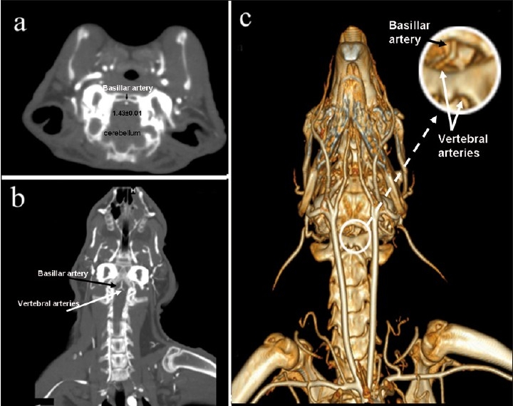
Demonstration of (a) axial, (b) coronal, (c) three-dimensional cranial CT angiograms before basilar artery vasospasm in rabbit (black arrows, basilar artery; white arrows, vertebral artery)
The luminal diameters of the basilar artery were measured by a single observer. The CT angiograms were converted on a computer. Using public domain imaging software, the diameters of the basilar artery were measured on 3 randomly selected points along each artery above the bifurcation of the vertebral arteries. We used the average of the 3 diameters to calculate the final diameter. The luminal relative diameter was used to estimate arterial narrowing. To produce comparable data, the points were the same for all animals.
Preparation of basilar artery for histopathologic analyses
Basilar arteries from all groups were fixed for histopathology in 10% buffered paraformaldehyde for a minimum of 24 h. Tissue samples were prepared in an autotechnicon and embedded in paraffin. The specimens were sectioned (5 μm) with a microtome and deparaffinated 3 times with xylene in a 60°C incubator. The tissue samples were dehydrated with alcohol, washed with water, and stained with Elastica van Gieson. The morphometric characteristics of the tissue samples were evaluated under a light microscope (100× magnification).
Statistical analyses
All data are expressed as mean ± standard deviation (SD). Data were analyzed by one-way ANOVA test using the “Graph Pad Prism 5” statistical program, and the individual comparisons of groups were obtained using Bonferroni's multiple comparison test. P values of less than 0.05 were selected as the levels of significance.
RESULTS
Analyses of serum cholesterol and Vitamin E
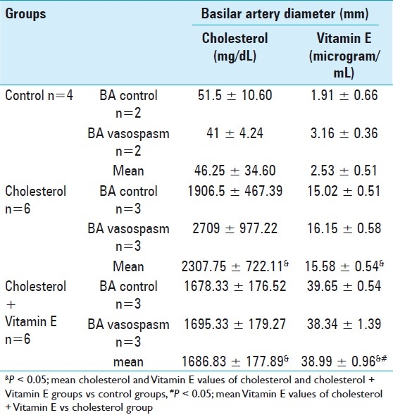
Blood analysis of the animals was performed after 8 weeks of treatment, just before SAH induction. Cholesterol and Vitamin E serum concentrations of the 3 animal groups are shown in Table 1. Two percent cholesterol diet supplementation for 8 weeks resulted in an approximate 50-fold increase of serum cholesterol. In the Vitamin E-treated group a 15-fold increase of the plasma level of Vitamin E was found.
Table 1.
Blood analysis results following cholesterol and cholesterol + Vitamin E treatment
Histological analyses of basilar artery
Rabbit basilar arteries from the 3 groups were examined by light microscopy. Rabbits in control group, which were fed a pure diet, exhibited normal arterial wall layers [Figure 2]. Rabbits in the cholesterol group, which were fed a cholesterol-rich diet, developed atherosclerosis like lesions in the basilar artery, and endothelial cell integrity was impaired relative to the control group. Light microscope analysis (100× magnifications) of the basilar artery showed disturbances in the subendothelial with neutrophil infiltration, degeneration of elastic fibers in the medial layer from endothelial cell desquamation, and a reduction of waves in the endothelial layer [Figure 2]. However, the cholesterol + Vitamin E group did not exhibit these changes [Figure 2].
Figure 2.
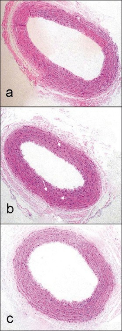
Histological analyses of basilar artery, (a) control group, (b) cholesterol group, (c) cholesterol + Vitamin E (100× magnifications). In the cholesterol group (b), there were disturbances in the subendothelial with neutrophil infiltration (white arrows)
CT angiographic evaluation of basilar artery vasospasm
CT angiography was used to measure the basilar artery diameter before and after SAH in all rabbits. Table 2 shows the effects of 8 weeks of treatment and before and after SAH, and the effects of cholesterol and Vitamin E treatment on the rabbits’ basilar artery diameter. After 8 weeks of the control diet and before production of SAH, the mean basilar artery diameter of the rabbits in control group was 1.43 ± 0.01 (n=4). The mean basilar artery diameter in cholesterol group was 1.34 ± 0.00 (n=6), indicating that the mean basilar artery diameter was somewhat affected by hypercholesterolemia. In the control group, it was decreased 7% compared with the mean diameter. However, the mean diameter of the basilar artery in rabbits in cholesterol + Vitamin E group was 1.43 ± 0.01, similar to control group and greater than the cholesterol group (1.34 ± 0.01). We also measured the changes in the average basilar artery diameter of rabbits in both BA vasospasm and BA control subgroups 3 days after induction of SAH. As shown in Table 2 and Figure 3, there was a statistically significant difference in the mean basilar arterial diameters in the subgroups′ BA vasospasm and BA control within all groups (P < 0.05). Therefore, vasospasm after SAH in the cisterna magna was successfully established in the basilar artery.
Table 2.
Basilar artery diameter results following the cholesterol and cholesterol + Vitamin E treatment
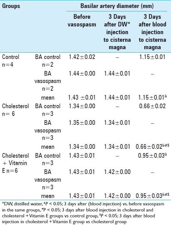
Figure 3.
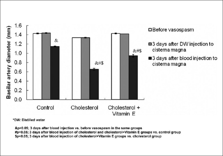
Diameter of basilary artery before and after vasospasm in control, cholesterol, and cholesterol + Vitamin E groups
The average diameter of the atherosclerotic basilar artery after vasospasm induction in the control, cholesterol, and cholesterol + Vitamin E groups (was 1.20 ± 0.01, 0.66 ± 0.02, and 0.95 ± 0.03, respectively. These values were compared by ANOVA test and found to be significantly different (P < 0.05). The mean diameter of the basilar artery in the cholesterol-treated groups was decreased approximately 47% compared to the mean diameter of the control group. This value was less affected in the cholesterol + Vitamin E-treated rabbits, which was decreased 18% compared with the mean diameter of the control group. The mean diameter of the basilar artery after SAH induction decreased in the following order: control group > cholesterol + Vitamin E group > cholesterol group [Figure 4]. The results show that vasospasm resulting from basilar artery subarachnoid hemorrhage is attenuated by Vitamin E in hypercholesterolemic rabbits.
Figure 4.

CT angiograms demonstration of basilar artery after vasospasm induction in rabbits, (a) control group, (b) cholesterol group, and (c) cholesterol + Vitamin E group
DISCUSSION
The development of atherosclerosis is a multifactorial process in which elevated plasma cholesterol levels play a major role; however, most patients with SAH have atherosclerotic vessels. Numerous studies have shown the involvement of the oxidative processes in the pathogenesis of atherosclerosis and the protective effect of Vitamin E against atherosclerosis.[19] Previously, Ozer et al. showed that Vitamin E protects against the development of atherosclerosis in cholesterol-fed rabbits.[15,19,20,22] We studied the same cholesterol-fed rabbit model to test the effects of hypercholesterolemia on vasospasm after SAH by using computed tomography (CT) angiograms of the rabbit basilar artery. In addition we also tested the effect of Vitamin E on these conditions, which have not been studied up to now. We have shown that cholesterol + Vitamin E serum concentrations depend on nutritional supplementation. Two percent cholesterol diet supplementation for 8 weeks resulted in an approximate 50-fold increase of serum cholesterol, a value that was not significantly affected by additional supplementation with Vitamin E. Serum Vitamin E concentrations were higher in the cholesterol-fed rabbits, which agrees with data in the literature.[5,20] This event is caused by a higher Vitamin E uptake (the diet contains low, but not zero, Vitamin E) resulting from increased lipid uptake produced by the high cholesterol diet. A corollary to this finding is that if Vitamin E were absent from the diet, the effect of cholesterol would be more disruptive and the effect of tocopherol supplementation could be exaggerated.
Rabbit basilar arteries from the 3 groups were examined by light microscopy. Rabbits in cholesterol group developed atherosclerosis similar lesions in the basilar artery, and endothelial cell integrity was impaired relative to the control group. They showed disturbances in the subendothelial and medial layers, degeneration of elastic fibers in the medial layer from endothelial cell desquamation, and reduced waves in the endothelial layer. However, the cholesterol + Vitamin E group did not exhibit these changes.
In fact, the atherosclerotic plaque could not be demonstrated in histological examination. Elastica van Gieson stain was used to demonstrate histopathologic analyses. However, histological analysis of the basilar artery after acute SAH using Elantica Van Gieson staining might be more effective for showing degeneration of the tunica muscularis. Nevertheless, initial changes in the subendothelial degeneration of elastic fibers and a reduction of waves in the endothelial layer were observed.
Our results are the first to suggest the potential of Vitamin E as a treatment for vasospasm in hypercholesterolemia-related changes in the vessels after SAH. The present study used CT angiography to measure changes in basilar artery diameter after SAH, while similar studies have used conventional angiography and/or solely histological examination to determine experimental basilar artery vasospasm after SAH.[4,13,26] Although conventional angiography via an arterial catheter is the standard in imaging the arterial system of the brain, reference, CT angiography offers the potential for a rapid, minimally invasive means of accurately diagnosing and monitoring cranial vessels vasospasm after subarachnoidal hemorrhage.[17,25,27] High resolution of multisection CT angiography (64-slice CT) provides quantitative data for measuring vasospasm in small arteries. CT spatial resolution refers to the ability to separate 2 structures and is scientifically determined by measuring the ability of a CT system to separate line pairs between 0.35 and 0.5 mm; the diameter of basilar arteries in each group was measured over these intervals.[8,18] Likewise, the luminal diameter of the basilar artery was measured on the EBW workstation CT scanner using magnification and the appropriate window setting. This provides reliable and semi-quantitative data to measure vasospasm. In addition, CT angiography allows the measurement of vasospasm in a live animal, as opposed to histological examinations of arterial diameter after euthanasia.
We could study only acute stage vasospasm; studies of chronic vasospasm would require a more prolonged study. Cerebral vasospasm after subarachnoid hemorrhage occurs on different time scales among species. Angiographic vasospasm appears 2 days after injecting autologous blood in the rabbit and after about 4-14 days in humans.[7,12,21] In this study, CT angiography was performed on day 3 after SAH. It agrees with the literature. Rabbits fed a 2% cholesterol diet plus 50 mg/kg of Vitamin E experienced decreased vasospasm in the basilar artery, effects that are mediated by Vitamin E.
Early or late period vasospasm following SAH is known. Although various spasmogens and their origins have been studied, the pathogenesis of vasospasm following SAH remains unclear.[28] Since we considered cholesterol and how Vitamin E treatments affect basilar artery diameter, in this study we compared basilar artery diameter before and after vasospasm in each group and after vasospasm; groups were compared with control and cholesterol groups. The average diameter of the basilar artery before vasospasm decreased 7% due to hypercholesterolemia. The mean diameter of the basilar artery after vasospasm was decreased 47% compared with the mean diameter of the control group. In other words, vasospasm following SAH in the artery of hypercholesterolemic rabbits was aggravated compared with the normocholesterolemic rabbits. Based on the literature, increased vessel wall rigidity and a decreased capacity to develop active tone are the dominant features of cerebral arteries in experimental cerebral vasospasm.[2,10,14] We presume that histopathologic changes in subendothelial and medial layers, such as degeneration of elastic fibers in the medial layer, accumulation of lipid (foam cells), endothelial desquamation, decreased waves in the endothelial layer, and vascular degeneration of tunica muscularis, caused increased artery wall rigidity and decreased capacity to develop active tone. These changes consequently caused a large degree of vasospasm in atherosclerotic vessels without plaque. In this study, we examined the role of hypercholesterolemia-related changes on vasospasm following SAH. It is also known that vasospasm can be influenced by many factors and that the sympathetic nervous system may also be involved.[4]
In conclusion, hypercholesterolemia increases vasospasm resulting in basilar artery subarachnoid hemorrhage; however, we found that supplementation with Vitamin E decreased the degree of vasospasm in hypercholesterolemic animals following SAH in the rabbit basilar artery SAH model. For the first time, we suggest that Vitamin E supplements and a low cholesterol diet may potentially diminish SAH complicated by vasospasm in hypercholesterolemia patients who are at a high risk.
Footnotes
Available FREE in open access from: http://www.surgicalneurologyint.com/text.asp?2011/2/1/29/77600
Contributor Information
Mehdi Sasani, Email: sasanim@gmail.com.
Burak Yazgan, Email: byazgan@hotmail.com.
Irfan Celebi, Email: irfancelebi@gmail.com.
Nurgul Aytan, Email: naytan@gmail.com.
Betul Catalgol, Email: betulcatalgol @gmail.com.
Tunc Oktenoglu, Email: tuncoktenoglu@gmail.com.
Tuncay Kaner, Email: tkaner2002@yahoo.com.
Nesrin Kartal Ozer, Email: nkozer@marmara.edu.tr.
Ali Fahir Ozer, Email: alifahirozer@gmail.com.
REFERENCES
- 1.Baker KF, Zervas NT, Pile-Spellman J, Vacanti FX, Miller D. Angiographic evidence of basilar artery constriction in the rabbit a new model of vasospasm. Surg Neurol. 1987;27:107–12. doi: 10.1016/0090-3019(87)90280-1. [DOI] [PubMed] [Google Scholar]
- 2.Bevan JA, Bevan RD, Frazee JG. Functional arterial changes in chronic cerebrovasospasm in monkeys an in vitro assessment of the contribution to arterial narrowing. Stroke. 1987;18:472–81. doi: 10.1161/01.str.18.2.472. [DOI] [PubMed] [Google Scholar]
- 3.Bocan TM, Mueller SB, Mazur MJ, Uhlendorf PD, Brown EQ, Kieft KA. The relationship between the degree of dietary-induced hypercholesterolemia in the rabbit and atherosclerotic lesion formation. Atherosclerosis. 1993;102:9–22. doi: 10.1016/0021-9150(93)90080-e. [DOI] [PubMed] [Google Scholar]
- 4.Cosar M, Iplikcioglu AC, Aytan N, Ozcan D, San T, Kartal-Ozer N, et al. The effect of temporary aneurysm clip on the common carotid artery of atherosclerotic rabbits. Surg Neurol. 2008;69:483–8. doi: 10.1016/j.surneu.2007.01.053. [DOI] [PubMed] [Google Scholar]
- 5.Godfried SL, Combs GF, Saroka JM, Dillingham LA. Potentiation of atherosclerotic lesions in rabbits by a high dietary level of vitamin E. Br J Nutr. 1989;61:607–17. doi: 10.1079/bjn19890148. [DOI] [PubMed] [Google Scholar]
- 6.Hirashima Y, Endo S, Kato R, Takaku A. Prevention of cerebrovasospasm following subarachnoid hemorrhage in rabbits by the platelet-activating factor antagonist, E5880. J Neurosurg. 1996;84:826–30. doi: 10.3171/jns.1996.84.5.0826. [DOI] [PubMed] [Google Scholar]
- 7.Isotani E, Azuma H, Suzuki R, Hamasaki H, Sato J, Hirakawa K. Impaired endothelium-dependent relaxation in rabbit pulmonary artery after subarachnoid hemorrhage. J Cardiovasc Pharmacol. 1996;28:639–44. doi: 10.1097/00005344-199611000-00005. [DOI] [PubMed] [Google Scholar]
- 8.Judy PF. The line spread function and modulation transfer function of a computed tomographic scanner. Med Phys. 1976;3:233–6. doi: 10.1118/1.594283. [DOI] [PubMed] [Google Scholar]
- 9.Kassell NF, Helm G, Simmons N, Phillips CD, Cail WS. Treatment of cerebral vasospasm with intra-arterial papaverine. J Neurosurg. 1992;77:848–52. doi: 10.3171/jns.1992.77.6.0848. [DOI] [PubMed] [Google Scholar]
- 10.Kim P, Sundt TM, Vanhoutte PM. Alterations of mechanical properties in canine basilar arteries after subarachnoid hemorrhage. J Neurosurg. 1989;71:430–6. doi: 10.3171/jns.1989.71.3.0430. [DOI] [PubMed] [Google Scholar]
- 11.Mayberg MR, Okada T, Bark DH. The significance of morphological changes in cerebral arteries after subarachnoid hemorrhage. J Neurosurg. 1990;72:626–33. doi: 10.3171/jns.1990.72.4.0626. [DOI] [PubMed] [Google Scholar]
- 12.Mizuno Y, Azuma H, Ito Y, Isotani E, Ohno K, Hirakawa K. Inhibitory effect of activated protein C on cerebral vasospasm after subarachnoid hemorrhage in the rabbit. J Cardiovasc Pharmacol. 2002;39:729–38. doi: 10.1097/00005344-200205000-00014. [DOI] [PubMed] [Google Scholar]
- 13.Nakai K, Morimoto Y, Wada K, Nawashiro H, Shima K, Kikuchi M. Pretreatment with continuous-wave ultraviolet irradiation to prevent the development of delayed vasospasm in the rabbit common carotid artery model. J Neurosurg. 2000;92:671–5. doi: 10.3171/jns.2000.92.4.0671. [DOI] [PubMed] [Google Scholar]
- 14.Naredi S, Lambert G, Edén E, Zäll S, Runnerstam M, Rydenhag B, et al. Increased sympathetic nervous activity in patients with nontraumatic subarachnoid hemorrhage. Stroke. 2000;31:901–6. doi: 10.1161/01.str.31.4.901. [DOI] [PubMed] [Google Scholar]
- 15.Negis Y, Aytan N, Ozer N, Ogru E, Libinaki R, Gianello R, et al. The effect of tocopheryl phosphates on atherosclerosis progression in rabbits fed with a high cholesterol diet. Arch Biochem Biophys. 2006;450:63–6. doi: 10.1016/j.abb.2006.02.027. [DOI] [PubMed] [Google Scholar]
- 16.Nierenberg DW, Nann SL. A method for determining concentrations of retinol tocopherol and five carotenoids in human plasma and tissue samples. Am J Clin Nutr. 1992;56:417–26. doi: 10.1093/ajcn/56.2.417. [DOI] [PubMed] [Google Scholar]
- 17.Ochi RP, Vieco PT, Gross CE. CT angiography of cerebral vasospasm with conventional angiographic comparison. Am J Neuroradiol. 1997;18:265–9. [PMC free article] [PubMed] [Google Scholar]
- 18.Otero HJ, Steigner ML, Rybicki FJ. e “post-64” era of coronary CT angiography: Understanding new technology from physical principles. Radiol Clin North Am. 2009;47:79–90. doi: 10.1016/j.rcl.2008.11.001. [DOI] [PMC free article] [PubMed] [Google Scholar]
- 19.Ozer NK, Azzi A. Effect of vitamin E on the development of atherosclerosis. Toxicology. 2000;148:179–85. doi: 10.1016/s0300-483x(00)00209-2. [DOI] [PubMed] [Google Scholar]
- 20.Ozer NK, Negis Y, Aytan N, Villacorta L, Ricciarelli R, Zingg JM, et al. Vitamin E inhibits CD36 scavenger receptor expression in hypercholesterolemic rabbits. Atherosclerosis. 2006;18:415–20. doi: 10.1016/j.atherosclerosis.2005.03.050. [DOI] [PubMed] [Google Scholar]
- 21.Shishido T, Suzuki R, Qian L, Hirakawa K. The role of superoxide anions in the pathogenesis of cerebral vasospasm. Stroke. 1994;25:864–8. doi: 10.1161/01.str.25.4.864. [DOI] [PubMed] [Google Scholar]
- 22.Sirikci O, Ozer NK, Azzi A. Dietary cholesterol-induced changes of protein kinase C and the effect of vitamin E in rabbit aortic smooth muscle cells. Atherosclerosis. 1996;126:253–63. doi: 10.1016/0021-9150(96)05909-6. [DOI] [PubMed] [Google Scholar]
- 23.Solomon RA, Antunes JL, Chen RY, Bland L, Chien S. Decrease in cerebral blood flow in rats after experimental subarachnoid hemorrhage a new animal model. Stroke. 1985;16:58–64. doi: 10.1161/01.str.16.1.58. [DOI] [PubMed] [Google Scholar]
- 24.Steinberg D, Parthasarathy S, Carew TE, Khoo JC, Witztum JL. Beyond cholesterol. Modifications of low-density lipoprotein that increase its atherogenicity. N Engl J Med. 1989;320:915–24. doi: 10.1056/NEJM198904063201407. [DOI] [PubMed] [Google Scholar]
- 25.Tins B, Oxtoby J, Patel S. Comparison of CT angiography with conventional arterial angiography in aortoiliac occlusive disease. Br J Radiol. 2001;74:219–25. doi: 10.1259/bjr.74.879.740219. [DOI] [PubMed] [Google Scholar]
- 26.Tsurutani H, Ohkuma H, Suzuki S. Effects of thrombin inhibitor on thrombin-related signal transduction and cerebral vasospasm in the rabbit subarachnoid hemorrhage model. Stroke. 2003;34:1497–1500. doi: 10.1161/01.STR.0000070424.38138.30. [DOI] [PubMed] [Google Scholar]
- 27.Wilkins RH. Attempted prevention or treatment of intracranial arterial spasm a survey. Neurosurgery. 1980;61:98–210. doi: 10.1227/00006123-198002000-00016. [DOI] [PubMed] [Google Scholar]
- 28.Zubkov AY, Ogihara K, Tumu P, Patlolla A, Lewis AI, Parent AD, et al. Mitogen-activated protein kinase mediation of hemolysate-induced contraction in rabbit basilar artery. J Neurosurg. 1999;90:1091–7. doi: 10.3171/jns.1999.90.6.1091. [DOI] [PubMed] [Google Scholar]


