Abstract
The carpal boss is an osseous overgrowth that is occasionally mistaken for a ganglion cyst. This report highlights the case a 36-year-old patient who was originally diagnosed by his primary care physician with a ganglion cyst and was sent to an orthopaedist for aspiration. Upon further evaluation with a plain radiograph, the dorsal wrist mass was found to be a carpal boss. The patient was treated with rest and a wrist brace, and was informed that a corticosteroid injection or surgical excision would be necessary if conservative treatment failed. The patient was asymptomatic on follow-up and invasive procedures were not necessary.
Background
Wrist pain is a common complaint to both primary care and orthopaedic providers. Although the most frequent cause of a wrist mass is a ganglion cyst, other aetiologies should be considered. Diagnosis was evident following radiographic evaluation. A carpal boss should always be considered in the differential diagnosis of a patient with a dorsal wrist mass.
Case presentation
A 36-year-old male presented to his primary care provider with a solid mass on the dorsum of his left hand. The mass had been present for 7 months and had recently increased in size. The patient denied a history of injury to the area. His is right-hand dominant and described a mild, dull and aching pain that worsened with prolonged typing. His medical and family history were unremarkable.
Physical examination revealed a 6 mm firm mass on the dorsal aspect of the hand along the proximal second metacarpal. The mass was hard and unmovable. Wrist flexion, extension and ulnar and radial deviation were all normal, and the patient demonstrated normal motion at the metacarpophalangeal (MCP) and interphalangeal joints. There was normal MCP joint contour with a clenched fist and no snuffbox tenderness. He had a normal elbow exam and a normal neuro-vascular exam of the wrist and hand. The patient was diagnosed with a ganglion cyst and referred to an orthopaedist for aspiration.
Investigations
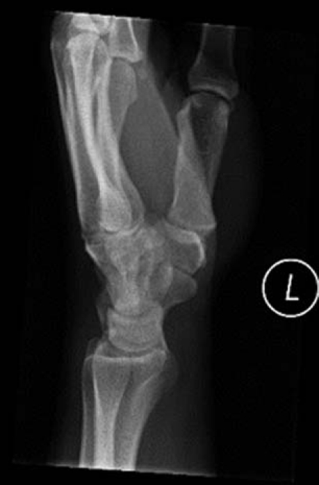
Dorsal-palmar and lateral radiographs of the left wrist were ordered and revealed no fractures and normal joint spaces. However, the radiographs did reveal a bony protuberance at the carpometacarpal (CMC) joint of the index finger (figure 1).
Figure 1.

Lateral radiograph of the wrist.
Differential diagnosis
The differential diagnosis should include ganglion cyst, subcutaneous calcification, giant cell tumours from the synovial tissue, lipomas, neuromas, neurofibromas, enchondromas, osteochondromas and osteoid osteomas.
Treatment
The diagnosis of a carpal boss at the second CMC joint was clear from this imaging. The patient was reassured that this was a benign finding and that the initial approach would be to use a cock-up wrist splint for 4 weeks and take NSAIDs as needed. Injection of a corticosteroid and surgery was discussed as an option if conservative treatment failed.
Outcome and follow-up
At the 4-week follow-up, the patient was pain free and had no complaints. He had been compliant with using his brace and taking NSAIDs. He did not feel that a corticosteroid injection was necessary and was not interested in surgery and would follow-up if pain returned.
Discussion
The carpal boss is described as a small protuberance of bone frequently found at the base of the second or third CMC joint. It may also be found fused to the surface of the trapezoid or capitate bones.1 2 The carpal boss can be asymptomatic but commonly causes pain similar to that of osteoarthritis. The pain is most frequently located near the base of the second or third metacarpals, the capitate or the scapholunate joint, and increases with activity and decreases with rest.2 A ganglion cyst or an os styloideum may be present as well.2 Carpal bosses are more common in females and occur more frequently in the right hand.
The focal degenerative disease of the carpal boss is thought to be caused by an abnormal configuration of bones and their inability to withstand repetitive stress and contact within the joint. It may be caused by periostitis at the insertion of the radial wrist extensor tendon, peritendinitis calcarea or non-union of a chip fracture at the base of the second or third metacarpals. It is most commonly thought to be a result of hypertrophic changes from post-traumatic laxity of the involved bony contacts.3 Additionally, degeneration of the MCP joint can lead to an osteophyte or a ganglion cyst that can lead to a carpal boss. If the carpal boss is already present, one of these other problems can increase irritation in the area.2
Primary treatment of the carpal boss involves rest, NSAIDs and immobilisation. Local steroid injection can be helpful for the periostitis or if peritendonitis calcarea is thought to be a contributing factor.4 If the carpal boss remains symptomatic after conservative treatment, surgical intervention should be considered. There are two procedures described for this condition. The first procedure is surgical excision of the bony protuberance. The second is arthrodesis of the affected CMC joint.1 2 A study conducted by Artz et al5 found that with no specific injury causing the carpal boss, only 11.4% of patients required surgery, while when there was a history of injury, 45.5% of patients required surgery. After a follow-up of 3.5 years, Cuono reported that there were no recurrences after complete excision.6
Learning points.
-
▶
A carpal boss is a treatable condition that may be mistaken for a ganglion cyst.
-
▶
This condition may signify localised degenerative arthritis at the second and third carpometacarpal articulations and should be considered in any patient with a hard bony mass on the dorsum of the hand.
-
▶
Diagnosis is made after a characteristic radiograph shows a dorsal overgrowth of the bones involved.
Footnotes
Competing interests None.
Patient consent Obtained.
References
- 1.Clarke AM, Wheen DJ, Visvanathan S, et al. The symptomatic carpal boss. Is simple excision enough? J Hand Surg Br 1999;24:591–5 [DOI] [PubMed] [Google Scholar]
- 2.Conway WF, Destouet JM, Gilula LA, et al. The carpal boss: an overview of radiographic evaluation. Radiology 1985;156:29–31 [DOI] [PubMed] [Google Scholar]
- 3.Joseph RB, Linscheid RL, Dobyns JH, et al. Chronic sprains of the carpometacarpal joints. J Hand Surg Am 1981;6:172–80 [DOI] [PubMed] [Google Scholar]
- 4.Lewis DD. Carpal boss and the differential diagnosis of dorsal hand masses. J Am Board Fam Pract 1994;7:248–9 [PubMed] [Google Scholar]
- 5.Artz TD, Posch JL. The carpometacarpal boss. J Bone Joint Surg Am 1973;55:747–52 [PubMed] [Google Scholar]
- 6.Cuono CB, Watson HK. The carpal boss: surgical treatment and etiological considerations. Plast Reconstr Surg 1979;63:88–93 [PubMed] [Google Scholar]


