Abstract
The molecular mechanisms that achieve homeostatic stabilization of neural function remain largely unknown. To help understand how neural function is stabilized during development and throughout life we used an electrophysiology-based forward genetic screen and assessed the function of over 250 neuronally expressed genes for a role in the homeostatic modulation of synaptic transmission in Drosophila. This screen ruled out the involvement of numerous synaptic proteins and identified a critical function for dysbindin, a gene linked to schizophrenia in human. Dysbindin was essential, presynaptically, for the retrograde, homeostatic modulation of neurotransmission. Dysbindin functioned in a dose-dependent manner downstream or independently of calcium influx. Thus, Dysbindin is essential for adaptive neural plasticity, and may link altered homeostatic signaling with a complex neurological disease.
At glutamatergic synapses of species ranging from Drosophila to human, disruption of postsynaptic neurotransmitter receptor function can be precisely offset by an increase in presynaptic neurotransmitter release to homeostatically maintain normal postsynaptic excitation (1–3). The Drosophila neuromuscular junction (NMJ) is a glutamatergic synapse that is used as a model for this form of homeostatic signaling in the nervous system (1, 4, 5). Efficient homeostatic modulation of presynaptic release at the Drosophila NMJ can occur in ten min following bath application of philanthotoxin-433 (PhTx), which persistently and specifically inhibits postsynaptic glutamate receptors (fig. S1)(4).
We have systematically screened for mutations that block the rapid, PhTx-dependent induction of synaptic homeostasis (Fig. 1). Mutations in 276 genes were screened electrophysiologically (see Supporting Text). For each mutant, we calculated an average value for the amplitude of both the spontaneous miniature excitatory junctional potential (mEJP) and evoked excitatory junctional potential (EJP) following treatment of the dissected neuromuscular preparation with PhTx for 10 min (4). We isolated 14 mutants with average EJP amplitudes more than two standard deviations smaller than the distribution mean (Fig. 1C, red filled bars). From these candidates we identified 7 mutants that block synaptic homeostasis without an obvious effect on NMJ morphology or baseline synaptic transmission. We conclude that the molecular mechanisms of synaptic homeostasis can be genetically separated from the mechanisms responsible for normal neuromuscular development and baseline synaptic transmission.
Figure 1. Electrophysiology-based screen for homeostatic mutations.
(A) Flow diagram of screen strategy and outcome. (B) Histogram of average mEJP amplitude per genotype after PhTx application. Wild-type average mEJP (blue arrow) and wild-type average mEJP after PhTx application (black arrow) are indicated. (C) Histogram of average EJP amplitudes per genotype after PhTx application. Arrows as in (B). Red columns indicate values greater than 2 standard deviations from the mean. (D,E) Homeostatic increases in quantal content observed in published genetic mutations. Data are normalized to the same genotype without PhTx treatment in D, and to wild-type values in E. Full genotypes, n values, and references are shown in Tables S1,2.
A fraction of the mutants we assayed (19.5%) are previously published genetic lesions. This allows us to rule out the involvement of numerous genes and associated biochemical processes. Mutations that disrupt RNA-interference/micro-RNA processing, retrograde trans-synaptic signaling, synaptic transmission, active zone assembly, synaptic vesicle endocytosis and mitochondria all showed reliable homeostatic compensation (Fig. 1, D and E, and fig. S1). Therefore, synaptic homeostasis is a robust phenomenon, unperturbed by a broad spectrum of synaptic mutations. In addition, significant homeostatic compensation in synaptojanin and endophilin mutants argues against the involvement of synaptic vesicle endocytosis and indicates that the size of the recycling synaptic vesicle pool is not a limiting factor for synaptic homeostasis. These data also emphasize the importance and specificity of those mutations we identified that do block synaptic homeostasis. These include four ion channels, two of which are of unknown function, and two calcium-binding proteins of unknown function. Thus, homeostatic signaling at the NMJ may include previously unexplored mechanisms of synaptic modulation.
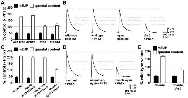
One mutation that was identified with a specific defect in homeostatic compensation is a transposon insertion that resides in the Drosophila homologue of dysbindin (CG6856; fig. S2). The DTNBP1 (dysbindin) locus is linked with schizophrenia in humans (6–11). We have identified a transposon insertion within the dysbindin locus (pBace01028, referred to as dysb1; fig. S2) that showed a complete absence of homeostatic compensation following application of PhTx (Fig. 2, A and B). A similar effect was observed when dysb1 was placed in trans to a deficiency that uncovers the dysb locus, indicating that the dysb1 mutant was a strong loss of function or null mutation (Fig. 2, A and B). No significant change in baseline synaptic transmission in dysb1 mutant animals (0.5 mM extracellular calcium) was observed. Thus, under these recording conditions, this mutation disrupted synaptic homeostasis without altering baseline neurotransmission (Fig. 2B). As a control, synaptic homeostasis was normal in animals in which the pBace01028 transposon was precisely excised (Fig. 2, C and D).
Figure 2. Dysbindin is required presynaptically for synaptic homeostasis.
(A) Mutations in dysbindin block the homeostatic increase in quantal content following PhTx application. Data are normalized to each genotype in the absence of PhTx. (B) Representative traces for data in (A). (C) Precise excision of the e01028 transposon (revertant) restores compensation. Neuronal-specific expression of dysb (with or without a venus tag; c155-GAL4/+; UAS-dysb/+; dysb1) restores compensation. Muscle-specific expression (UAS-dysb/+; mhc-GAL4, dysb1/dysb1) does not. (D) Representative traces for data in (C). (E) Sustained homeostatic compensation is blocked in GluRIIASP16; dysb1 double mutants.
The dysb gene is ubiquitously expressed in Drosophila embryos (fig. S2) consistent with widespread expression in vertebrates (12, 13). Therefore, we generated and expressed a dysbindin transgene in the dysb1 mutant. Presynaptic expression of dysb fully restored homeostatic compensation in the dysb1 mutant background, whereas muscle-specific expression of dysb did not (Fig. 2, C and D, and fig. S3). Thus, Dysbindin is necessary presynaptically for the rapid induction of synaptic homeostasis.
We next asked whether Dysbindin is also required for the sustained expression of synaptic homeostasis. We generated double mutant animals harboring both the dysb1 mutation and a mutation in a gene encoding a postsynaptic glutamate receptor (GluRIIA). GluRIIA mutant animals normally show robust homeostatic compensation (14). However, homeostatic compensation was blocked in GluRIIA; dysb1 double mutant animals (Fig. 2E). Thus, dysbindin was also necessary for the sustained expression of synaptic homeostasis over several days of larval development.
Next we demonstrated that synapse morphology was qualitatively normal in dysb mutants (Fig. 3A) including both the shape of the presynaptic nerve terminal and the levels, localization and organization of synaptic markers including futsch-positive microtubules, synapsin and synaptotagmin (fig. S4). Bouton number and active zone density are also normal in dysb mutants (Fig. 3, C to E)(15). Thus, the disruption of synaptic homeostasis in dysb1 mutants is not a secondary consequence of altered or impaired NMJ development.
Figure 3. NMJ morphology and Dysbindin localization.
(A) Wild-type (w1118) and dysb1 mutant NMJs co-immunostained for anti-HRP (red; motoneuron membrane) and anti-DLG (green; postsynaptic muscle membrane). (B) ven-dysb expressed in neurons co-localizes with the synaptic vesicle associated protein synapsin (green). (C) Quantification of bouton number at muscles 4 and 6/7 comparing wild-type (n=19) and dysb (n=17) (p>0.41; Student’s t-test). (D) Quantification of nc82 puncta per NMJ comparing wild-type (n=15) and dysb (n=16) (p>0.97; Student’s t-test). (E) Density of nc82 puncta comparing wild-type (n=15) and dysb (n=16) (p>0.32; Student’s t-test). Scale bar is 5 microns.
In the vertebrate nervous system, Dysbindin is associated with synaptic vesicles (16). We examined the localization of a Venus-tagged dysb transgene (ven-dysb) that rescues the dysb1 mutant (Fig. 2, C and D). Ven-Dysb showed extensive overlap with synaptic vesicle associated proteins when expressed in neurons (Fig. 3B). Thus, Dysbindin functions presynaptically, potentially at or near the synaptic vesicle pool.
To further define the function of Dysbindin, we investigated baseline synaptic transmission in the dysb mutant in greater detail. At 0.5 mM extracellular calcium, synaptic transmission in dysb1 mutant animals was in distinguishable from wild type (Fig. 2, A and B). However, when extracellular calcium was reduced (0.2 – 0.4 mM Ca2+), baseline synaptic transmission was significantly impaired in dysb compared to wild type (Fig. 4A) and this defect was rescued by presynaptic expression of dysb (fig. S5). Thus, there is an alteration of the calcium dependence of synaptic transmission in the dysb mutant. Indeed, at reduced extracellular calcium, both paired-pulse facilitation and facilitation that occurs during a prolonged stimulus train were increased in dysb mutants (Fig. 4B and fig. S6).
Figure 4. Dysbindin modulates the calcium-dependence of vesicle release.
(A) Quantal content as a function of extracellular calcium concentration. (B) Enhanced paired-pulse facilitation in dysb1 mutants. (C) Quantal content is increased by neuronal overexpression of dysb (dysb OE; c155-GAL4/Y; UAS-dysb/+). (D) Homeostatic compensation is calculated as in Fig. 1E. Compensation is suppressed in cacS/+ (n=15) compared with wild type (n=25; p<0.001; Student’s t-test) or dysb1/+ (n=17; p<0.001; Student’s t-test) but is not further suppressed in cacS/+; dysb1/+ mutants (n=19; p>0.27; Student’s t-test). (E) Compensation remains blocked when cac is neuronally overexpressed in a dysb1 mutant (n=9, dysb;cac OE; c155-GAL4/Y; UAS-cacGFP/+; dysb1). (F) Quantal content is increased by neuronal overexpression of dysb (n=17, dysb OE; c155-GAL4/Y; UAS-dysb/+; p<0.0002; Student’s t-test) and when dysb is overexpressed in a cacS mutant (n=19, cacS/+; dysb OE; cacS/c155-GAL4; UAS-dysb/+; p<0.006; Student’s t-test).
In vertebrates, the levels of dysb expression correlate with parallel changes in extracellular glutamate concentration (17). Therefore, we tested whether dysb overexpression might increase presynaptic release. In wild-type animals overexpressing dysb in neurons, synaptic transmission is normal at low extracellular calcium (0.2 and 0.3 mM Ca2+) but was enhanced at relatively higher extracellular calcium (0.5 mM Ca2+; Fig. 4C and fig. S5). The complementary effects of dysb loss-of-function and overexpression confirm that Dysbindin has an important influence on calcium-dependent vesicle release.
The presynaptic CaV2.1 calcium channel, encoded by cacophony (cac), is required for synaptic vesicle release at the Drosophila NMJ (18). cac mutations decrease presynaptic calcium influx (19) and also block synaptic homeostasis (4). We therefore tested for a genetic interaction between dysb and cac during synaptic homeostasis. Because homozygous cac and dysb mutations individually block synaptic homeostasis, analysis of double mutant combinations would not be informative. We resorted to an analysis of heterozygous mutant combinations and gene overexpression (5). Synaptic homeostasis was suppressed by a heterozygous mutation in cac (Fig. 4D). However, this suppression was not enhanced by the presence of a heterozygous mutation in dysb (Fig. 4 and fig. S7). In addition, neuronal overexpression of cac did not restore homeostatic compensation in dysb mutant animals and the enhancement of presynaptic release caused by neuronal dysb overexpression still occurs in a heterozygous cac mutant background (Fig. 4, E and F). Thus, Dysbindin may function downstream or independently of Cac during synaptic homeostasis.
To further explore the relationship between Dysbindin and Cac, we asked whether dysb mutations might directly influence presynaptic calcium influx. The spatially averaged calcium signal in dysb1 was in distinguishable from wild type, indicating no difference in presynaptic calcium influx (fig. S8). Thus, Dysbindin appears to function downstream or independently of calcium influx to control synaptic homeostasis.
Through a systematic electrophysiological analysis of more than 250 mutants we were able to rule out the involvement of numerous synaptic proteins and biochemical processes in the mechanisms of synaptic homeostasis and demonstrate that this phenomenon is separable from the molecular mechanisms that specify structural and functional synapse development. We then identify Dysbindin as an essential presynaptic component within a homeostatic signaling system that regulates and stabilizes synaptic efficacy. Dysbindin functions downstream or independently of the presynaptic CaV2.1 calcium channel in the mechanisms of synaptic homeostasis.
Emerging lines of evidence suggest that glutamate hypofunction could be related to the etiology of schizophrenia (20–24). Likewise, reduced levels of dysbindin expression were associated with schizophrenia (25, 26). The sandy mouse, which lacks Dysbindin, has a decreased rate of vesicle release (~30% decrease), a correlated decrease in vesicle pool size and an increased thickness of the postsynaptic density (27). We confirm a modest, facilitatory function for Dysbindin during baseline transmission. However, numerous mutations with similar or more severe defects in baseline transmission show normal synaptic homeostasis (Fig. 1, D and E). By contrast, loss of Dysbindin completely blocks the adaptive, homeostatic modulation of vesicle release, suggesting that the potential contribution of dysbindin mutations to schizophrenia may be derived from altered homeostatic plasticity as opposed to decreased baseline glutamatergic transmission.
Supplementary Material
Acknowledgments
We thank M. Muller for assistance with data analysis and members of the Davis laboratory for comments and discussion. This work was supported by postdoctoral fellowships from the A.P. Giannini Foundation and Jane Coffin Childs Memorial Fund to DKD and by NIH grant number NS39313 to GWD.
Footnotes
Materials and Methods
Supplemental Text
Tables S1 and S2
Figs. S1- S8
References
References
- 1.Davis GW. Annu Rev Neurosci. 2006;29:307. doi: 10.1146/annurev.neuro.28.061604.135751. [DOI] [PubMed] [Google Scholar]
- 2.Branco T, Staras K, Darcy KJ, Goda Y. Neuron. 2008 Aug 14;59:475. doi: 10.1016/j.neuron.2008.07.006. [DOI] [PMC free article] [PubMed] [Google Scholar]
- 3.Turrigiano GG, Nelson SB. Nat Rev Neurosci. 2004 Feb;5:97. doi: 10.1038/nrn1327. [DOI] [PubMed] [Google Scholar]
- 4.Frank CA, Kennedy MJ, Goold CP, Marek KW, Davis GW. Neuron. 2006 Nov 22;52:663. doi: 10.1016/j.neuron.2006.09.029. [DOI] [PMC free article] [PubMed] [Google Scholar]
- 5.Frank CA, Pielage J, Davis GW. Neuron. 2009 Feb 26;61:556. doi: 10.1016/j.neuron.2008.12.028. [DOI] [PMC free article] [PubMed] [Google Scholar]
- 6.Ross CA, Margolis RL, Reading SA, Pletnikov M, Coyle JT. Neuron. 2006 Oct 5;52:139. doi: 10.1016/j.neuron.2006.09.015. [DOI] [PubMed] [Google Scholar]
- 7.Norton N, Williams HJ, Owen MJ. Curr Opin Psychiatry. 2006 Mar;19:158. doi: 10.1097/01.yco.0000214341.52249.59. [DOI] [PubMed] [Google Scholar]
- 8.Benson MA, Sillitoe RV, Blake DJ. Trends Neurosci. 2004 Sep;27:516. doi: 10.1016/j.tins.2004.06.004. [DOI] [PubMed] [Google Scholar]
- 9.Kendler KS. Am J Psychiatry. 2004 Sep;161:1533. doi: 10.1176/appi.ajp.161.9.1533. [DOI] [PubMed] [Google Scholar]
- 10.Owen MJ, Williams NM, O’Donovan MC. J Clin Invest. 2004 May;113:1255. doi: 10.1172/JCI21470. [DOI] [PMC free article] [PubMed] [Google Scholar]
- 11.Williams NM, O’Donovan MC, Owen MJ. Schizophr Bull. 2005 Oct;31:800. doi: 10.1093/schbul/sbi061. [DOI] [PubMed] [Google Scholar]
- 12.Benson MA, Newey SE, Martin-Rendon E, Hawkes R, Blake DJ. J Biol Chem. 2001 Jun 29;276:24232. doi: 10.1074/jbc.M010418200. [DOI] [PubMed] [Google Scholar]
- 13.Nazarian R, Starcevic M, Spencer MJ, Dell’Angelica EC. Biochem J. 2006 May 1;395:587. doi: 10.1042/BJ20051965. [DOI] [PMC free article] [PubMed] [Google Scholar]
- 14.Petersen SA, Fetter RD, Noordermeer JN, Goodman CS, DiAntonio A. Neuron. 1997 Dec;19:1237. doi: 10.1016/s0896-6273(00)80415-8. [DOI] [PubMed] [Google Scholar]
- 15.Wagh DA, et al. Neuron. 2006 Mar 16;49:833. doi: 10.1016/j.neuron.2006.02.008. [DOI] [PubMed] [Google Scholar]
- 16.Talbot K, et al. Hum Mol Genet. 2006 Oct 15;15:3041. doi: 10.1093/hmg/ddl246. [DOI] [PubMed] [Google Scholar]
- 17.Numakawa T, et al. Hum Mol Genet. 2004 Nov 1;13:2699. doi: 10.1093/hmg/ddh280. [DOI] [PubMed] [Google Scholar]
- 18.Kawasaki F, Felling R, Ordway RW. J Neurosci. 2000 Jul 1;20:4885. doi: 10.1523/JNEUROSCI.20-13-04885.2000. [DOI] [PMC free article] [PubMed] [Google Scholar]
- 19.Macleod GT, et al. Eur J Neurosci. 2006 Jun;23:3230. doi: 10.1111/j.1460-9568.2006.04873.x. [DOI] [PubMed] [Google Scholar]
- 20.Collier DA, Li T. Eur J Pharmacol. 2003 Nov 7;480:177. doi: 10.1016/j.ejphar.2003.08.105. [DOI] [PubMed] [Google Scholar]
- 21.Konradi C, Heckers S. Pharmacol Ther. 2003 Feb;97:153. doi: 10.1016/s0163-7258(02)00328-5. [DOI] [PMC free article] [PubMed] [Google Scholar]
- 22.Coyle JT. Cell Mol Neurobiol. 2006 Jun 14; doi: 10.1007/s10571-006-9062-8. [DOI] [PMC free article] [PubMed] [Google Scholar]
- 23.Sodhi M, Wood KH, Meador-Woodruff J. Expert Rev Neurother. 2008 Sep;8:1389. doi: 10.1586/14737175.8.9.1389. [DOI] [PubMed] [Google Scholar]
- 24.Patil ST, et al. Nat Med. 2007 Sep;13:1102. doi: 10.1038/nm1632. [DOI] [PubMed] [Google Scholar]
- 25.Weickert CS, Rothmond DA, Hyde TM, Kleinman JE, Straub RE. Schizophr Res. 2008 Jan;98:105. doi: 10.1016/j.schres.2007.05.041. [DOI] [PMC free article] [PubMed] [Google Scholar]
- 26.Talbot K, et al. J Clin Invest. 2004 May;113:1353. doi: 10.1172/JCI20425. [DOI] [PMC free article] [PubMed] [Google Scholar]
- 27.Chen XW, et al. J Cell Biol. 2008 Jun 2;181:791. doi: 10.1083/jcb.200711021. [DOI] [PMC free article] [PubMed] [Google Scholar]
Associated Data
This section collects any data citations, data availability statements, or supplementary materials included in this article.


