
Figure 4a:
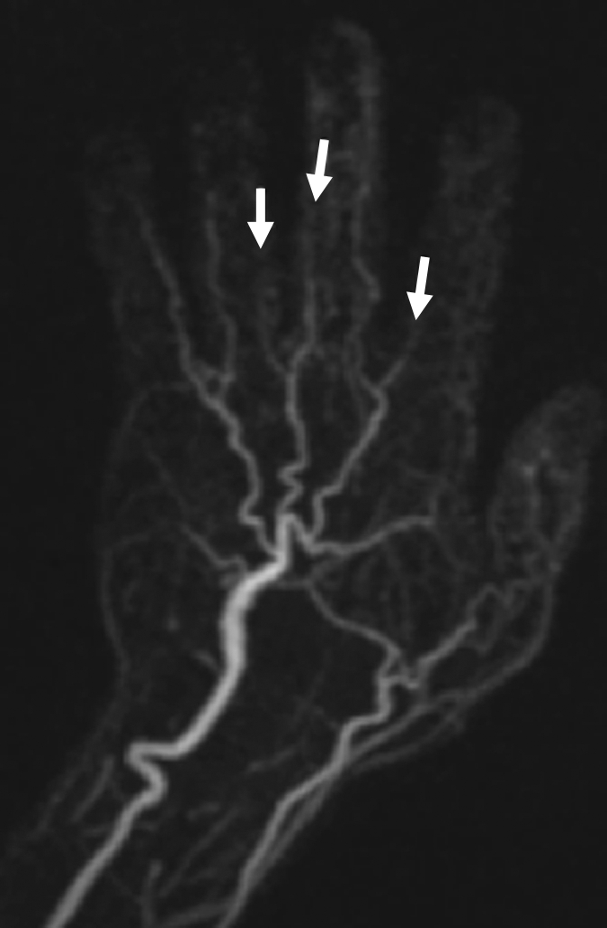
(a) Contrast-enhanced time-resolved, (b) contrast-enhanced high-spatial-resolution, and (c) nonenhanced FSD-prepared MR angiograms obtained in patient with focal mild stenosis (arrows) involving proper digital arteries of index, middle, and ring fingers of left hand.