Abstract
Erythropoietin (EPO) improves functional recovery after traumatic brain injury (TBI). This study was designed to investigate long-term (3 mo) effects of EPO on brain remodeling and functional recovery in rats after TBI. Young male Wistar rats were subjected to unilateral controlled cortical impact injury. TBI rats were divided into the following groups: 1) Saline group (n = 7); 2) EPO-6h group (n = 8); and 3) EPO-24h group (n = 8). EPO (5,000 U/kg in saline) was administered intraperitoneally at 6 h, and 1 and 2 days (EPO-6h group) or at 1, 2, and 3 days (EPO-24h group) post injury. Neurological function was assessed using a modified neurological severity score, footfault and Morris water maze tests. Animals were sacrificed at 3 mos after injury and brain sections stained for immunohistochemical analyses. Compared to the saline, EPO-6h treatment significantly reduced cortical lesion volume, while EPO-24h therapy did not affect the lesion volume (P<0.05). Both the EPO-6h and EPO-24h treatments significantly reduced hippocampal cell loss (P<0.05), promoted angiogenesis (P<0.05) and increased endogenous cellular proliferation (BrdU-positive cells) in the injury boundary zone and hippocampus (P<0.05) compared to saline controls. Significantly enhanced neurogenesis (BrdU/NeuN-positive cells) was seen in the dentate gyrus of both EPO groups compared to the saline group. Both EPO treatments significantly improved long-term sensorimotor and cognitive functional recovery after TBI. In conclusion, the beneficial effects of posttraumatic EPO treatment on injured brain persisted for at least 3 months. The long-term improvement in functional outcome may in part be related to the neurovascular remodeling induced by EPO.
Keywords: angiogenesis, cell proliferation, erythropoietin, neurogenesis, functional recovery, traumatic brain injury
1. Introduction
Trauma1tic brain injury (TBI) is a leading cause of mortality and morbidity in the United States, particularly among the young (Thurman et al., 1999). The most prevalent and debilitating features in survivors of TBI are cognitive deficits and sensorimotor dysfunctions (Davis, 2000). To date, there is no effective treatment identified to promote functional recovery after TBI except for routine medical intervention and care (Narayan et al., 2002; Royo et al., 2003).
Erythropoietin (EPO), essential for erythropoiesis, provides neuroprotection in animal models of stroke (Gonzalez et al., 2007; Wang et al., 2004), spinal cord injury (Celik et al., 2002; Grasso et al., 2005; Grasso et al., 2006), concussive brain injury (Brines et al., 2000), kainate-induced seizure activity (Brines et al., 2000), and autoimmune encephalomyelitis (Cerami, 2001; Sakanaka et al., 1998). Several lines of evidence demonstrate that EPO administered within 6 h post TBI reduces lesion volume (Cherian et al., 2007; Grasso et al., 2007; Hartley et al., 2008; Xiong et al., 2008a). Our recent work further demonstrates that delayed treatment (24 h post injury) with EPO provides robust benefits in rats up to one month after TBI (Lu et al., 2005; Mahmood et al., 2007), and after stroke (Wang et al., 2004). Therefore, EPO has promising potential for treatment of TBI. However, no studies to date have examined the long- term (3 months) effects of EPO treatment on sensorimotor, cognitive function and brain remodeling after TBI. In addition, there are no data comparing the long-term efficacy of multiple doses of EPO administered early (6 h post TBI) with late (24 h post TBI) for treatment of TBI. Therefore, using a controlled cortical impact (CCI) TBI rat model, we investigated whether posttraumatic multiple administration of EPO significantly promotes brain neurovascular remodeling and improves long-term functional recovery in rats after TBI.
2. Results
2.1. Body weight and hematocrit
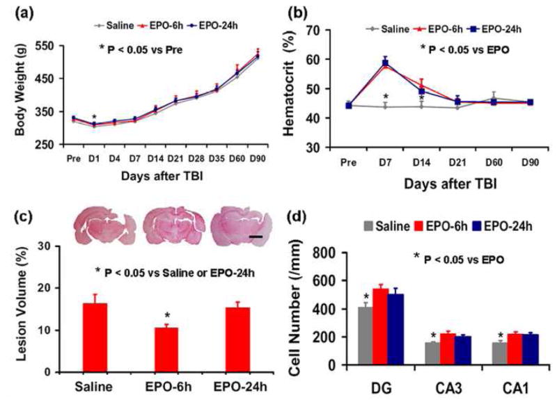
The animals in all three groups showed no significant difference in body weight before TBI. TBI rats treated with saline or EPO significantly lost some body weight at day 1 post injury (P<0.05) compared to their preinjury level and then gradually gained the body weight (Fig. 1a). There was no significant difference in body weight among TBI rats treated with EPO or saline at any time post injury. The preinjury baseline of hematocrit (HCT) was similar for all animals (Fig.1b). As compared to the saline treatment, EPO treatment significantly increased HCT up to 2 weeks (P<0.05), which then returned to normal.
Fig. 1.
Graphs showing body weight, hematocrit, cortical lesion volume and hippocampal neuron number in TBI rats treated with saline or EPO. “Pre” represents preinjury level. A significant body weight loss was observed at Day 1 after injury in all groups (a). EPO treatment significantly increased hematocrit up to 2 weeks (b). EPO treatment starting at 6 h significantly reduced cortical lesion volume (c). EPO treatment significantly increased the number of neurons in the hippocampus (d). Data represent mean ± SD. Scale bar = 3 mm (c). N (rats/group) = 7 (Saline); 8 (EPO-6 h); 8 (EPO-24 h).
2.2. Lesion volume
Rats were euthanized at 90 days post TBI for histological measurements. The lesion volume was 16.4 ± 2.1%, 10.5 ± 0.9%, and 15.4 ± 1.3% for TBI rats treated with saline, EPO-6h, and EPO-24h, respectively. Compared to the saline treatment, EPO treatment starting 6 h postinjury significantly reduced the lesion volume (P<0.05) while EPO treatment starting 24 h postinjury did not alter the lesion volume (Fig.1c).
2.3. Cell loss in the hippocampus
When examined at 90 days post TBI, neuron counts in the ipsilateral CA1, CA3 and DG significantly decreased by 22%, 45% and 38%, respectively, compared to the contralateral hemispheres (P<0.05). As compared to the saline controls, both EPO treatments significantly increased the neuron counts in the DG, CA3 and CA1 regions (Fig. 1d, P<0.05). There was no significant difference in neuron counts between the EPO-6h and EPO-24h groups in these regions. The DG distances in the images analyzed were 487.7 ± 36.8, 485.6 ± 44.1, and 461.3 ± 28.4 μm for the saline group, EPO-6 h group, and EPO-24 h group, respectively. The CA3 distances were 196.5 ± 2 7.2, 185.8 ± 19.6, and 184.9 ± 20.3 μm for the saline group, EPO-6 h group, and EPO-24 h group, respectively. The CA1 distances were 187.7 ± 12.6, 171.6 ± 12.7, and 181.8 ± 20.1 μm for the saline group, EPO-6 h group, and EPO-24 h group, respectively. There was no significant difference in distances among treatment groups (P<0.05), indicating that there were no significant systematic differences in tissue shrinkage.
2.4. Spatial learning test
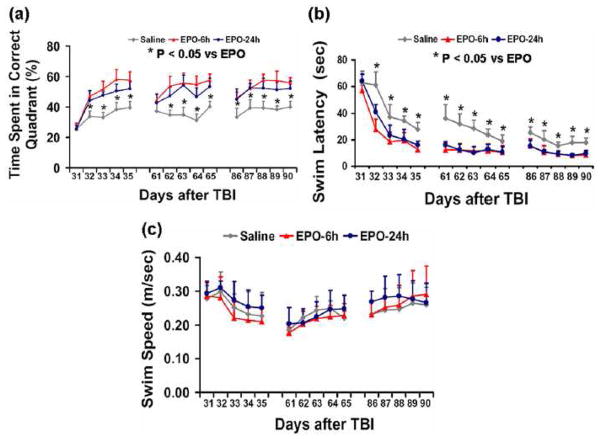
The Morris water maze protocol in the present study was used to detect spatial learning and memory deficits. To analyze day-by-day differences in the MWM, repeated measures ANOVA was performed followed by Student-Newman-Keuls tests for multiple comparisons. The more time the rats spent in the correct quadrant, the better the spatial learning and memory. As shown in Figure 2a, the percentage of time spent in the correct quadrant (Northeast) by saline-treated TBI rats was significantly less than that of EPO-treated TBI rats at Days 32–35 (P<0.05) after injury. Examined at 2 and 3 months after injury, saline-treated TBI rats did not show significant recovery in spatial learning while TBI rats treated with EPO either starting 6 or 24 h after injury showed improved spatial learning (P<0.05 vs the saline group). However, at the time points studied (ie, 1, 2, and 3 months) there was no significant difference in spatial learning between the EPO-24h and EPO-6h groups.
Fig. 2.
Effect of EPO on spatial learning function 1, 2 and 3 months after TBI. Treatment with EPO (EPO-6 h and EPO-24 h) significantly improves spatial learning performance (increased percentage of time spent in the correct quadrant) measured by a recent version of the Morris water maze test at Days 32-35, 62-65 and 86–90 compared with the saline-treated group (a). EPO treatment also significantly reduced the latency to find the hidden escape platform in the Morris water maze at Days 32–35, 61–65 and 86–90 compared to the saline treatment (b). No significant difference in swim speed among the 3 groups was observed (c). Data represent mean ± SD. N (rats/group) = 7 (Saline); 8 (EPO-6 h); 8 (EPO-24 h).
Compared to EPO-treated TBI animals, injured saline-treated rats displayed a substantial learning dysfunction, indicated by an increased latency to find a hidden platform in the water maze during Days 32–35 after TBI (Fig. 2b, P < 0.05). Compared to the saline-treated group, EPO-treated rats showed a significant reduction in latency to find the hidden platform at 1 month post TBI. They also maintained spatial memory later on, that is, they remembered spatial cues used for navigation at 1 month post injury to quickly find the hidden platform even at 2 and 3 months post injury (P<0.05 vs the saline controls, Fig. 2b). There was no significant difference in latency between the two EPO treatment groups. The injured saline-treated rats spent a significantly longer time finding the hidden platform and had poorer spatial memory at 2 and 3 months post injury than EPO-treated animals.
No significant group differences in swim speed were observed in the present study (Fig. 2c), indicating that physical disability does not play a significant role in the observed spatial learning/memory deficits. Taken together, the Morris water maze data indicate that EPO treatment has a significant beneficial effect on cognitive functional recovery after TBI (Fig. 2a and 2b).
2.5. Footfault test
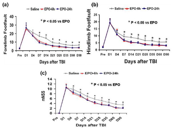
As compared to preinjury baseline, the incidence of contralateral right forelimb footfaults significantly increased after TBI (Fig. 3a). Treatment with EPO significantly reduced the number of contralateral forelimb footfaults at 7 to 90 days after TBI compared to treatment with saline (P<0.05). Similar results were found for the contralateral hindlimb (Fig. 3b). As compared to preinjury baseline, TBI significantly increased the incidence of contralateral hindlimb footfaults at 1 to 90 days post-injury (P<0.05). Treatment with EPO significantly reduced the number of contralateral hindlimb footfaults at 7 to 90 days after TBI compared to treatment with saline (P<0.05). However, no significant differences in footfault testing were observed in EPO-6h and EPO-24h groups.
Fig. 3.
Effect of EPO on sensorimotor function (footfault) before and after TBI. “Pre” represents pre-injury level. EPO treatment significantly reduces forelimb (a) and hindlimb (b) foot faults at Days 7–90 compared with the saline treatment. EPO treatment significantly improves neurological functional recovery (reduced mNSS scores) at Days 4–90 (c). Data represent mean ± SD. N (rats/group) = 7 (Saline); 8 (EPO-6 h); 8 (EPO-24 h).
2.6. Modified neurological severity score
The mNSS is a composite of the motor (muscle status, abnormal movement), sensory (visual, tactile and proprioceptive) and reflex tests and has been employed in previous studies (Lu et al., 2007). Figure 3c shows that significantly improved scores were measured at Days 4–90 after TBI in both EPO groups compared to the saline-treated group (P<0.05). There was no significant difference in the mNSS scores between the EPO-6h and EPO-24h groups.
2.7. Angiogenesis
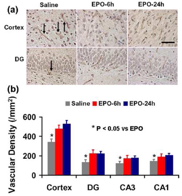
vWF-staining has been used to identify vascular structure in the brain after TBI (Lu et al., 2003). EPO treatment significantly increased the vascular density in the injured cortex and ipsilateral hippocampus (P<0.05) compared to the saline treatment (Fig.4). There was no significant difference in the vascular density between the EPO-6h and EPO-24h groups (Fig. 4b).
Fig. 4.
The effect of EPO on vWF-staining vascular structure in the injured cortex and ipsilateral hippocampus 90 days after TBI (a, b). EPO treatment significantly increased vasculature (brown-stained vessels, arrows as example) in the injured brain after TBI compared to the saline-treated group (a). The number of vWF-stained vasculature is shown in (b). Data represent mean ± SD. Scale bar = 50 μm (a). N (rats/group) = 7 (Saline); 8 (EPO-6 h); 8 (EPO-24 h).
2.8. Cell proliferation
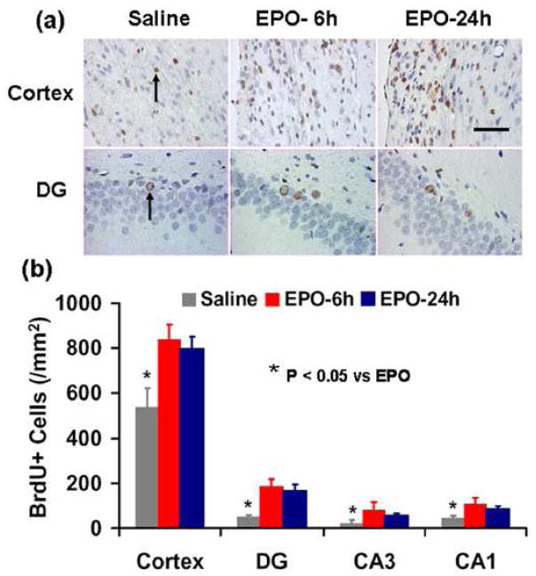
BrdU, an analog of thymidine, can be incorporated into the newly synthesized DNA of replicating cells during the S phase of the cell cycle, substituting for thymidine during DNA replication. BrdU immunostaining is commonly used to detect cell proliferation. The number of BrdU-positive cells found in the ipsilateral cortex and hippocampus was significantly increased in the EPO-treated rats examined at 90 days after TBI, compared to the saline controls (Fig. 5, P<0.05). There was no significant difference in the number of BrdU-positive cells seen in the hippocampus of the EPO-6h and EPO-24h groups (Fig. 5b).
Fig. 5.
Effect of EPO on cell proliferation in the injured cortex and ipsilateral hippocampus 90 days after TBI (a, b). The cells with BrdU (brown stained, arrows as example) that clearly localized to the nucleus (hematoxylin stained) were counted as BrdU-positive cells (a). EPO treatment significantly increased the number of BrdU- positive cells in these regions compared to the saline treatment (b). Data represent mean ± SD. Scale bar = 50 μm (a). N (rats/group) = 7 (Saline); 8 (EPO-6 h); 8 (EPO-24 h).
2.9. Neurogenesis
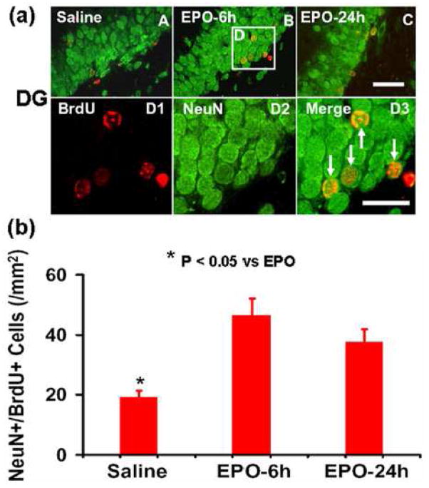
Newborn mature neurons were identified by double labeling for BrdU (proliferating marker) and NeuN (mature neuronal marker). EPO treatment significantly increased the number of newborn neurons in the ipsilateral DG (Fig. 6, P<0.05) compared to the saline controls. However, there was no significant difference in the number of newborn neurons in the DG between EPO-6h and EPO-24h groups.
Fig. 6.
Double fluorescent staining for BrdU (red) and NeuN (green) to identify newborn neurons (yellow after merge). EPO treatment significantly increases the number of newborn neurons in the DG (a, b). BrdU-positive cells (D1) differentiate into new neurons (D3, arrows) expressing NeuN (D2). The number of NeuN/BrdU-colabeled cells is shown in (b). Data represent mean ± SD. Scale bars = 50 μm (A-C); 25 μm (D1-D3). N (rats/group) = 7 (Saline); 8 (EPO-6 h); 8 (EPO-24 h).
3. Discussion
The main findings of the present study are: 1) posttraumatic (starting either 6 or 24 h) EPO treatment provides similar and significant long-term (at least 3 months) improvements in sensorimotor functional recovery and spatial learning, as evaluated by footfault, mNSS score, and MWM tests; 2) the long-term improvements in spatial learning and sensorimotor function may mainly derive from EPO-promoted neurovascular remodeling including increased angiogenesis, cell proliferation and neurogenesis; and 3) the effect of EPO on reducing cortical lesion volume may not play a major role in long-term functional recovery.
Many treatments including EPO in experimental TBI initiated immediately or within 6 hours after TBI show neuroprotection (Cherian et al., 2007; Grasso et al., 2007; Hartley et al., 2008; Liao et al., 2008; Xiong et al., 2008a). However, our recent short-term study (14 or 35-day survival) after CCI in rats demonstrates that delayed (24 h) treatment with EPO not only significantly improves spatial memory and learning after CCI but also promotes sensorimotor functional recovery (reduced footfaults and mNSS score) (Lu et al., 2005; Xiong et al., 2010), indicating that the therapeutic time window may not be limited to early hours but can be extended to later time periods after TBI. In the present study, we investigated the long-term (3 months) beneficial effects of two EPO treatment paradigms (starting at 6 or 24 h with two additional injections) on both sensorimotor and cognitive functional recovery after TBI. We also observed significant brain remodeling including increased cell proliferation, angiogenesis and neurogenesis promoted by EPO treatment. The EPO-promoted brain remodeling may play an important role in functional recovery after TBI. In our previous studies, we did not observe any significant effects of EPO on functional outcomes and angiogenesis and neurogenesis at Day 35 in sham-operated rats (Xiong et al., 2008a; Zhang et al., 2009). It remains unknown, however, whether early EPO treatment would affect these parameters at 3 months in sham animals. Even if there were some effects, they would not affect our conclusion that EPO treatment significantly improves long-term functional outcomes and angiogenesis and neurogenesis compared to saline-treatment in TBI rats.
Although the nervous and vascular systems are functionally different, they show a high degree of anatomic parallelism and cross-talk (Segura et al., 2009). The realization that both systems use common genetic pathways not only forms a link between vascular biology and neuroscience, but also promises to accelerate the discovery of new mechanistic insights and therapeutic opportunities (Carmeliet, 2003). Under normal conditions, adult neurogenesis in the subgranular zone (SGZ) of the DG and subventricular zone (SVZ) takes place within an angiogenic microenvironment (Zhang and Chopp, 2009). In vivo, neurogenesis and angiogenesis are highly interdependent and work together to promote brain remodeling and subsequent improvement of neurological functional after brain injury (Zhang and Chopp, 2009). Our previous short-term study indicated that angiogenesis is coupled with neurogenesis. This was shown by increased vasculature in the DG where NeuN/BrdU-positive cells (newborn neurons) are also enhanced after EPO treatment (Xiong et al., 2010). The present long- term study further supports the coupling of angiogenesis and neurogenesis. In our previous short-term (35-day) study, newborn mature neurons were found in the injury boundary zone (Xiong et al., 2010), which probably came from neuroblasts migrating from the SVZ to the injury boundary zone after injury and treatment (Petraglia et al., 2010). In the present study, we did not observe newborn neurons in the injury boundary zone, indicating that newborn neurons died by 3 months in this region. However, significantly increased endogenous cell proliferation was observed at 3 months after EPO treatment in the injury boundary zone, indicating that these newborn non-neuronal cells in this region may play an important role in functional recovery. Our recent data show that increased expression of glial cell derived neurotrophic factor and tissue plasminogen activator in the astrocytes creates a hospitable environment for brain remodeling which may contribute to the long-term functional improvement from ischemic brain injury (Li et al., 2005; Shen et al., 2010; Xin et al., 2010). Further studies are necessary to determine if similar mechanisms underlie the long-term beneficial effect of EPO treatment. Our previous study shows that most of the BrdU-positive cells in the DG differentiate into granule neurons and that cell-lineage commitment patterns do not change in the DG after TBI and EPO therapy (Xiong et al., 2010). Our present study demonstrates that significantly more newborn neurons are present in the DG in the EPO-treated groups than in the saline controls 3 months after TBI. However, the number of newborn neurons seen at 3 months in the present study is much less than that observed at 35 days in our earlier research (Xiong et al., 2010), indicating that many newborn neurons die during a period of 1-3 months after TBI and only those integrating into existing functional circuitry may survive. This finding is in agreement with the report that the majority of BrdU-positive cells in the DG that survive for 10 weeks become granule neurons after TBI, and these neurons exhibit extensive anatomical integration into the CA3 region at the time cognitive recovery is observed (Sun et al., 2007). Our present data indicate that newborn neurons in the DG may participate in brain repair and functional recovery. It is unknown whether additional doses of EPO increase the survival rate of newborn neurons in the injured brain, and further studies are necessary.
EPO and its receptors are weakly expressed in the normal adult central nervous system (Grasso et al., 2004). TBI transiently upregulates expression of EPO and its receptors in the rat brain (Liao et al., 2008). Endogenous EPO level though increased, may not provide enough neuroprotection. Previous studies from us and others show that EPO administered within 6 h significantly reduces cortical lesion volume after CCI and improves functional recovery (Cherian et al., 2007; Xiong et al., 2008a). In our recent short-term 35-day study, a single dose of delayed (24 h) EPO therapy shows substantial benefits (i.e., significantly reducing hippocampal cell loss, increasing angiogenesis and neurogenesis, and improving functional recovery), while three doses of EPO (24, 48, and 72 h) provide better outcomes (Xiong et al., 2010). These delayed treatments did not reduce the lesion volume. In the present study, we investigated whether the long-term (3 month) effects of early multiple dose EPO treatment strategy differ from those of late multiple dose EPO treatment strategy in terms of lesion volume, cell loss, cell proliferation, angiogenesis and neurogenesis as well as functional recovery after TBI. At 3 months after TBI, the lesion volume in the EPO-24h group is comparable to that in the saline group while the lesion volume in the EPO-6h group is significantly reduced compared to the saline or EPO-24h group. However, the functional recovery was significantly improved in these two EPO groups and no significant difference was observed between the EPO-6h and EPO-24h groups, indicating that the EPO effect on moderately reducing lesion volume may not contribute to the functional improvement. The precise mechanism whereby recovery in sensorimotor function occurs for the delayed EPO treatment group without change in contusion volume is unclear. Recent studies demonstrated that after focal injury to the cerebral cortex the remaining, intact tissue undergoes structural and functional changes that could play a substantial role in neurological recovery (Nudo, 2006). Our studies demonstrate that cortical neurons surviving in the peri-infarct motor cortex undergo axonal sprouting to restore connections between different cerebral areas, and bone marrow stromal cells (MSCs) enhance inter- and intracortical axonal connections and improve functional recovery after ischemic stroke in adult rats (Liu et al., 2010). In addition, the contralateral somatosensory cortex can compensate for the loss of the ipsilateral somatosensory cortex by remodeling neuronal circuits and establishing new sensory processing (Takatsuru et al., 2009). Our recent studies show that delayed MSC therapy-enhanced contralesional axonal remodeling of the corticospinal system in adult rats partially contributes to functional recovery after stroke (Liu et al., 2008). Furthermore, our recent studies show that EPO treatment promotes corticospinal tract fibers (originating from the contralesional intact cerebral hemisphere) sprouting into the denervated spinal cord, which may at least partially contribute to functional recovery (Zhang et al., 2010). Further work is required to investigate the role of axonal sprouting in the long term-effect of EPO treatment for TBI.
In conclusion, posttraumatic EPO therapy for TBI significantly reduces hippocampal cell loss, enhances angiogenesis and neurogenesis, and improves long-term (3 month) sensorimotor function and spatial learning recovery, suggesting that treatment with EPO provides both neuroprotective and neurorestorative (that is, neurovascular remodeling) effects. Our data also indicate that EPO-promoted brain neurovascular remodeling may play a more significant role in improving long-term functional recovery than the EPO-derived lesion reduction effect in the present study.
4. Experimental procedures
4.1. TBI Model
All experimental procedures were approved by the Institutional Animal Care and Use Committee (IACUC) of Henry Ford Health System. Investigators blinded to the treatment status performed all functional tests and cell counting.
A CCI model of TBI in the rat was utilized for the present study (Dixon et al., 1991; Mahmood et al., 2004). Young adult male Wistar rats (300–400 g) were anesthetized intraperitoneally with chloral hydrate (350 mg/kg body weight). Rectal temperature was maintained at 37°C using a feedback-regulated water-heating pad. A CCI device was used to induce injury. Rats were placed in a stereotactic frame. Two 10-mm-diameter craniotomies were performed adjacent to the central suture, midway between lambda and bregma. The second craniotomy allowed for lateral movement of cortical tissue. The dura mater was kept intact over the cortex. Injury was delivered by impacting the left cortex (ipsilateral cortex) with a pneumatic piston containing a 6-mm-diameter tip at a rate of 4 m/second and 2.5 mm of compression. Velocity was measured with a linear velocity displacement transducer.
4.2 Experimental groups and treatment
TBI was induced by CCI over the left parietal cortex in young adult male Wistar rats. TBI rats were randomly divided into 3 groups: 1) Saline group (n = 7); 2) EPO-6h group (n = 8); and 3) EPO-24h group (n = 8). EPO at a dose of 5000 U/kg body weight (Epoetin alpha, AMGEN, Thousand Oaks, CA) was administered intraperitoneally at 6 h, 1 day and 2 days (for the EPO-6h group) or at 1, 2 and 3 days (for the EPO-24h group) after TBI. The dose of EPO was selected based on previous studies and dissolved in saline (Lu et al., 2005; Zhang et al., 2004). Animals in the saline-treated group received an equal volume of saline at 6h, 1 and 2 days after injury. For labeling proliferating cells, 5-bromo-2’-deoxyuridine (BrdU, 100 mg/kg; Sigma, St. Louis, MO) was injected intraperitoneally into rats daily for 10 days, starting 1 day after TBI. All rats were sacrificed at 90 days after TBI.
4.3. Hematocrit and body weight
To determine the effects of EPO on hematocrit (HCT), a blood sample (50 μl) was collected via tail vein before injury, and at Days 4, 14, 21, 60 and 90 after TBI. HCT was measured in micro-HCT capillary tubes (Fisher Scientific, Pittsburgh, PA) using standard procedures (Readacrit Centrifuge, Clay Adams, Parsippany, NJ) (Xiong et al., 2008b). Body weight was recorded just before injury, and at Days 1, 4, 7, 14, 21, 28, 35, 60 and 90 after injury.
4.4. Morris water maze test
To detect spatial learning impairments, a recent version of the Morris water maze (MWM) test was used (Choi et al., 2006). The procedure was modified from previous versions (Day et al., 1999; Morris, 1984; Morris et al., 1982; Sutherland et al., 1982), and has been found to be useful for chronic spatial memory assessment in rats and mice with brain injury (Choi et al., 2006; Lu et al., 2005). Spatial learning was tested at 1, 2, and 3 months postinjury. Each rat received a block of four daily trials for five consecutive days (i.e., Days 31–35, 60–65, 86–90) to locate the hidden platform. The inter-trial interval was 30 min. Data collection was automated by the HVS Image 2020 Plus Tracking System (US HVS Image, San Diego, CA.). For data collection, a blue pool (1.8 m in diameter) was subdivided into four equal quadrants formed by imaging lines. At the start of a trial, the rat was placed at one of four fixed starting points, randomly facing toward a wall (designated North, South, East and West) and allowed to swim for 90 sec or until it found the platform. If the animal found the platform, it was allowed to remain on it for 10 sec. If the animal failed to find the platform within 90 sec, it was placed on the platform for 10 sec. Throughout the test period the platform was located in the Northeast quadrant 2 cm below water in a randomly changing position, including locations against the wall, toward the middle of the pool or off-center but always within the target quadrant. If the animal was unable to find the platform within 90 sec, the trial was terminated and a maximum score of 90 sec was assigned. If the animal reached the platform within 90 sec, the percentage of time traveled within the Northeast (correct) quadrant was calculated relative to the total amount of time spent swimming before reaching the platform and employed for statistical analysis. The swim speed and latency in finding the hidden escape platform were recorded and analyzed. The advantage of this version of the water maze is that each trial takes on the key characteristics of a probe trial because the platform is not in a fixed location within the target quadrant (Schallert, 2006).
4.5. Footfault test
To evaluate sensorimotor function, the footfault test was carried out before TBI and at 1, 4, 7, 14, 21, 28, 35, 60 and 90 days after TBI or surgery. The rats were allowed to walk on a grid (Zhang et al., 2002). With each weight-bearing step, a paw might fall or slip between the wires and, if this occurred, it was recorded as a footfault (Barth et al., 1990; Baskin et al., 2003). A total of 50 steps were recorded for each right forelimb and hindlimb.
4.6. Modified neurological severity score (mNSS) test
Neurological functional measurement was performed using the mNSS score test (Chen et al., 2001). The test was carried out on all rats preinjury and on Days 1, 4, 7, 14, 21, 28, 35, 60 and 90 after TBI. In this TBI model, injury in the left hemispheric cortex of rats causes sensory and motor functional deficiency with elevated scores on motor, sensory, and Beam Balance Tests in the early phase after injury (Day 1 after injury). Absent reflexes and abnormal movements can be measured on rats with severe injury. Slow recovery in asymmetry deficiency as reflected by Beam Balance Test results has been reported in unilateral brain injuries including TBI (Lu et al., 2007) and ischemia (Chen et al., 2001). This test is suitable for evaluating long-term neurological function after unilateral brain injury.
4.7. Tissue preparation and measurement of lesion volume
At Day 90 after TBI, rats were anesthetized intraperitoneally with chloral hydrate and perfused transcardially first with saline solution, followed by 4% paraformaldehyde in 0.1 M PBS (pH 7.4). Their brains were removed and post-fixed in 4% paraformaldehyde for 2 days at room temperature. The brain tissue was cut into 7 equally spaced 2-mm coronal blocks and processed for paraffin sectioning. A series of adjacent 6-μm-thick sections were cut from each block in the coronal plane and stained with H & E. For lesion volume measurement, the 7 brain sections were traced by a microcomputer imaging device (MCID) (Imaging Research, St. Catharine’s, Ontario, Canada), as previously described (Chen et al., 2005). The exact distance between the analyzed sections was determined using the Rat Brain in Stereotaxic Coordinates (Paxinos and Watson, 2nd edition, 1986). To reduce differences in tissue shrinkage in each block, the indirect lesion area was calculated (i.e., the intact area of the ipsilateral hemisphere is subtracted from the area of the contralateral hemisphere) (Swanson et al., 1990), and the lesion volume presented as a volume percentage of the lesion compared with the contralateral hemisphere.
4.8. Immunohistochemistry
To examine the effect of EPO on cell proliferation and angiogenesis, coronal sections were histochemically stained with mouse anti-BrdU (Lu et al., 2005) and rabbit anti-human von Willebrand factor (vWF) (Lu et al., 2005), respectively. For BrdU detection, paraffin-embedded coronal sections (6 μm) were deparaffinized and rehydrated. Antigen retrieval was performed by boiling sections in 10-mM citrate buffer (pH 6.0) for 10 min (Chen et al., 2001). After washing with PBS, sections were incubated with 0.3 % H2O2 in PBS for 10 min, blocked with 1% BSA containing 0.3 % Triton-X 100 for 1 h at room temperature, and incubated with mouse anti-BrdU (1:200; Dako, Carpinteria, CA) at 4°C overnight. After washing, sections were incubated with biotinylated anti-mouse antibody (1:200; Vector Laboratories, Inc., Burlingame, CA) at room temperature for 30 min. After washing, sections were incubated with an avidin-biotin-peroxidase system (ABC kit, Vector Laboratories, Inc., Burlingame, CA). Diaminobenzidine (Sigma, St. Louis, MO) was then used as a sensitive chromogen for light microscopy. Sections were counterstained with hematoxylin.
To identify vascular structure, brain sections were deparaffinized and then incubated with 0.4% Pepsin solution at 37°C for 1 h. After washing, the sections were blocked with 1% BSA at room temperature for 1 h, and then incubated overnight at 4°C in rabbit anti-human vWF (1:200; DakoCytomation, Carpinteria, CA). After washing, sections were incubated at room temperature for 30 min in biotinylated anti-rabbit antibody (1:200; Vector Laboratories, Inc., Burlingame, CA). The subsequent procedures were the same as for BrdU staining.
BrdU-positive cells and vWF-stained vascular structures in the DG, CA3, and the cortex of ipsilateral hemispheres were examined at 20x magnification and counted. The cells with BrdU (brown stained) that clearly localized to the nucleus (hematoxylin stained) were counted as BrdU-positive cells.
4.9. Immunofluorescent staining
Newly generated neurons were identified by double labeling for BrdU and NeuN. After dehydration, tissue sections were boiled for 10 min in 10 mM citric acid buffer (pH 6). After washing with PBS, sections were incubated for 20 min at 37°C in 2.4 N HCl. Sections were incubated with 1% BSA containing 0.3% Triton-X-100 in PBS. Sections were then incubated overnight at 4°C in mouse anti-NeuN antibody (1:200; Chemicon, Temecula, CA). FITC-conjugated anti-mouse antibody (1:400; Jackson ImmunoResearch, West Grove, PA) was added to sections at room temperature for 2 h. Sections were then incubated at 4°C overnight in rat anti-BrdU antibody (1:200; Dako, Glostrup, Denmark). Sections were then incubated with Cy3-conjugated anti-rat antibody for 2 h at room temperature (1:400; Jackson ImmunoResearch, West Grove, PA). Each of the steps was followed by three 5-min rinses in PBS. Tissue sections were mounted with Vectashield mounting medium (Vector laboratories, Burlingame, CA). Images were collected with fluorescent microscopy. NeuN/BrdU-colabeled cells in the DG and the cortex were counted at 40x magnification.
4.10. Cell counting and quantitation
Cell counts were performed by observers blinded to the individual treatment status of the animals. For cell counting and vascular density measurement, images were acquired from eight fields of view in the lesion boundary zone of each section (five sections with 400-μm intervals at Bregma level of −2.3 to −4.3 mm). The lesion boundary zone is defined as pericontusional regions or sites closer to the primary injury site (Harris et al., 2010). Images were acquired from three fields of view in the ipsilateral CA3 and CA1, and nine fields of view in the ipsilateral dorsal DG (i.e., DG in the dorsal portion of the hippocampus, five sections with 50-μm intervals with the center at Bregma −3.3 mm). All images were analyzed with a microscope (Nikon i80) at 400x magnification via the MCID system (Lu et al., 2005). The numerical aperture of the Nikon Plan Apochromat 40x Objective is 0.95. All counting was performed on a computer monitor to improve visualization and in one focal plane to avoid oversampling (Zhang et al., 2002). To evaluate whether intraperitoneally administered EPO reduces neuronal damage after TBI, the number of cells was counted in the hippocampus using the MCID system. Although H & E staining is not neuron-specific, the morphological characteristics of neuronal cells in the DG, CA1 and CA3 region aid in counting them. Counts were averaged and normalized by measuring the linear distance (in mm) of the DG, CA1 and CA3 for each section. Although it is just an estimate of the cell number, this method permits a meaningful comparison of differences between groups. For cell proliferation, the total number of BrdU-positive cells was counted in the lesion boundary zone and the hippocampus, using the MCID system. The cells with BrdU (brown stained) that clearly localized to the nucleus (hematoxylin stained) were counted as BrdU-positive cells. The number of BrdU-positive cells was expressed in cells/mm2 in the lesion boundary zone and the hippocampus. For analysis of neurogenesis, additional sections used in the above studies were used to evaluate neurogenesis in the DG by calculating the density of BrdU-labeled cells and BrdU/NeuN-colabeled cells (Popoli et al., 2007). We mainly focused on the ipsilateral DG and its subregions, including the SGZ, granular cell layer, and the molecular layer. The number of BrdU-positive cells (red stained) and NeuN/BrdU-colabeled cells (yellow after merge) were counted in the DG (Xiong et al., 2008b).
4.11. Statistical Analyses
All data are presented as means ± SD. Data on mNSS were first evaluated for normality. The rank data were used for the analysis since data were not normal. Analysis of variance (ANCOVA), PROC MIXED with CONTRAST statement in SAS, was employed to test the group differences on mNSS. The analysis began testing the overall group effect, followed by pair-wise group comparisons if the overall group effect was detected at the 0.05 level; otherwise the pair-wise group comparisons would be considered as exploratory analyses. Data on body weight, HCT, footfault and water maze tests were analyzed by ANOVA for repeated measurements. For lesion volume, cell counting, and vWF-stained vascular density, a one-way ANOVA followed by post hoc Student-Newman-Keuls tests was used to compare the differences between the EPO-6h, EPO-24h, and saline-treated groups. Statistical significance was set at P < 0.05.
Acknowledgments
This work was supported by NIH grants RO1 NS62002 (YX) and PO1 NS42345 (AM and MC). Special thanks to Susan MacPhee-Gray for editorial assistance.
Sources of financial support: NINDS grants RO1 NS62002 (Ye Xiong) and PO1 NS42345 (Asim Mahmood, Michael Chopp).
Footnotes
Disclosure
There is no conflict of interest reported.
Author contributions to the study and manuscript preparation include the following. Conception and design: Xiong, Mahmood, Chopp. Acquisition of data: Ning, Zhang, Meng, Xiong, Qu. Analysis and interpretation of data: Xiong, Ning, Mahmood, Chopp. Drafting of the manuscript: Xiong, Ning, Chopp. Critical review of the manuscript: Xiong, Chopp. Review of final version of the manuscript and approved for submission: all authors.
Publisher's Disclaimer: This is a PDF file of an unedited manuscript that has been accepted for publication. As a service to our customers we are providing this early version of the manuscript. The manuscript will undergo copyediting, typesetting, and review of the resulting proof before it is published in its final citable form. Please note that during the production process errors may be discovered which could affect the content, and all legal disclaimers that apply to the journal pertain.
References
- Barth TM, Jones TA, Schallert T. Functional subdivisions of the rat somatic sensorimotor cortex. Behav Brain Res. 1990;39:73–95. doi: 10.1016/0166-4328(90)90122-u. [DOI] [PubMed] [Google Scholar]
- Baskin YK, Dietrich WD, Green EJ. Two effective behavioral tasks for evaluating sensorimotor dysfunction following traumatic brain injury in mice. J Neurosci Methods. 2003;129:87–93. doi: 10.1016/s0165-0270(03)00212-7. [DOI] [PubMed] [Google Scholar]
- Brines ML, Ghezzi P, Keenan S, Agnello D, de Lanerolle NC, Cerami C, Itri LM, Cerami A. Erythropoietin crosses the blood-brain barrier to protect against experimental brain injury. Proc Natl Acad Sci U S A. 2000;97:10526–10531. doi: 10.1073/pnas.97.19.10526. [DOI] [PMC free article] [PubMed] [Google Scholar]
- Carmeliet P. Blood vessels and nerves: common signals, pathways and diseases. Nat Rev Genet. 2003;4:710–720. doi: 10.1038/nrg1158. [DOI] [PubMed] [Google Scholar]
- Celik M, Gokmen N, Erbayraktar S, Akhisaroglu M, Konakc S, Ulukus C, Genc S, Genc K, Sagiroglu E, Cerami A, Brines M. Erythropoietin prevents motor neuron apoptosis and neurologic disability in experimental spinal cord ischemic injury. Proc Natl Acad Sci U S A. 2002;99:2258–2263. doi: 10.1073/pnas.042693799. [DOI] [PMC free article] [PubMed] [Google Scholar]
- Cerami A. Beyond erythropoiesis: novel applications for recombinant human erythropoietin. Semin Hematol. 2001;38:33–39. doi: 10.1016/s0037-1963(01)90128-3. [DOI] [PubMed] [Google Scholar]
- Chen J, Sanberg PR, Li Y, Wang L, Lu M, Willing AE, Sanchez-Ramos J, Chopp M. Intravenous administration of human umbilical cord blood reduces behavioral deficits after stroke in rats. Stroke. 2001;32:2682–2688. doi: 10.1161/hs1101.098367. [DOI] [PubMed] [Google Scholar]
- Chen J, Zhang C, Jiang H, Li Y, Zhang L, Robin A, Katakowski M, Lu M, Chopp M. Atorvastatin induction of VEGF and BDNF promotes brain plasticity after stroke in mice. J Cereb Blood Flow Metab. 2005;25:281–290. doi: 10.1038/sj.jcbfm.9600034. [DOI] [PMC free article] [PubMed] [Google Scholar]
- Cherian L, Goodman JC, Robertson C. Neuroprotection with erythropoietin administration following controlled cortical impact injury in rats. J Pharmacol Exp Ther. 2007;322:789–794. doi: 10.1124/jpet.107.119628. [DOI] [PubMed] [Google Scholar]
- Choi SH, Woodlee MT, Hong JJ, Schallert T. A simple modification of the water maze test to enhance daily detection of spatial memory in rats and mice. J Neurosci Methods. 2006;156:182–193. doi: 10.1016/j.jneumeth.2006.03.002. [DOI] [PubMed] [Google Scholar]
- Davis AE. Mechanisms of traumatic brain injury: biomechanical, structural and cellular considerations. Crit Care Nurs Q. 2000;23:1–13. doi: 10.1097/00002727-200011000-00002. [DOI] [PubMed] [Google Scholar]
- Day LB, Weisand M, Sutherland RJ, Schallert T. The hippocampus is not necessary for a place response but may be necessary for pliancy. Behav Neurosci. 1999;113:914–924. doi: 10.1037//0735-7044.113.5.914. [DOI] [PubMed] [Google Scholar]
- Dixon CE, Clifton GL, Lighthall JW, Yaghmai AA, Hayes RL. A controlled cortical impact model of traumatic brain injury in the rat. J Neurosci Methods. 1991;39:253–262. doi: 10.1016/0165-0270(91)90104-8. [DOI] [PubMed] [Google Scholar]
- Gonzalez FF, McQuillen P, Mu D, Chang Y, Wendland M, Vexler Z, Ferriero DM. Erythropoietin enhances long-term neuroprotection and neurogenesis in neonatal stroke. Dev Neurosci. 2007;29:321–330. doi: 10.1159/000105473. [DOI] [PubMed] [Google Scholar]
- Grasso G, Sfacteria A, Cerami A, Brines M. Erythropoietin as a tissue-protective cytokine in brain injury: what do we know and where do we go? Neuroscientist. 2004;10:93–98. doi: 10.1177/1073858403259187. [DOI] [PubMed] [Google Scholar]
- Grasso G, Sfacteria A, Passalacqua M, Morabito A, Buemi M, Macri B, Brines ML, Tomasello F. Erythropoietin and erythropoietin receptor expression after experimental spinal cord injury encourages therapy by exogenous erythropoietin. Neurosurgery. 2005;56:821–827. doi: 10.1227/01.neu.0000156493.00904.7e. discussion 821–827. [DOI] [PubMed] [Google Scholar]
- Grasso G, Sfacteria A, Erbayraktar S, Passalacqua M, Meli F, Gokmen N, Yilmaz O, La Torre D, Buemi M, Iacopino DG, Coleman T, Cerami A, Brines M, Tomasello F. Amelioration of spinal cord compressive injury by pharmacological preconditioning with erythropoietin and a nonerythropoietic erythropoietin derivative. J Neurosurg Spine. 2006;4:310–318. doi: 10.3171/spi.2006.4.4.310. [DOI] [PubMed] [Google Scholar]
- Grasso G, Sfacteria A, Meli F, Fodale V, Buemi M, Iacopino DG. Neuroprotection by erythropoietin administration after experimental traumatic brain injury. Brain Res. 2007;1182:99–105. doi: 10.1016/j.brainres.2007.08.078. [DOI] [PubMed] [Google Scholar]
- Harris NG, Mironova YA, Hovda DA, Sutton RL. Chondroitinase ABC enhances pericontusion axonal sprouting but does not confer robust improvements in behavioral recovery. J Neurotrauma. 2010;27:1971–1982. doi: 10.1089/neu.2010.1470. [DOI] [PMC free article] [PubMed] [Google Scholar]
- Hartley CE, Varma M, Fischer JP, Riccardi R, Strauss JA, Shah S, Zhang S, Yang ZJ. Neuroprotective effects of erythropoietin on acute metabolic and pathological changes in experimentally induced neurotrauma. J Neurosurg. 2008;109:708–714. doi: 10.3171/JNS/2008/109/10/0708. [DOI] [PubMed] [Google Scholar]
- Li Y, Chen J, Zhang CL, Wang L, Lu D, Katakowski M, Gao Q, Shen LH, Zhang J, Lu M, Chopp M. Gliosis and brain remodeling after treatment of stroke in rats with marrow stromal cells. Glia. 2005;49:407–417. doi: 10.1002/glia.20126. [DOI] [PubMed] [Google Scholar]
- Liao ZB, Zhi XG, Shi QH, He ZH. Recombinant human erythropoietin administration protects cortical neurons from traumatic brain injury in rats. Eur J Neurol. 2008;15:140–149. doi: 10.1111/j.1468-1331.2007.02013.x. [DOI] [PubMed] [Google Scholar]
- Liu Z, Li Y, Zhang X, Savant-Bhonsale S, Chopp M. Contralesional axonal remodeling of the corticospinal system in adult rats after stroke and bone marrow stromal cell treatment. Stroke. 2008;39:2571–2577. doi: 10.1161/STROKEAHA.107.511659. [DOI] [PMC free article] [PubMed] [Google Scholar]
- Liu Z, Li Y, Zhang ZG, Cui X, Cui Y, Lu M, Savant-Bhonsale S, Chopp M. Bone marrow stromal cells enhance inter- and intracortical axonal connections after ischemic stroke in adult rats. J Cereb Blood Flow Metab. 2010 doi: 10.1038/jcbfm.2010.8. [DOI] [PMC free article] [PubMed] [Google Scholar]
- Lu D, Mahmood A, Zhang R, Copp M. Upregulation of neurogenesis and reduction in functional deficits following administration of DEtA/NONOate, a nitric oxide donor, after traumatic brain injury in rats. J Neurosurg. 2003;99:351–361. doi: 10.3171/jns.2003.99.2.0351. [DOI] [PubMed] [Google Scholar]
- Lu D, Mahmood A, Qu C, Goussev A, Schallert T, Chopp M. Erythropoietin enhances neurogenesis and restores spatial memory in rats after traumatic brain injury. J Neurotrauma. 2005;22:1011–1017. doi: 10.1089/neu.2005.22.1011. [DOI] [PubMed] [Google Scholar]
- Lu D, Mahmood A, Qu C, Hong X, Kaplan D, Chopp M. Collagen scaffolds populated with human marrow stromal cells reduce lesion volume and improve functional outcome after traumatic brain injury. Neurosurgery. 2007;61:596–602. doi: 10.1227/01.NEU.0000290908.38438.B2. discussion 602-593. [DOI] [PMC free article] [PubMed] [Google Scholar]
- Mahmood A, Lu D, Chopp M. Marrow stromal cell transplantation after traumatic brain injury promotes cellular proliferation within the brain. Neurosurgery. 2004;55:1185–1193. doi: 10.1227/01.neu.0000141042.14476.3c. [DOI] [PubMed] [Google Scholar]
- Mahmood A, Lu D, Qu C, Goussev A, Zhang ZG, Lu C, Chopp M. Treatment of traumatic brain injury in rats with erythropoietin and carbamylated erythropoietin. J Neurosurg. 2007;107:392–397. doi: 10.3171/JNS-07/08/0392. [DOI] [PubMed] [Google Scholar]
- Morris R. Developments of a water-maze procedure for studying spatial learning in the rat. J Neurosci Methods. 1984;11:47–60. doi: 10.1016/0165-0270(84)90007-4. [DOI] [PubMed] [Google Scholar]
- Morris RG, Garrud P, Rawlins JN, O'Keefe J. Place navigation impaired in rats with hippocampal lesions. Nature. 1982;297:681–683. doi: 10.1038/297681a0. [DOI] [PubMed] [Google Scholar]
- Narayan RK, Michel ME, Ansell B, Baethmann A, Biegon A, Bracken MB, Bullock MR, Choi SC, Clifton GL, Contant CF, Coplin WM, Dietrich WD, Ghajar J, Grady SM, Grossman RG, Hall ED, Heetderks W, Hovda DA, Jallo J, Katz RL, Knoller N, Kochanek PM, Maas AI, Majde J, Marion DW, Marmarou A, Marshall LF, McIntosh TK, Miller E, Mohberg N, Muizelaar JP, Pitts LH, Quinn P, Riesenfeld G, Robertson CS, Strauss KI, Teasdale G, Temkin N, Tuma R, Wade C, Walker MD, Weinrich M, Whyte J, Wilberger J, Young AB, Yurkewicz L. Clinical trials in head injury. J Neurotrauma. 2002;19:503–557. doi: 10.1089/089771502753754037. [DOI] [PMC free article] [PubMed] [Google Scholar]
- Nudo RJ. Mechanisms for recovery of motor function following cortical damage. Curr Opin Neurobiol. 2006;16:638–644. doi: 10.1016/j.conb.2006.10.004. [DOI] [PubMed] [Google Scholar]
- Petraglia AL, Marky AH, Walker C, Thiyagarajan M, Zlokovic BV. Activated protein C is neuroprotective and mediates new blood vessel formation and neurogenesis after controlled cortical impact. Neurosurgery. 2010;66:165–171. doi: 10.1227/01.NEU.0000363148.49779.68. discussion 171–162. [DOI] [PMC free article] [PubMed] [Google Scholar]
- Popoli P, Pepponi R, Martire A, Armida M, Pezzola A, Galluzzo M, Domenici MR, Potenza RL, Tebano MT, Mollinari C, Merlo D, Garaci E. Neuroprotective effects of thymosin beta4 in experimental models of excitotoxicity. Ann N Y Acad Sci. 2007;1112:219–224. doi: 10.1196/annals.1415.033. [DOI] [PubMed] [Google Scholar]
- Royo NC, Schouten JW, Fulp CT, Shimizu S, Marklund N, Graham DI, McIntosh TK. From cell death to neuronal regeneration: building a new brain after traumatic brain injury. J Neuropathol Exp Neurol. 2003;62:801–811. doi: 10.1093/jnen/62.8.801. [DOI] [PubMed] [Google Scholar]
- Sakanaka M, Wen TC, Matsuda S, Masuda S, Morishita E, Nagao M, Sasaki R. In vivo evidence that erythropoietin protects neurons from ischemic damage. Proc Natl Acad Sci U S A. 1998;95:4635–4640. doi: 10.1073/pnas.95.8.4635. [DOI] [PMC free article] [PubMed] [Google Scholar]
- Schallert T. Behavioral tests for preclinical intervention assessment. NeuroRx. 2006;3:497–504. doi: 10.1016/j.nurx.2006.08.001. [DOI] [PMC free article] [PubMed] [Google Scholar]
- Segura I, De Smet F, Hohensinner PJ, Ruiz de Almodovar C, Carmeliet P. The neurovascular link in health and disease: an update. Trends Mol Med. 2009;15:439–451. doi: 10.1016/j.molmed.2009.08.005. [DOI] [PubMed] [Google Scholar]
- Shen LH, Li Y, Chopp M. Astrocytic endogenous glial cell derived neurotrophic factor production is enhanced by bone marrow stromal cell transplantation in the ischemic boundary zone after stroke in adult rats. Glia. 2010;58:1074–1081. doi: 10.1002/glia.20988. [DOI] [PMC free article] [PubMed] [Google Scholar]
- Sun D, McGinn MJ, Zhou Z, Harvey HB, Bullock MR, Colello RJ. Anatomical integration of newly generated dentate granule neurons following traumatic brain injury in adult rats and its association to cognitive recovery. Exp Neurol. 2007;204:264–272. doi: 10.1016/j.expneurol.2006.11.005. [DOI] [PubMed] [Google Scholar]
- Sutherland RJ, Kolb B, Whishaw IQ. Spatial mapping: definitive disruption by hippocampal or medial frontal cortical damage in the rat. Neurosci Lett. 1982;31:271–276. doi: 10.1016/0304-3940(82)90032-5. [DOI] [PubMed] [Google Scholar]
- Swanson RA, Morton MT, Tsao-Wu G, Savalos RA, Davidson C, Sharp FR. A semiautomated method for measuring brain infarct volume. J Cereb Blood Flow Metab. 1990;10:290–293. doi: 10.1038/jcbfm.1990.47. [DOI] [PubMed] [Google Scholar]
- Takatsuru Y, Fukumoto D, Yoshitomo M, Nemoto T, Tsukada H, Nabekura J. Neuronal circuit remodeling in the contralateral cortical hemisphere during functional recovery from cerebral infarction. J Neurosci. 2009;29:10081–10086. doi: 10.1523/JNEUROSCI.1638-09.2009. [DOI] [PMC free article] [PubMed] [Google Scholar]
- Thurman DJ, Alverson C, Dunn KA, Guerrero J, Sniezek JE. Traumatic brain injury in the United States: A public health perspective. J Head Trauma Rehabil. 1999;14:602–615. doi: 10.1097/00001199-199912000-00009. [DOI] [PubMed] [Google Scholar]
- Wang L, Zhang Z, Wang Y, Zhang R, Chopp M. Treatment of stroke with erythropoietin enhances neurogenesis and angiogenesis and improves neurological function in rats. Stroke. 2004;35:1732–1737. doi: 10.1161/01.STR.0000132196.49028.a4. [DOI] [PubMed] [Google Scholar]
- Xin H, Li Y, Shen LH, Liu X, Wang X, Zhang J, Pourabdollah-Nejad DS, Zhang C, Zhang L, Jiang H, Zhang ZG, Chopp M. Increasing tPA activity in astrocytes induced by multipotent mesenchymal stromal cells facilitate neurite outgrowth after stroke in the mouse. PLoS One. 2010;5:e9027. doi: 10.1371/journal.pone.0009027. [DOI] [PMC free article] [PubMed] [Google Scholar]
- Xiong Y, Lu D, Qu C, Goussev A, Schallert T, Mahmood A, Chopp M. Effects of erythropoietin on reducing brain damage and improving functional outcome after traumatic brain injury in mice. J Neurosurg. 2008a;109:510–521. doi: 10.3171/JNS/2008/109/9/0510. [DOI] [PMC free article] [PubMed] [Google Scholar]
- Xiong Y, Mahmood A, Lu D, Qu C, Kazmi H, Goussev A, Zhang ZG, Noguchi CT, Schallert T, Chopp M. Histological and functional outcomes after traumatic brain injury in mice null for the erythropoietin receptor in the central nervous system. Brain Res. 2008b;1230:247–257. doi: 10.1016/j.brainres.2008.06.127. [DOI] [PMC free article] [PubMed] [Google Scholar]
- Xiong Y, Mahmood A, Meng Y, Zhang Y, Qu C, Schallert T, Chopp M. Delayed administration of erythropoietin reducing hippocampal cell loss, enhancing angiogenesis and neurogenesis, and improving functional outcome following traumatic brain injury in rats: comparison of treatment with single and triple dose. J Neurosurg. 2010;113:598–608. doi: 10.3171/2009.9.JNS09844. [DOI] [PMC free article] [PubMed] [Google Scholar]
- Zhang R, Wang Y, Zhang L, Zhang Z, Tsang W, Lu M, Chopp M. Sildenafil (Viagra) induces neurogenesis and promotes functional recovery after stroke in rats. Stroke. 2002;33:2675–2680. doi: 10.1161/01.str.0000034399.95249.59. [DOI] [PubMed] [Google Scholar]
- Zhang R, Zhang Z, Zhang C, Zhang L, Robin A, Wang Y, Lu M, Chopp M. Stroke transiently increases subventricular zone cell division from asymmetric to symmetric and increases neuronal differentiation in the adult rat. J Neurosci. 2004;24:5810–5815. doi: 10.1523/JNEUROSCI.1109-04.2004. [DOI] [PMC free article] [PubMed] [Google Scholar]
- Zhang Y, Xiong Y, Mahmood A, Meng Y, Qu C, Schallert T, Chopp M. Therapeutic effects of erythropoietin on histological and functional outcomes following traumatic brain injury in rats are independent of hematocrit. Brain Res. 2009;1294:153–164. doi: 10.1016/j.brainres.2009.07.077. [DOI] [PMC free article] [PubMed] [Google Scholar]
- Zhang Y, Xiong Y, Mahmood A, Meng Y, Liu Z, Qu C, Chopp M. Sprouting of corticospinal tract axons from the contralateral hemisphere into the denervated side of the spinal cord is associated with functional recovery in adult rat after traumatic brain injury and erythropoietin treatment. Brain Res. 2010;1353:249–257. doi: 10.1016/j.brainres.2010.07.046. [DOI] [PMC free article] [PubMed] [Google Scholar]
- Zhang ZG, Chopp M. Neurorestorative therapies for stroke: underlying mechanisms and translation to the clinic. Lancet Neurol. 2009;8:491–500. doi: 10.1016/S1474-4422(09)70061-4. [DOI] [PMC free article] [PubMed] [Google Scholar]


