
Figure 5. AFC8-hu HSC/Hep mice develop liver fibrosis after HCV infection.
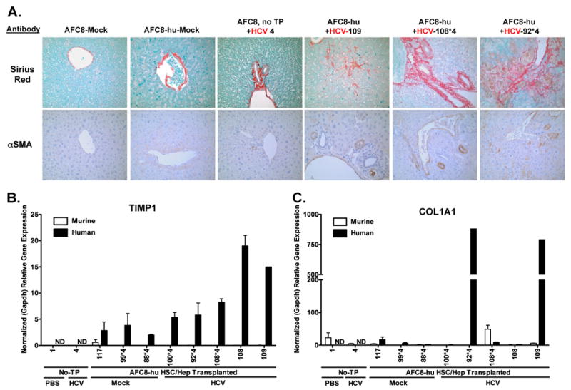
(A) Representative liver sections from AFC8/mock, AFC8-hu/mock, AFC8 (no transplant)/HCV, and three AFC8-hu/HCV mice were stained with Sirius Red/Fast Green (top panels) to visualize liver fibrosis. Liver sections were also stained with a mouse monoclonal antibody against human α-smooth muscle actin (αSMA, bottom panels) to visualize activated stellate cells. Human or murine specific gene expression of TIMP1 (B) and COL1A1 (C) in the liver was measured using species-specific PCR primers. Values shown are relative gene expression normalized to human or mouse GAPDH, respectively. Data represent mean ± s.e.m.