Abstract
The purpose of this study was to evaluate the long-term results of vascularised fibular graft for reconstruction of the wrist after excision of grade III giant cell tumour in the distal radius. From January 1998 to September 2003, 18 patients with wrist defects due to distal radius grade III giant cell tumour resection were treated with vascularised fibular graft and were followed-up. The limb function was restored to an average 80% of normal function and bone union was achieved within six months in 18 patients with vascularised fibular graft. MSTS score averaged 25.6 and ranged between 21 and 29; Mayo wrist score averaged 56 with a range from 40 to 65. It is appropriate to use the head of the fibula as a substitute for the distal radius. The healing of vascularised fibular graft is very quick and without bone resorption. Thus, in the procedure for reconstruction and limb salvage after bone tumour resection of distal radius, the free vascularised fibular graft with fibular head is an ideal substitute.
Introduction
Giant cell tumour (GCT) of distal radius is a rare and unpredictable lesion. Its standard treatment has ranged from surgical curettage to wide resection, and varying oncological and functional results have been reported for all modalities. Curettage alone or with bone grafting has been reported to be associated with high incidence of local recurrence in these tumours. Especially for grade III giant cell tumour, the high incidence of local recurrence is unacceptable. The purpose of this study was to evaluate the long-term results of the treatment of grade III giant cell tumour of distal radius with wide resection and reconstruction with vascularised fibula transfer at Qilu Hospital of Shandong University.
Patients and methods
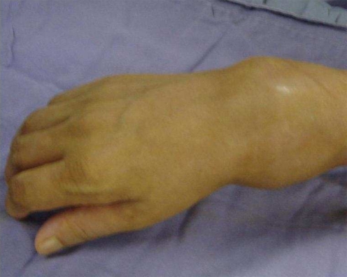
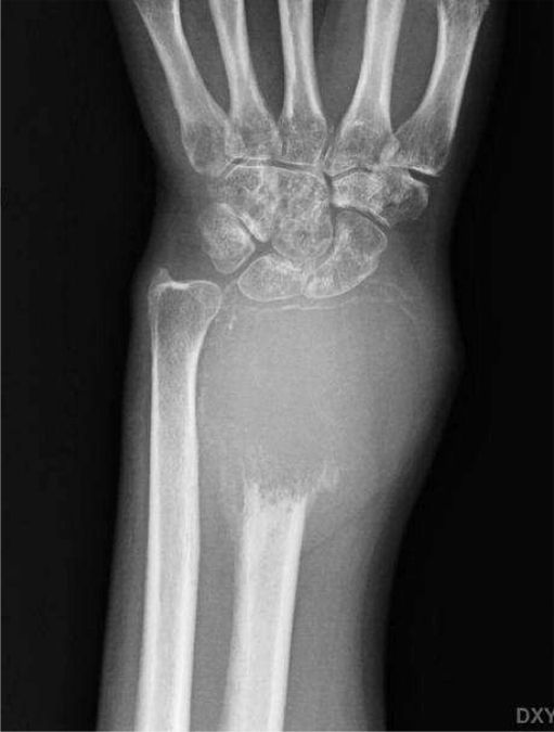
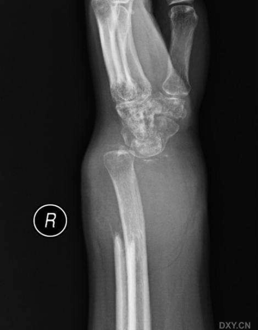
At Qilu Hospital of Shandong University, 26 patients with grade III giant cell tumour of distal radius were treated between 1998 and 2003. Of all these 26 grade III cases, six patients were followed-up for less than five years and two patients died from other diseases. These eight patients were excluded from the study. The remaining 18 patients were followed-up for at least five years. The average age at histological diagnosis was 32.2 years (range 15–66 years). All the slides of biopsied and excised tumours were reviewed and the diagnoses were confirmed by an experienced bone pathologist. All the tumours were Enneking Stage III [Figs. 1, 2, and 3]. The average length of follow-up was 72 months, which ranged from ten to 112 months. The surgical stage was determined according to the staging system of benign lesions of Enneking (1, latent—always intracapsular, surrounded by mature cortical rim, remains static or heals spontaneously; 2, active—remains intracapsular, surrounded by thin but intact reactive bone which may be deformed, slowly expands; 3, locally aggressive—often extracapsular, not limited by reactive bone or by natural barriers).
Fig. 1.
Preoperative giant cell tumor of the distal radius
Fig. 2.
The anteroposterior X-ray representation of giant cell tumor of the distal radius
Fig. 3.
The lateral X-ray representation of giant cell tumor of the distal radius
Methods
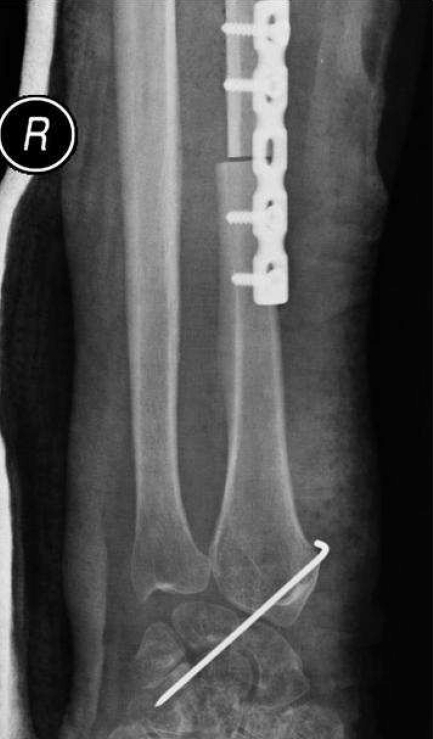
All patients underwent imaging studies for tumour screening including plain radiographs of the wrist and chest and total body bone scan [Figs. 2 and 3]. The diagnosis was confirmed by histological examination of the biopsy specimen in all cases. There was no evidence of metastatic disease in any of the patients at the time of diagnosis. Patients with grade III giant cell tumours involving the distal end of the radius were treated with en bloc resection and reconstruction with a free vascularised fibular graft. The lesion was approached through a dorsal incision, the extensor tendons were identified and protected throughout the procedure. An effort was made to remove all the neoplastic tissue. For fibular head transfer along with the shaft to replace the radial joint surface, it is very important to make sure that the length of reconstructed radial styloid is longer than that of ulnar styloid to restore the ulnar drift of radius. It is necessary to adjust the direction of the articular facet of the fibular head and fix it to the scaphoid and lunate [Fig. 4]. Fixation of the grafted fibula is very important, because inadequate fixation can influence the tension of the vascular anastomosis and result in nonunion. Blood circulation of the grafted fibula is reconstructed by a microsurgical technique, with radial artery anastomosis to the peroneal artery and fibular vein anastomosis to the cephalic vein, and the union of fracture is obviously shortened. The patients can perform functional exercises early so the function of wrist can be restored to the greatest extent. The patients were followed-up for a minimum of five years. Repeat X-rays of the wrist and chest were performed on each visit along with the clinical assessment. Findings of the clinical and radiological examination were documented. Wrist X-rays were assessed for evidence of local recurrence of the tumour and distal radio-ulnar impingement. Functional evaluation of patients was performed according to the most recent system of the Musculoskeletal Tumour Society (MSTS). This system involves six factors. Pain, function, emotional acceptance, hand positioning, dexterity and lifting ability are typical factors for the upper limbs. A maximum of five points for each factor results in a maximum score of 30 points. Functional analysis was performed at the most latest follow-up visit.
Fig. 4.
The postoperative X-ray representation of giant cell tumor of the distal radius
Results
There were ten males and eight females in this series. Mean age of the patients was 41 years (range, 16–73 years) and mean duration of symptoms was 37 weeks (range, 4–78 weeks). Eleven patients had the lesion on the right side and seven had left distal radius involvement. There was no local recurrence in the group of patients receiving wide resection as their initial surgery. The disease-free ratio of the patients treated with wide resection was significantly better than that of those treated with excision and curettage or intralesional excision reported in other articles. Functional results in this group were 28.2 (average) undergoing wide resection. All cases were evaluated with X-ray and excellent bone union of the grafted fibula with radius was found. The average duration to bone union was 32 months. The average range of motion of the wrist was: 67.0 ± 9.2 degrees of extension, 33.2 ± 5.1 degrees of flexion, 14.5 ± 4.7 degrees of radial deviation, 19.4± 3.9 degrees of ulnar deviation, 33.8 ± 6.6 degrees of pronation, and 13.3 ± 4.0 degrees of supination. Average grip strength was 33.1 kg which ranged from 15.7 to 50.1 kg. Compared with the contralateral side, this accounted for 75%. MSTS score averaged 25.6 and ranged from 21 to 29; Mayo wrist score averaged 56 ranging from 40 to 65. There was no wrist instability or deformation. All patients were satisfied with the clinical effect. According to the degree of alleviation of the clinical symptoms, the results were evaluated according to the Odom standard: clinical effect of eight cases was excellent, six cases were good, four cases satisfactory; thus, the portion of excellent and good results was 78%.
Discussion
Giant cell tumour is a benign locally aggressive tumour with a high tendency to recurrence, and a low rate of pulmonary metastases. In 90% of cases the tumour occurs in the long bones, especially near the epiphysis. Approximately 8% of all giant cell tumours involve the distal part of the radius [1–5]. The aim of treatment is complete removal of the tumour and preservation of the maximum function of the extremity. As for grade III giant cell tumour, the endosteal surface of the cortex is destroyed and a thin shell of reactive bone is formed. This neocortex appears thin and expanded on X-rays; in some cases, it may be difficult to see on plain X-rays. Curettage alone or with bone grafting has been associated with a high incidence of local recurrence in grade III giant cell tumour and rates of recurrence between 28 and 85% have been reported in some studies [2–6]. We agree that curettage alone may not be the best treatment option for grade III giant cell tumours, but it works very well in most of the grade I and II tumours [3, 7]. Lower rates of local recurrence have been noted after wide resection of the diseased bone of grade III; a complex reconstruction procedure (arthroplasty) or arthrodesis of the wrist is required. All these procedure have certain morbidity and complications associated with them.
The surgical approach to giant cell tumours in the 19th century was routine amputation. This was later changed by Joseph Bloodwood who suggested that simple curettage was usually sufficient; this has since been extensively used in clinical practice [4–9]. However, simple curettage of giant cell tumours has been criticised for its high rate of local recurrence. The techniques of curettage and grading of the tumour have not been included in previous analyses; therefore, it is difficult to comment on the causes of recurrence. It has also been said that curettage alone may not have completely removed the tumour, and a local recurrence may well be the residual tumour [6, 8]. The high rate of local recurrence was due to the indiscriminate use of curettage for all lesions including the highly aggressive tumours, for example, grade III giant cell tumours. En-bloc resection is strongly recommended, especially in high grade tumours, those which have recurred, have pathological fracture, have enlarged rapidly or are frankly malignant.
In this series, the recurrence rate was zero after wide resection. We considered that the adjunctive therapy should be performed in patients who received intralesional excision to avoid local recurrence, and wide excision is perhaps a much better choice for most patients with grade III and a selection of grade II giant cell tumour of distal radius. Vascularised fibular autograft was first used in 1945 for congenital absence of radius [10, 11]. Later, fibular transplants were used by various authors for tumours of the lower end radius. This reconstruction technique has yielded good functional results for giant cell tumour of the lower end of the radius in various series, although large series with longer follow-ups are few [5, 12]. This procedure also has problems such as delayed union, nonunion, stress fractures, bone resorption, deformities, ulnar impingement, carpal degenerative changes and donor site morbidity [5].
In our study, patients with tumours of the distal radius demonstrated good functional results, as reported by Campbell and Akbarnia [3, 9, 13]. Some doctors report that a high surgical stage correlates with poor functional results. This may be due to the frequent surgical procedures needed owing to the frequent recurrence of high-stage tumours. Most recurrences occurred within two years after operation. Some have reported that 16 out of 24 patients (67%) suffered local recurrence within the first two years, six (25%) suffered recurrence between two and five years later and two (8.3%) suffered recurrence more than five years later. Therefore, postoperative follow-up examinations of GCT patients are essential for at least the first five years [11–14], as recommended by Eckardt and Grogan [5, 7, 12].
Complete eradication of the diseased tissue while preserving normal bony architecture and joint function is the goal of treatment for any tumour. Several reconstructive procedures have been described for the complete defect of the distal radius that is created after a wide excision of a giant cell tumour of bone; our patients with giant cell tumours involving the distal end of the radius were treated with en bloc resection and reconstruction with a free vascularised fibular graft [11]. There was radiographic evidence of bone union at the host–graft junctions in all cases. In the newly reconstructed wrist joint, there was palmar subluxation of the carpal bones and degenerative changes in both patients. The postoperative function was satisfactory. Reconstruction with a vascularised fibular shaft graft has proven to be a useful and reliable procedure for reconstruction of the wrist after excision of giant cell tumour of the distal end of the radius.
While single extended curettage performed carefully by an experienced orthopaedic oncologist offers a good chance of eliminating the disease, the results are not so predictable in aggressive grade III tumours [4, 13, 14]. We recommend that treatment with curettage for grade I and II giant cell tumours offers a very good (90%) chance of cure. A high rate of local recurrence and complications is to be expected with curettage alone in grade III tumours. In the procedure for reconstruction and limb salvage after bone tumour resection of the distal radius, the vascularised fibular graft with the fibular head is an ideal substitute.
References
- 1.Bianchi G, Donati D, Staals EL, et al. Osteoarticular allograft reconstruction of the distal radius after bone tumor resection. J Hand Surg Br. 2005;30:369–373. doi: 10.1016/j.jhsb.2005.04.006. [DOI] [PubMed] [Google Scholar]
- 2.Cheng CY, Shih HN, Hsu KY, et al. Treatment of giant cell tumor of the distal radius. Clin Orthop Relat Res. 2001;383:221–228. doi: 10.1097/00003086-200102000-00026. [DOI] [PubMed] [Google Scholar]
- 3.Malek F, Krueger P, Hatmi ZN, et al. Local control of long bone giant cell tumour using curettage, burring and bone grafting without adjuvant therapy. Int Orthop. 2006;30:495–498. doi: 10.1007/s00264-006-0146-3. [DOI] [PMC free article] [PubMed] [Google Scholar]
- 4.Joo Han Oh, Yoon PW, Lee SH, et al. Surgical treatment of giant cell tumour of long bone with anhydrous alcohol adjuvant. Int Orthop. 2006;30:490–494. doi: 10.1007/s00264-006-0154-3. [DOI] [PMC free article] [PubMed] [Google Scholar]
- 5.Werner M. Giant cell tumour of bone: morphological, biological and histogenetical aspects. Int Orthop. 2006;30:484–489. doi: 10.1007/s00264-006-0215-7. [DOI] [PMC free article] [PubMed] [Google Scholar]
- 6.Kawamura K, Yajima H, Kobata Y, et al. Wrist arthrodesis with vascularized fibular grafting. J Reconstr Microsurg. 2009;25(8):501–505. doi: 10.1055/s-0029-1234022. [DOI] [PubMed] [Google Scholar]
- 7.Szendrıi MA. Bone tumours. Int Orthop. 2006;30:435–436. doi: 10.1007/s00264-006-0272-y. [DOI] [PMC free article] [PubMed] [Google Scholar]
- 8.Minami A, Kato H, Iwasaki N. Vascularized fibular graft after excision of giant-cell tumor of the distal radius: wrist arthroplasty versus partial wrist arthrodesis. Plast Reconstr Surg. 2002;110:112–117. doi: 10.1097/00006534-200207000-00020. [DOI] [PubMed] [Google Scholar]
- 9.Muramatsu K, Ihara K, Azuma E, et al. Free vascularized fibula grafting for reconstruction of the wrist following wide tumor incision. Microsurgery. 2005;25:101–106. doi: 10.1002/micr.20088. [DOI] [PubMed] [Google Scholar]
- 10.Natarajan MV, Chandra Bose J, Viswanath J, et al. Custom prosthetic replacement for distal radial tumours. Int Orthop. 2009;33(4):1081–1084. doi: 10.1007/s00264-009-0732-2. [DOI] [PMC free article] [PubMed] [Google Scholar]
- 11.Szabo RM, Anderson KA, Chen JL. Functional outcome of en bloc excision and osteoarticular allograft replacement with the Sauve-Kapandji procedure for Campanacci grade 3 giant-cell tumor of the distal radius. J Hand Surg Am. 2006;31:1340–1348. doi: 10.1016/j.jhsa.2006.06.004. [DOI] [PubMed] [Google Scholar]
- 12.Warnecke I, Brüner S, Frerichs O, et al. Functional reconstruction of the radial articular surface after giant cell tumor. Orthopade. 2007;36:679–682. doi: 10.1007/s00132-007-1091-6. [DOI] [PubMed] [Google Scholar]
- 13.Rtaimate M, Laffargue P, Farez E, et al. Reconstruction of the distal radius for primary bone tumors using a non-vascularized fibular graft (report of 4 cases) Chir Main. 2001;20:272–279. doi: 10.1016/S1297-3203(01)00046-4. [DOI] [PubMed] [Google Scholar]
- 14.Matejovsky Z, Jr, Matejovsky Z, Kofranek I. Massive allografts in tumour surgery. Int Orthop. 2006;30:478–483. doi: 10.1007/s00264-006-0223-7. [DOI] [PMC free article] [PubMed] [Google Scholar]


