Abstract
Background: Lymphangiomas occur most commonly in the head and neck region, while other sites are rarely affected. A combination of retroperitoneal and genital lymphangioma is very rare indeed. Though congenital, it may persist into adulthood due to missed diagnosis and inadequate or total lack of treatment. Materials and methods: A report of a 22-year-old male student who presented with recurrent multiloculated genital, thigh, groin and retroperitneal lymphangioma. He underwent surgical excision and adjuvant sclerotherapy using ethylene-diamine tetra acetic acid. Results and Conclusions: There was an initial recurrence after surgery which responded satisfactorily to sclerotherapy. Complete surgical excision of lymphangioma may be precluded by vital structures but sclerotherapy produces satisfactory resolution. The difficulties in management with limited facilities for diagnosis and treatment are highlighted.
Keywords: Lymphangioma, Genital, Retroperitoneal, Therapeutic challenges, Surgical excision, Sclerotherapy
Introduction
Lymphangiomas are uncommon harmatomatous congenital malformations of the lymphatic system that may involve any part of the body [1, 2]. They are benign, and are classified into three types based on the depth and the size of abnormal lymphatic vessels. These are Cystic (macro cystic), Simple (supermicrocystic) and Cavernous (micro cystic). However, most of such lesions contain varying proportions of the different types with one predominating.
Ninety-five percent of lymphangiomas occur in the head and neck and axillae [3], though any part of the body may be affected [2]. About 1 to 5% of lymhangiomas are retroperitoneal [4, 5, 6]. Penile lymphangiomas are also rare [7]. Though benign, lymphangiomas have the potential to infiltrate surrounding tissues, and unlike congenital haemangioma, spontaneous involution is rare.
The diagnosis and treatment of such lesions have been enhanced with the advent of imaging techniques such as ultrasonography, computerized tomography and magnetic resonance imaging [8, 9, 10].
Lymphoscintigraphy is helpful in characterizing the functional aspects of the lymphangiomas and for providing information on the direction of lymphatic flow and the relation to normal lymphatic drainage pathways [11]. The mainstay of treatment has been surgical resection. Difficulties with an inability to clearly separate and spare normal structures from the abnormal tissue, as well as high rates of recurrence after incomplete resection, have resulted in many unsatisfactory outcomes, particularly with diffuse lymphangiomas [11].
This case report is presented to highlight the uncommon extensive cavernous lymphangioma in the retroperitoneum and the pelvis, in association with diffuse genital, groin and perineal lymphangioma; and the challenges faced by surgeons practicing in a developing country where imaging and treatment facilities are limited. Awareness of this type of presentation will resolve diagnostic puzzle and lead to proper treatment planning.
Case report
A 22-year-old male student presented to the plastic surgery clinic of our hospital with recurrent left groin and thigh swelling of 9 years duration. He also had multiple verrucous skin and subcutaneous masses with foul smelling, milky discharge, which caused him aesthetic embarrassment among his peers. He had a history of similar swellings since birth for which he had surgical excision at 7 years of age and had repeat surgeries 3 and 6 years later, but the lesions never completely disappeared. Histopathology of the lesion confirmed the diagnosis of lymphangioma. There was no family history of similar illness.
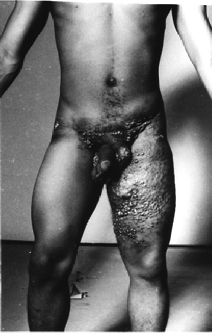
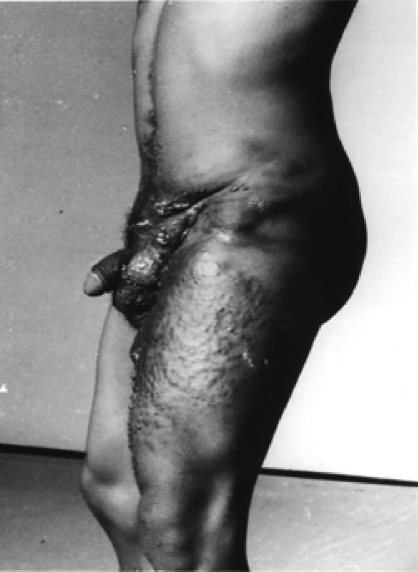
Physical examination revealed an otherwise healthy young man with multiloculated swellings involving the left thigh, the suprapubic region, left groin and the external genitalia (Figures 1 & 2). The medial left upper thigh swelling empties and the skin of the upper thigh and external genitalia had multiple warty lesions with foul smelling milky discharge.
Figure 1.
Left Lateral view (Note scrotal, groin and flank swellings)
Figure 2.

Anterior view (Note pelvic bulges, groin and left thigh verrucous swellings)
A diagnosis of recurrent infected lymphangioma was made and treatment commenced with penicillin V and metronidazole. Baseline hematocrit and blood urea and electrolytes were normal. Abdomino-pelvic ultrasound revealed a left sided retroperitoneal cavity with smaller surrounding vesicles. It consisted of an elongated mass of mixed echoic pattern in the left lumbar region, infero-posterior to the kidney and extending to the left iliac fossa; within the mass are areas of irregular sonolucency. Computerized tomography and magnetic resonance imaging were not available.
Surgery was done to excise the lesions. There were multiple cystic dilatations ramifying in the subcutaneous tissue and extending into the intermuscular planes in the thigh, around the femoral neurovascular bundles and superiorly into the retroperitoneal space and surrounding nerve roots. Lymphangiomatous tissues around the groin, root of the penis and the thigh were excised. Incision was carried up to the left iliac fossa postero-laterally towards the flanks and the peritoneum reflected anteriorly. A near total excision of lymph sacs was done with some perineural and perivascular lesions being left behind. A drain was placed in the wound for one week.
Six months postoperatively, recurrence of the thigh lesion occurred. This was then treated with injection sclerotherapy under ultrasonographic guidance, using sodium tetradecyl sulphate injection administered monthly. There was progressive symptomatic and clinical relief. A repeat ultrasound seven months after commencement of sclerotherapy showed about 90% resolution. The patient was being followed up in the surgical outpatient until he was lost to follow-up 18 months later.
Discussion
The origin of lymphangiomas is unclear. They are believed to result from sequestration of lymphatic channels from the main lymphatic vessels in embryonic development and retain their proliferative potential. Retroperitoneal and genital lymphangiomas are rare, but are usually complicated by infections such as recurrent cellulitis [12]. Other complications include lymphoedema, lymphorrhea and lymphatic effusions [11]. Retroperitoneal lesions may present as a mass in the iliac fossa or flanks [1]. Prior to the advent of modern radiologic imaging techniques, definitive diagnosis used to be established intraoperatively as an unexpected finding but radiological imaging techniques have made this obsolete. However these imaging facilities are not readily available or affordable in many developing countries.
Image guided sclerotheapy using ultrasonography and fluoroscopy reported complete resolution in 66.6% of patients without serious complications [10]. The use of sclerotherapy has been suggested as a preferred primary mode of treatment in childhood [13]. Recent reports on the use of OK 432 (a streptococcal derivative used as a biologic response modifier) in the treatment of retroperitoneal lymphangiomas is giving promising results [14, 15]. Other sclerosants used include bleomycin, doxycycline, sodium morrhuate or dextrose [11].
Surgical excision is regarded as the treatment of choice by many surgeons [9, 16, 17], but this is not always possible either due to inaccessibility, or neural and vascular involvement [5] as was the situation in this case report. Expectant treatment is sometimes utilized in young children as some lesions are known to undergo spontaneous regression without treatment, but this is rare and may be adopted till the child is 6 years old.
Radiation therapy is generally considered ineffective for lymphangiomas, but there is a report of good response to lymphangiolipoma [18]. Cryotherapy and electrocautery is used on small lesions of lymphangioma circumscriptum. Carbon dioxide laser vaporization [19] and combined laser light and radiofrequency energy [20] are reported to be effective and relatively safe. Cryosurgery and laser surgery were not available to be considered in the treatment of this patient.
Recurrences usually appear within the first 9 months of treatment (6months in this case). The recurrence rate is higher with cavernous lymphangiomas.
Prognosis of lymphangiomas depends on the location and extent of the lesion and the presence of other associated abnormalities [3]. Treatment and outcome should be better as newer imaging techniques become more commonly available as well as access to the other forms of therapies such as laser. In difficult cases sclerotherapy combined with surgery may result in satisfactory resolution.
References
- 1.Rifki Jai S, Adraoui J, Khaiz D, Chehad F, Lakhloufi A, Bouzidi A. Retroperitoneal cystic lymphangioma. Prog Urol. 2004;14(4):548–50. [PubMed] [Google Scholar]
- 2.Shah A, Meacock I, More B, Chandran H. Lymphangioma of the penis; a rare anomaly. Pediatr. Surg Int. 2005 Apr;21(4):329–30. doi: 10.1007/s00383-004-1346-9. [DOI] [PubMed] [Google Scholar]
- 3.Mohite PN, Bhatnagar AM, Parikh SN. A huge omental lymphangioma with extension into labia majorae: a case report. BMC Surgery. 2006;6:18. doi: 10.1186/1471-2482-6-18. [DOI] [PMC free article] [PubMed] [Google Scholar]
- 4.Cherk M, Nikfarjam M, Christophi C. Retroperitoneal Lymphangioma. Asian J Surg. 2006;29(1):51–4. doi: 10.1016/S1015-9584(09)60297-9. [DOI] [PubMed] [Google Scholar]
- 5.Kobayashi D, Kumagai H, Satsuma S. Cavernous lymphangioma of the leg in children treated by the injection of OK-432 after resection. J Bone Joint surg Br. 2003;85(6) 89:1–4. [PubMed] [Google Scholar]
- 6.Huseyin O, Ercan K, Zilkif B, Bengu C. Recurrent Retroperitoneal Cystic Lymphangioma. Yonsei Med J. 2005;46(5):715–8. doi: 10.3349/ymj.2005.46.5.715. [DOI] [PMC free article] [PubMed] [Google Scholar]
- 7.Sheu JY, Chung HJ, Chen KK, Lin AT, Chang YH, Wu HH, Huang WJ, Hsu YS, Kuo JY, Chang LS. Lymphangioma of male exogenital organs. J Chin Med Assoc. 2004;67(4):204–6. [PubMed] [Google Scholar]
- 8.Taro O, Toshiaki M, Eisei J, Yoshikazu K, Hiroyuki K. Five cases of lymphangioma of the mediastinum in adults. Ann Thorac Cardiovasc Surg. 2001;7:103–5. [PubMed] [Google Scholar]
- 9.Gomez P J A, Martin M A, Bonilla P R, Alvarado R A, Blanco R F, Rodero G P, Baena G V. Retroperitoneal Cystic lymphangioma; A silent disease in Adults. Actas Urol Esp. 2002 May;26(5):356–60. doi: 10.1016/s0210-4806(02)72790-3. [DOI] [PubMed] [Google Scholar]
- 10.Won J H, Kim B M, Park S W, Kim M D. Percutaneous sclerotherapy of lymphangiomas with acetic acid. J Vasc Interv Radiol. 2004;15(6):595–600. doi: 10.1097/01.rvi.0000127899.31047.0e. [DOI] [PubMed] [Google Scholar]
- 11.Molitch I Howard, Unger C Evan, Witte L Charles, vanSonnemberg Eric. Percutaneous Sclerotherapy of lymphangiomas. Radiology. 1995;194:343–47. doi: 10.1148/radiology.194.2.7529933. [DOI] [PubMed] [Google Scholar]
- 12.Swanson D L. Genital lymphangioma with recurrent cellulitis in men. Int Journal of Dermatology. 2006;45(7):800–4. doi: 10.1111/j.1365-4632.2006.02782.x. [DOI] [PubMed] [Google Scholar]
- 13.Sanlialp I, Karnak I, Tanyel F C, Senocak M F, Buyukpamukru N. Sclerotherapy for lymphangioma in children. Int J Pediatr Otorhinolaryngol. 2003;67(7):795–800. doi: 10.1016/s0165-5876(03)00123-x. [DOI] [PubMed] [Google Scholar]
- 14.Everaldo R, Jr, Elvis T V, Francisco V, Luiz G T. OK-432 therapy for lymphangioma in children. J Pediatr (Rio J) 2004;80(2):154–8. doi: 10.2223/1156. [DOI] [PubMed] [Google Scholar]
- 15.Uchida K, Inoue M, Araki T, Miki C, Kusunoki M. Huge scrotal, flank and retroperitoneal lymphangioma successfully treated by OK-432 sclerotherapy. J Urology. 2002;60(6):1112. doi: 10.1016/s0090-4295(02)01957-x. [DOI] [PubMed] [Google Scholar]
- 16.Bloom D C, Perkins J A, Manning S C. Management of lymphatic malformations. Curr Opin in Otolaryngol Head Neck Surg. 2004;12(6):500–4. doi: 10.1097/01.moo.0000143971.19992.2d. [DOI] [PubMed] [Google Scholar]
- 17.Al Salem A H. Lymphangiomas in infancy and Childhood. Saudi Med J. 2004 Apr;25(4):466–9. [PubMed] [Google Scholar]
- 18.Bruns F, Steitz W, Schieller R, Schaefer U, Willich N, Micke O. Lympangioma of the lower extremity: 5-year radiological follow-up after radiotherapy. Brit J Radiol. 2002;75:767–71. doi: 10.1259/bjr.75.897.750767. [DOI] [PubMed] [Google Scholar]
- 19.Anthony F, Mortimer PS, Harland CC. Acquired scrotal lymphangiomas: successful treatment with cutting diathermy and carbon dioxide laser. Clin Exp Dermatol. 2002;27(3):192–4. doi: 10.1046/j.1365-2230.2002.00993.x. [DOI] [PubMed] [Google Scholar]
- 20.Lapidoth M., Ackerman L, Amitai D B, Raveh E, Kalish E, David M. Treatment of Lymphangioma circumscriptum with combined Radiofrequency Current and 900nm Diode Laser. Dermatologic Surgery. 2006;32(6):790–94. doi: 10.1111/j.1524-4725.2006.32162.x. [DOI] [PubMed] [Google Scholar]


