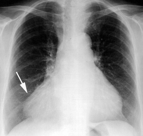
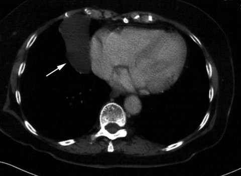
A 76-year-old hypertensive woman presented at our cardiology department with dyspnea and palpitations. Upon auscultation, we found dysrhythmia and a systolic murmur in the apical zone. Atrial fibrillation was diagnosed on electrocardiography. The chest radiograph showed an abnormal shadow at the right side of the heart (Fig. 1). A computed tomogram confirmed the presence of a pericardial mass, and a 4 × 8-cm pericardial cyst was identified at the right cardiophrenic angle (Fig. 2). On echocardiography, severe fibrotic mitral stenosis and insufficiency were observed.
Fig. 1 Chest radiograph shows an abnormal shadow at the right cardiophrenic sinus (arrow).
Fig. 2 Contrast-enhanced computed tomographic scan of the chest with mediastinal window settings shows a slightly enlarged, well-circumscribed water attenuation lesion (arrow).
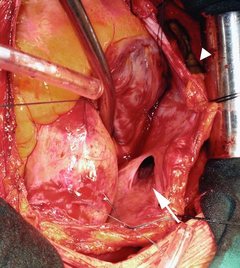
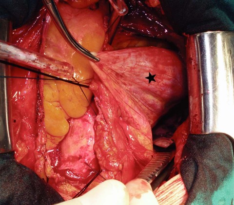
Subsequently, the patient underwent mitral valve replacement surgery via median sternotomy. Routine pericardiotomy revealed an aperture at the mid-right pericardial cavity, and the diverticulum led down to a right cardiophrenic recess (Figs. 3 and 4). After a partial pericardiectomy and complete resection of the pericardial diverticulum with its orifice, we performed standard mitral valve replacement with a 27-mm mechanical valve. The lesion was diagnosed as pericardial diverticulum with mild infiltration of inflammatory cells (seen upon histologic examination). The patient was discharged from the hospital on postoperative day 7, with no complication.
Fig. 3 Intraoperative photograph shows the diverticular orifice (arrow) within the pericardial cavity and the enlarged pericardial cyst (arrowhead).
Fig. 4 Intraoperative photograph of the enlarged pericardial cyst (star).
Comment
Pericardial cysts and diverticula are rare conditions that usually are detected upon routine chest radiography. They are estimated to occur in approximately 1 in 100,000 cases and to account for 13% to 17% of all mediastinal cysts.1 Most pericardial cysts and diverticula are found in the right costophrenic angle, approximately 25% are found in the left costophrenic angle, and the remaining 8% are found in the posterior and anterior superior mediastinum.2 They present with fewer symptoms than other mediastinal tumors. Le Roux and colleagues3 reported that only 20% of all pericardial cysts are symptomatic, usually with dyspnea, recurrent chest pain, cough, and odynophagia. The differential diagnosis includes all conditions associated with abnormal intrathoracic shadows.
Footnotes
Address for reprints: Mehmet Ali Sahin, MD, GATA Kalp Damar Cerrahisi A.D., Etlik, 06018 Ankara, Turkey. E-mail: mali_irem@yahoo.com
References
- 1.Akiba T, Marushima H, Masubuchi M, Kobayashi S, Morikawa T. Small symptomatic pericardial diverticula treated by video-assisted thoracic surgical resection. Ann Thorac Cardiovasc Surg 2009;15(2):123–5. [PubMed]
- 2.Borges AC, Gellert K, Dietel M, Baumann G, Witt C. Acute right-sided heart failure due to hemorrhage into a pericardial cyst. Ann Thorac Surg 1997;63(3):845–7. [DOI] [PubMed]
- 3.le Roux BT, Kallichurum S, Shama DM. Mediastinal cysts and tumors. Curr Probl Surg 1984;21(11):1–77. [DOI] [PubMed]


