Abstract
Spotted fever group rickettsiae are known to produce distinct plaque phenotypes. Strains that cause lytic infections in cell culture form clear plaques, while nonlytic strains form opaque plaques in which the cells remain intact. Clear plaques have historically been associated with more-virulent species or strains of spotted fever group rickettsiae. We have selected spontaneous mutant pairs from two independent strains of Rickettsia rickettsii, the virulent R strain and the avirulent Iowa strain. A nonlytic variant of R. rickettsii R, which typically produces clear plaques, was isolated and stably maintained. A lytic variant of the Iowa strain, which characteristically produces opaque plaques, was also selected and maintained. Genomic resequencing of the variants identified only a single gene disrupted in each strain. In both cases, the mutation was in a gene annotated as relA/spoT-like. In the Iowa strain, a single mutation introduced a premature stop codon upstream from region encoding the predicted active site of RelA/SpoT and caused the transition to a lytic plaque phenotype. In R. rickettsii R, the nonlytic plaque phenotype resulted from a single-nucleotide substitution that shifted a tyrosine residue to histidine near the active site of the enzyme. The intact relA/spoT gene thus occurred in variants with the nonlytic plaque phenotype. Complementation of the truncated relA/spoT gene in the Iowa lytic plaque variant restored the nonlytic phenotype. The relA/spoT mutations did not affect the virulence of either strain in a Guinea pig model of infection; R strain lytic and nonlytic variants both induced fever equally, and the mutation in Iowa to a lytic phenotype did not cause them to become virulent.
Rickettsia rickettsii is a member of the spotted fever group of rickettsiae and is the etiologic agent of Rocky Mountain spotted fever (RMSF). R. rickettsii is a small obligate intracellular Gram-negative bacterium maintained in its tick host through transovarial and transtadial transmission (4, 20, 25). In the United States, Rocky Mountain spotted fever is the most severe and most frequently reported rickettsial disease, although there is considerable variation in virulence between strains from different geographic areas and even within a given locale (1-3, 12, 24, 31).
Transmission of R. rickettsii to mammals occurs through the bite of an infected tick. Once the organism gains access to the host, it replicates within vascular endothelial cells and can directly spread from cell to cell by actin-based motility (15). Damage to the vascular endothelium by R. rickettsii leads to increased vascular permeability and leakage of fluid into the interstices, causing the characteristic rash observed in RMSF (14). The ultrastructural changes that occur during replication within a cell include marked dilation of internal membranes, particularly the rough endoplasmic reticulum (28). R. rickettsii causes direct cytopathology as it replicates intracellularly (32); however, the exact mechanism of cell lysis has not been defined. Proposed mechanisms include protease activity (34), free radical-induced lipid peroxidation (27, 29), and phospholipase A activity (33). Strains of R. rickettsii differ in their virulence for animal models (3), serological properties (1), plaque morphologies (2, 12, 17), and cytopathic effects on human endothelial cells (11). Historically, avirulent strains of the spotted fever group have been described as producing opaque (turbid) plaques when grown in culture as a result of reduced host cell destruction, whereas virulent strains have been reported to cause clear plaques resulting from lysis of the host cells (2, 11, 12, 17).
With limited genetic systems, it has been difficult to definitively identify virulence factors in R. rickettsii. We have taken a comparative genomic approach in an attempt to identify genomic differences between closely related R. rickettsii strains that exhibit differences in virulence. A number of genomic differences have been identified between the virulent R. rickettsii Sheila Smith strain and the avirulent R. rickettsii Iowa strain, including the absence of rickettsial OmpA (rOmpA) from the avirulent R. rickettsii Iowa strain (10). While this comparison revealed important differences between R. rickettsii Sheila Smith and R. rickettsii Iowa, the number of polymorphisms makes it difficult to ascertain which are responsible for the variation in virulence between the two strains. Therefore, the continued comparison of multiple genomes of R. rickettsii that differ in their virulence may identify unique differences between strains which are involved in the pathogenesis of R. rickettsii.
Here, we describe the isolation of plaque variants of the R. rickettsii R strain and R. rickettsii Iowa. The variants spontaneously arose after repeated passage in Vero cells from single plaque-cloned isolates. Both strains displayed the clear- and opaque-plaque types, suggesting a difference in the abilities of these plaque variants to lyse host cells. Here, we describe phenotypic and genomic differences between these independently derived plaque variants.
MATERIALS AND METHODS
Rickettsia.
R. rickettsii strains were propagated in Vero cells with Medium 199 and purified by Renografin density gradient centrifugation (35).
Plaque cloning.
Vero cells were seeded at 3 × 10⁵ cells/ml into Falcon 6-well plates and allowed to adhere overnight. The cell monolayers were infected with serial dilutions of R. rickettsii in brain heart infusion (BHI) broth for 30 min in a humidified 34°C chamber. Each well was then overlaid with 5 ml of Medium 199 containing 5% fetal bovine serum and 0.5% agarose (GenePure ME; ISC Bioexpress). To allow formation of clonable plaques, plates were incubated for 8 days at 34°C and 5% CO2. Morphologically variant plaques (clear and opaque) were marked and isolated by removing the plug of agarose over the plaque. The intact plaque was scraped and aspirated in 10 μl of BHI and used to inoculate T25 flasks of Vero cells for clonal expansion.
LDH release assay.
Vero cells were seeded as described above into Falcon 6-well plates and allowed to adhere overnight. Vero cells were infected with either R. rickettsii R strain or R. rickettsii Iowa plaque variants at a multiplicity of infection (MOI) of 0.025. On days 1 and 3 after infection, 100 μl of medium was removed to determine the relative amount of lactate dehydrogenase (LDH) released into the medium by use of a model II LDH cytotoxicity assay kit (BioVision) as directed by the manufacturer. The remaining medium was removed and replaced with 1 ml of K36 (0.1 M KCl, 0.015 M NaCl, 0.05 M K2HPO4, 0.05 M KH2PO4, pH 7.0) to determine the number of PFU. The monolayer was removed by scraping, and the host cells were lysed by bead beating 2 times for 4 s with (1-mm) glass beads. The cell suspension was then frozen at −80°C until plaque assays were performed as previously described.
Microarray analysis.
Monolayers of Vero cells seeded at 2.5 × 10⁶/ml in T25 flasks were allowed to adhere overnight and infected with either the R. rickettsii R strain clear-plaque variant or the R strain opaque-plaque variant at an MOI of 0.025. After 2 days of growth (late log phase), the medium was removed and replaced with 1 ml of TRIzol. The monolayer was disrupted by scraping, and the suspension was frozen at −80°C until RNA was harvested. For GeneChip analysis, RNA was harvested as previously described (9, 10). Biotinylated cDNA was hybridized to custom Affymetrix GeneChips (no. 3 RMLchip3a520351F) containing 1,991 probe sets from two different R. rickettsii strains (Sheila Smith and Iowa) and analyzed as previously described (9).
Genomic DNA purification.
To isolate R. rickettsii genomic DNA, freshly purified R. rickettsii bacteria were first lysed by incubation in 50 mM Tris-HCl pH 8.0, 50 mM EDTA, 1% SDS, 10 mM dithiothreitol (DTT), and 4 mg/ml RNase A (Invitrogen) for 10 min at 37°C. The temperature was then increased to 60°C, and 3 μl of 20 mg/ml proteinase K was added at 20-min intervals for 2 h. After 2 h, 1 volume of phenol-chloroform-isoamyl alcohol (25:24:1) was added and the mixture was centrifuged for 3 min at 13,000 rpm. The aqueous phase was removed and subjected to two more phenol-chloroform-isoamyl alcohol extractions. The aqueous phase was then removed and extracted twice with equal volumes of chloroform-isoamyl alcohol (24:1). DNA was precipitated with a 1/10 volume of 3 M sodium acetate (pH 5.0) plus a 0.6 volume of isopropanol and resuspended in Tris-EDTA (TE; pH 8.0).
CGS.
Purified rickettsial DNA was sent to NimbleGen, Inc., for comparative genome sequencing (CGS). Their protocol is available from NimbleGen, Inc. (Madison, WI).
Solid sequencing and data analysis.
Mate-paired libraries constructed from the R. rickettsii R strain clear- and opaque-plaque variants with insert sizes between 2 and 3 kb were generated in accordance with the SOLiD System 2.0 user guide (Applied Biosystems, Foster City, CA). The clear-plaque sample generated 10,171,929 paired 25-nucleotide reads with insert sizes of 1,690 to 3,210 nucleotides, and the opaque-plaque sample generated 12,781,088 paired 25-nucleotide reads with insert sizes of 1,750 to 3,440 nucleotides. The reads were mapped to the reference strain R. rickettsii Sheila Smith (NC_009882) by use of Corona Lite, version 0.4r2.0 (Applied Biosystems), and ZOOM, version 1.0.5 (Bioinformatics Solutions Inc.). All mappings were done allowing up to 2 mismatches and resulted in approximately 50% of the reads being mapped to the reference genome. This equated to 200-fold coverage for the clear-plaque strain and 240-fold coverage for the opaque-plaque strain. A total of 22 single nucleotide polymorphisms (SNPs) were identified for the clear-plaque strain, and 21 SNPs were identified for the opaque-plaque strain, compared to the level for the reference genome. The results generated by Corona Lite and ZOOM were compared to each other and were found to be identical. The resulting SNPs were mapped to the R. rickettsii Sheila Smith genome to identify genes and amino acid changes.
Guinea pig infections.
Six-week-old female Hartley guinea pigs were inoculated intraperitoneally (i.p.) or intradermally (i.d.) with 10 to 1.0 × 10⁵ PFU of either the R. rickettsii R strain clear-plaque variant or the R. rickettsii R strain opaque-plaque variant and rectal temperatures monitored for 14 days after infection.
To determine if the R. rickettsii R strain clear- and opaque-plaque variants were stable during an infection. Guinea pigs were infected i.p. with ∼1,000 PFU of either the clear-plaque variant or the opaque-plaque variant. The spleens from 2 guinea pigs were aseptically removed on day 4 after infection, and a single cell suspension was made by passing the spleens through stainless steel 120-mesh gauze in 4 ml of BHI. The spleen cells were disrupted using 7 ml Wheaton glass tissue grinders, and the released rickettsial homogenate was centrifuged at 1,000 rpm to pellet nuclei and debris. Serial dilutions of the supernatants were made in BHI and plaqued on Vero cell monolayers as described above.
To determine if there was a competitive advantage for either the R. rickettsii R strain clear-plaque variant or the R strain opaque-plaque variant, a mixed sample containing 1,000 PFU of the clear-plaque variant and 1,000 PFU of the opaque-plaque variant was inoculated i.p. into guinea pigs, and after 4 days, spleens were harvested as described above. The resulting mixture was then plaqued on Vero cells to determine the ratio of the R. rickettsii R strain clear-plaque variant to the R strain opaque-plaque variant.
All experimentation involving animals was conducted under a protocol approved by the Rocky Mountain Laboratories Animal Care and Use Committee.
Cloning.
The relA/spoT-like gene (A1G_06035/RrIowa_1296) was amplified from the R. rickettsii R strain opaque-plaque variant by use of Phusion Hot Start II polymerase (New England BioLabs) according to the manufacturer's instructions with primers TCspoTphusF1 (5′-TATCTAGAGTCGACCTGCAGAAGCTCTTGATCCTGAGAATCCTGCCACC-3′) and TCspoTphusR1 (5′-CCCAGCTTGCATGCCTGCAGCTAATCTTTGTTAAATATTAGCACTTCAGCA-3′). The resulting PCR product was gel purified alongside a PstI (New England BioLabs) linearized transposon vector, pMW1650 (19). The gel products were column purified and mixed at a 2-to-1 ratio of insert to vector by use of an In-Fusion (Clontech) cloning reaction mixture for 30 min. Each reaction mixture was transformed into Escherichia coli strain Fusion Blue (Clontech) and grown on Luria-Bertani plates containing 50 μg/ml each of kanamycin and rifampin. The resulting clones were sequenced to ensure accuracy.
Transposon mutagenesis.
The R. rickettsii R strain was transformed, and clonal isolates were sequenced using previously described protocols for the electroporation and characterization of rickettsial species (18, 19).
Microarray data accession number.
The GeneChip data are posted on the Gene Expression Omnibus (GEO) (http://www.ncbi.nlm.nih.gov/geo/) under accession no. GPL4692.
RESULTS
Isolation of the clear- and opaque-plaque variants form the R. rickettsii R strain and R. rickettsii Iowa.
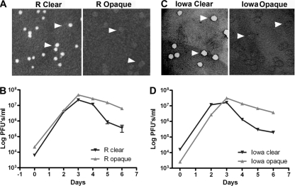
The R. rickettsii R strain was originally isolated in Western Montana from a Dermacentor andersoni tick suspension (24). This strain produces clear plaques on Vero cell monolayers (2, 10, 13) and is virulent in a guinea pig model of infection. The R strain had been passed 10 times in embryonated eggs and 3 times in Vero cells. Mice were subsequently inoculated, and the strain was reisolated from the spleen and plaque purified. The plaque phenotype at this point was the original clear-plaque type. The strain was expanded over three serial passages in Vero cells. In examination of the third passage, a distinct plaque type (opaque) was observed. The two plaque types (the R clear- and R opaque-plaque variants) were biologically cloned and expanded as stable variants which retained the clear- and opaque-plaque morphologies (Fig. 1A). To determine if the growth rates of these strains were similar, growth curves were performed using the R clear- and R opaque-plaque variants (Fig. 1B). The data indicate no difference in the growth rates of the clear- and opaque-plaque variants of the R. rickettsii R strain. By 5 to 7 days postinfection, the numbers of the clear-plaque variant declined relative to those of the opaque-plaque variant, presumably due to lysis of the host cell, which releases the rickettsiae into the medium, with a concurrent loss of viability.
FIG. 1.
R. rickettsii R strain and R. rickettsii Iowa plaque variants. (A) R. rickettsii R strain clear- and opaque-plaque variants. (B) Representative growth curves for the R. rickettsii R strain clear- and opaque-plaque variants. (C) R. rickettsii Iowa clear- and opaque-plaque variants. (D) Representative growth curves for the R. rickettsii Iowa clear- and opaque-plaque variants. Error bars represent standard errors of the means (SEM).
R. rickettsii Iowa was isolated from guinea pigs infected with a suspension made from Dermacentor variabilis (6). The original isolate was then passed over 271 times in eggs. Clonal isolates from high egg passages demonstrate a reduced ability to lyse Vero cells and form opaque plaques (13). Two different plaque types, identical to the R clear- and R opaque-plaque types described above, were also isolated from the avirulent R. rickettsii Iowa strain after passage through mice and plaque purification as described above for the R strain (Fig. 1C). The growth rates of the Iowa plaque variants were also indistinguishable (Fig. 1D).
R. rickettsii clear-plaque variants release greater amounts of LDH than R. rickettsii opaque-plaque variants.
Historically, plaque size and morphology have been correlated with the virulence of R. rickettsii strains; large, clear plaques have been thought to indicate virulent strains, whereas small, opaque, or turbid plaques were thought to represent less virulent or avirulent strains (2, 11, 12, 17). The plaque morphology of the isolated clear-plaque variants suggests that these strains cause more cellular lysis than those of opaque-plaque variants. Microscopically, the clear plaques are completely devoid of cells while the opaque plaques retain an almost complete monolayer. To confirm that clear-plaque variants cause more cellular destruction than the opaque-plaque variants, the amount of LDH released from Vero cells infected with either the R clear-plaque or the Iowa clear-plaque variant was compared to that released from Vero cells infected with either the R opaque-plaque or the Iowa opaque-plaque variant, respectively. LDH released into the medium was measured on days 1 and 3 postinfection. The clear-plaque variants cause more cellular destruction than the opaque-plaque variants, as indicated by increased LDH release from Vero cells on day 3 postinfection (Fig. 2). Determination of PFU confirmed that there was no difference in growth rate between the clear- and opaque-plaque cultures on day 1 or 3 (data not shown). These data confirm that the clear-plaque variants cause greater cell lysis than the opaque-plaque variants.
FIG. 2.
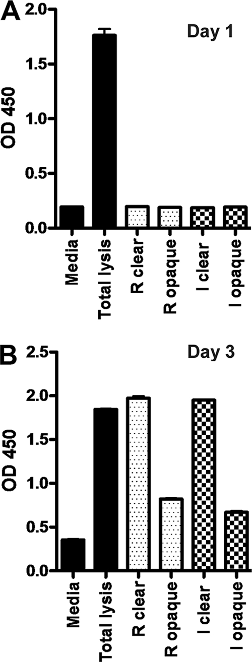
Comparison of LDH released from Vero cells infected with the clear- and opaque-plaque variants from R. rickettsii R strain and R. rickettsii Iowa. (A) LDH release on day 1 after infection. OD 450, optical density at 450 nm. (B) LDH release on day 3 after infection. Error bars represent SEM.
The R. rickettsii R clear-plaque variant and the Iowa clear-plaque variant have mutations in a RelA/SpoT-like protein.
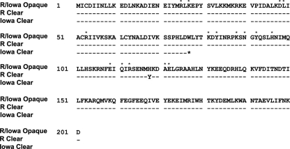
To identify the genetic differences associated with the phenotypes of clear- and opaque-plaque variants, R. rickettsii R strain and R. rickettsii Iowa strain clear- and opaque-plaque variants were subjected to SOLiD resequencing and comparative genome sequencing (CGS) (NimbleGen). Resequencing of the R. rickettsii R strain clear- and opaque-plaque variants revealed only a single gene disrupted. This mutation (C to T) is located at bp 354 of the A1G_06035 gene (RrIowa_1296), which is annotated as a guanosine polyphosphate pyrophosphohydrolase/synthetase-like (RelA/SpoT-like) protein gene. This protein has homology to the synthetase, metal binding, and nucleotidyltransferase domains of RelA/SpoT-like bifunctional enzymes which are involved the synthesis and hydrolysis of the bacterial alarmone (p)ppGpp (16). The mutation in the R. rickettsii R strain causes a nonsynonymous amino acid substitution at amino acid 118, changing a tyrosine to histidine (Y to H) in the opaque-plaque variant (Fig. 3). Analysis of the R. rickettsii Iowa opaque-plaque and clear-plaque variants also revealed disruption of only a single gene. This mutation (G to A) is located at bp 231 of the same gene (the A1G_06035/RrIowa_1296 gene) and introduces a premature stop codon at the region encoding amino acid 77 upstream of the active site of the enzyme (Fig. 3). Based upon this truncation of the RelA/SpoT protein, the clear-plaque variants are thus predicted to have a catalytically inactive form of SpoT in both the R. rickettsii R and the Iowa strains. The relA/spoT genes from the opaque-plaque variants of the R and Iowa strains are identical.
FIG. 3.
Amino acid sequences of the RelA/SpoT-like proteins, which are identical in the opaque-plaque variants of both the R. rickettsii R and the R. rickettsii Iowa strains. The position of the mutation in the clear-plaque variant of the R. rickettsii R strain (H118Y) is shown below, as is the position of the mutation in the clear-plaque variant of the R. rickettsii Iowa strain (W77*). Conserved amino acids shared with the active site of the synthetase domain of the Streptococcus equisimilis Rel protein (16) are identified with asterisks.
Complementation of RelA/SpoT.
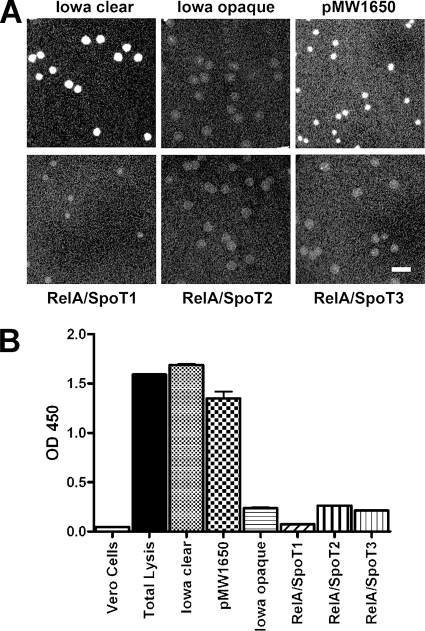
Because of the relatively low numbers of rickettsiae present in infected cells and the extensive purification procedures necessary to obtain cell-free organisms, we were unable to assess rickettsial (p)ppGpp content. To confirm a role for SpoT in the clear- and opaque-plaque phenotypes, we expressed wild-type relA/spoT in a clear-plaque-forming strain and analyzed the plaque phenotype. relA/spoT was PCR amplified from the R. rickettsii R strain opaque-plaque variant and cloned into a mariner-based Himar1 transposon system (19), which was then transformed into the clear-plaque variant of R. rickettsii Iowa and plated on Vero cells. Opaque-plaque variants comprised a majority of the resultant plaques; transformation with the parent pMW1650 plasmid yielded only clear plaques (Fig. 4A). Three opaque plaques were picked, recloned twice, and expanded for further analysis. The nonlytic phenotype in transformants bearing a functional relA/spoT gene was confirmed in LDH assays (Fig. 4B). Sequencing of purified genomic DNA from each of the clones by use of primers within the transposon identified each of the unique insertion sites (Table 1). Complementation with a functional relA/spoT gene thus restored a nonlytic phenotype to rickettsiae.
FIG. 4.
Complementation of RelA/SpoT confers an opaque-plaque phenotype on R. rickettsii Iowa. (A) Vero cell monolayers showing plaques formed by the R. rickettsii Iowa clear-plaque variant, the opaque-plaque variant, a clear-plaque variant clone transformed with the parent pMW1650 plasmid, and three representative clonal isolates after transformation of the R. rickettsii Iowa clear-plaque variant with intact relA/spoT showing restoration of the opaque-plaque phenotype. Bar = 5 mm. The insertion site of the transposon for each of the transformants is given in Table 1. (B) LDH release from Vero cells at 3 days postinfection with each of the R. rickettsii Iowa plaque variants and transformants shown above. Error bars represent SEM.
TABLE 1.
Insertion sites of relA/spoT
| Clone | Insertion site (bp) | Sequence | NCBI protein accession no. | Annotation |
|---|---|---|---|---|
| pMW1650 vector control | 780329 | CAATTCTTGTTAAAGTACTAAT | ABY72788.1 | N utilization substance protein A |
| RelA/SpoT1 | 453231 | TAATAATTAATAAAGTAATTTT | Intergenic | |
| RelA/SpoT2 | 1088618 | TTTATTACATTAAAGACGGTAA | ABY73129.1 | ABC transporter ATP-binding protein Uup |
| RelA/SpoT3 | 793818 | GCATTACTACTATGCATAGTTT | Intergenic |
The R. rickettsii R clear- and opaque-plaque variants show no difference in gene expression in the late log phase of growth.
In other organisms, mutations that disrupt the synthesis of (p)ppGpp dramatically alter the gene expression profile of the organism during stress, producing effects such as amino acid starvation (8, 30). To determine if the mutation in the SpoT-like protein resulted in differences in gene regulation between the R strain clear- and opaque-plaque variants, microarray analysis was performed on cultures grown to late log phase. By this time, at 3 days postinfection, the host cells are heavily infected with R. rickettsii. Differences in gene expression of ≥2-fold were not detected, suggesting that the mutation in the Rel/SpoT-like protein does not affect gene expression under these experimental conditions. The data are posted on the Gene Expression Omnibus (GEO) (http://www.ncbi.nlm.nih.gov/geo/) under accession no. GPL4692.
R. rickettsii R clear-plaque variants and R opaque-plaque variants show no difference in virulence when an intraperitoneal route of infection is used.
The R. rickettsii R strain was chosen to examine virulence between the clear- and opaque-plaque variants. Guinea pigs were infected intraperitoneally with 1,000 PFU of either the R strain clear-plaque variant or the R strain opaque-plaque variant, and fevers were monitored for 14 days as previously described (10). The fevers were similar in magnitude and duration, indicating that the opaque-plaque variant was not attenuated for virulence in this model (see Fig. S1 in the supplemental material). Guinea pigs infected intraperitoneally with 1,000 PFU of the Iowa strain clear- or opaque-plaque variant and monitored for 14 days showed no evidence of a fever response (Fig. S1).
One possible explanation for the results is that the plaque variants were reverting after inoculation into animals. To test this, guinea pigs were infected i.p. with 1,000 PFU of either the R strain clear-plaque variant or the R strain opaque-plaque variant, and spleens were harvested at the peak of fever on day 4 postinfection. Plaques recovered from the spleens of animals infected with either the R. rickettsii R strain clear-plaque variant or the R strain opaque-plaque variant showed no change in plaque phenotype during infection (data not shown). Similar numbers of viable organisms were recovered from the spleens of animals inoculated with the clear- and opaque-plaque variants (3.6 × 10⁴ and 4.2 × 10⁴ PFU/g, respectively), suggesting that there was no substantial growth difference between the two variants in Guinea pigs.
To determine if either plaque variant displayed a competitive growth advantage, two guinea pigs were infected with equal amounts of the R strain clear- and opaque-plaque variants. Infected spleens were harvested from infected animals on day 4 after infection, and plaque assays were performed to enumerate clear- and opaque-plaque types and determine a competitive index. The ratio of the clear-plaque variant to the opaque-plaque variant used to infect the guinea pigs was 0.97. After 4 days of infection, the ratios from the two guinea pigs were 0.85 and 0.75 indicating that there is little or no growth advantage for either the clear-plaque or the opaque-plaque strains over this interval up to the peak of fever. Taken together, the data confirm that there is no difference in virulence between R clear-plaque and R opaque-plaque variants when the i.p. route of infection is used.
R. rickettsii R strain clear-plaque variant induces fevers in guinea pigs with as little as 10 PFU given either intraperitoneally or intradermally.
The natural route of infection for R. rickettsii is through inoculation into the skin via the bite of an infected tick (20). We hypothesized that a more natural route of infection may provide a more sensitive model for the evaluation of virulence differences. We therefore compared intraperitoneal and intradermal routes of infection with decreasing doses (105, 104, 103, 102, and 101 PFU) of the R. rickettsii R strain clear-plaque variant. The ability to induce fevers in guinea pigs was monitored for 14 days after infection (see Fig. S2 in the supplemental material). By either route of infection, as little as 10 PFU was sufficient to induce fever in guinea pigs.
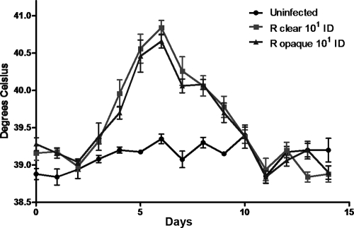
To determine if there was a difference in virulence between R clear-plaque and R opaque-plaque variants when administered through an intradermal route of infection, guinea pigs were infected with 103, 102, or 101 PFU of either the R clear-plaque variant or the R opaque-plaque variant and temperatures monitored over 14 days (Fig. 5). Again, no detectable difference in the abilities of the R strain clear- and opaque-plaque variants to induce fevers in guinea pigs with as little as 10 PFU was observed. These data, along with our previous results, suggest that in these models of infection there is no difference in the abilities of the R clear- and R opaque-plaque variants to induce fevers or replicate within guinea pigs.
FIG. 5.
Comparison of fever responses of Guinea pigs inoculated intradermally with 10 PFU of the R. rickettsii R strain clear- and opaque-plaque variants. Mean temperatures ± SEM are shown.
DISCUSSION
Spotted fever group rickettsiae differ in their association with disease in humans and their ability to cause disease in animal model systems (1, 2, 12, 20, 22, 24). Although not extensively investigated, this had been correlated with an increased cytolytic activity of virulent strains on host cell monolayers (2, 11, 12, 17). The ability of virulent strains to produce clear plaques while avirulent strains produce opaque plaques implies that there are stable genomic differences that account for differences in virulence. The genomes of virulent R. rickettsii Sheila Smith and avirulent R. rickettsii Iowa have recently been compared and indicate a number of interesting differences between the two strains (10). However, with 492 SNPs and deletions between the two strains, it has been difficult to definitively assign specific mutations to virulence differences. Isolation of plaque variants from plaque-cloned R. rickettsii R strain and R. rickettsii Iowa has offered an opportunity to discover differences between potential isogenic variants.
The clear- and opaque-plaque variants were independently isolated from two different strains of R. rickettsii, the virulent R stain and the avirulent Iowa strain. The clear- and opaque-plaque variants showed no growth defect in tissue culture, although the lytic nature of the strains producing the clear-plaque phenotype was confirmed visually in plaque assays and by demonstration of increased LDH release from infected cells. Genomic resequencing identified a single gene that contained mutations in both the R. rickettsii R strain and the R. rickettsii Iowa clear-plaque variants. These mutations were in a gene annotated as a RelA/SpoT-like protein gene. RelA/SpoT proteins are bifunctional enzymes responsible for the synthesis and hydrolysis of the bacterial alarmones guanosine tetraphosphate and guanosine pentaphosphate (p)ppGpp (5, 21, 23, 30). Synthesis of (p)ppGpp is typically induced under conditions of amino acid starvation or other nutrient deprivation and induces a number of adaptations, collectively known as the stringent response. The stringent response includes downregulation of stable RNA synthesis, transcription, translation, and ultimately growth arrest. A role for (p)ppGpp in pathogenesis is observed in multiple bacterial pathogens, with a trend toward decreased pathogenicity in the absence of (p)ppGpp (5, 23).
The limitations imposed by the obligate intracellular nature of rickettsiae precluded direct measurement of (p)ppGpp in the R. rickettsii clear- and opaque-plaque variants under different conditions by nucleotide extraction and high-performance liquid chromatography (HPLC). Similarly, microarrays did not demonstrate transcriptional differences between the R. rickettsii R strain clear- and opaque-plaque variants under the conditions examined. Therefore, the actual role of the relA/spoT gene product remains to be verified. To confirm a role for this gene in the lytic and nonlytic phenotypes, we used a mariner-based Himar1 transposon system (19) to reintroduce the wild-type relA/spoT gene and restore a nonlytic plaque phenotype. Interestingly, a closely related rickettsial species, Rickettsia conorii, the agent of Mediterranean spotted fever, has been shown to transcribe at least 5 genes annotated as spoT paralogs (26). All are present in R. rickettsii as well. The relA/spoT gene identified here is not, however, annotated in R. conorii. Upon inspection, the DNA sequence is present in R. conorii but displays a single-nucleotide deletion that causes a premature termination at amino acid 52 of the predicted enzyme. In contrast to the rickettsial small SpoT proteins identified in R. conorii, the intact R. rickettsii RelA/SpoT-like protein overlaps the synthetase domain of RelSeq predicted from the crystal structure (16, 26).
Surprisingly, the R. rickettsii R strain variants showed no growth defect in guinea pigs or a reduced ability of the nonlytic opaque-plaque mutants to cause fever in the guinea pig model when given by either the i.p. or the i.d. route of infection. Similarly, Iowa strain clear-plaque variants remained avirulent in the Guinea pig model. However, there was a difference in the amount of LDH released from Vero cells when infected with the variants, confirming the differences seen in plaque types. Rickettsiae are well known for their natural cycle between ticks and mammalian hosts. It is possible that any advantage conferred by relA/spoT is restricted to the arthropod host and not detected in the animal model used here.
The recent development of the mariner-based Himar1 transposon system for genetic manipulation of rickettsiae (19) has opened new possibilities for the application of molecular methods for better understanding of rickettsial biology. Transposon-based mutagenesis and directed knockouts have defined or confirmed virulence factors in both spotted fever and typhus group rickettsiae (7, 18). The ability to complement disrupted genes in rickettsiae adds to the available repertoire of tools for further exploring virulence determinants in this important group of pathogens.
Supplementary Material
Acknowledgments
This work was supported by the Division of Intramural Research of the National Institute for Allergy and Infectious Diseases, National Institutes of Health.
We thank the Genomics Unit of the NIAID Research Technologies Branch for the SOLiD resequencing and assistance with the microarray analysis. We thank E. Lutter, L. Bauler, J. Mital, and A. Omsland for critical review of the manuscript.
Editor: R. P. Morrison
Footnotes
Published ahead of print on 7 February 2011.
Supplemental material for this article may be found at http://iai.asm.org/.
REFERENCES
- 1.Anacker, R. L., R. H. List, R. E. Mann, and D. L. Wiedbrauk. 1986. Antigenic heterogeneity in high- and low-virulence strains of Rickettsia rickettsii revealed by monoclonal antibodies. Infect. Immun. 51:653-660. [DOI] [PMC free article] [PubMed] [Google Scholar]
- 2.Anacker, R. L., T. F. McCaul, W. Burgdorfer, and R. K. Gerloff. 1980. Properties of selected rickettsiae of the spotted fever group. Infect. Immun. 27:468-474. [DOI] [PMC free article] [PubMed] [Google Scholar]
- 3.Anacker, R. L., R. N. Philip, J. C. Williams, R. H. List, and R. E. Mann. 1984. Biochemical and immunochemical analysis of Rickettsia rickettsii strains of various degrees of virulence. Infect. Immun. 44:559-564. [DOI] [PMC free article] [PubMed] [Google Scholar]
- 4.Azad, A. F., and C. B. Beard. 1998. Rickettsial pathogens and their arthropod vectors. Emerg. Infect. Dis. 4:179-186. [DOI] [PMC free article] [PubMed] [Google Scholar]
- 5.Braeken, K., M. Moris, R. Daniels, J. Vanderleyden, and J. Michiels. 2006. New horizons for (p) ppGpp in bacterial and plant physiology. Trends Microbiol. 14:45-54. [DOI] [PubMed] [Google Scholar]
- 6.Cox, H. R. 1941. Cultivation of rickettsiae of the rocky mountain spotted fever, typhus and Q fever groups in the embryonic tissues of developing chicks. Science 94:399-403. [DOI] [PubMed] [Google Scholar]
- 7.Driskell, L. O., et al. 2009. Directed mutagenesis of the Rickettsia prowazekii pld gene encoding phospholipase D. Infect. Immun. 77:3244-3248. [DOI] [PMC free article] [PubMed] [Google Scholar]
- 8.Durfee, T., A. M. Hansen, H. Zhi, F. R. Blattner, and D. J. Jin. 2008. Transcription profiling of the stringent response in Escherichia coli. J. Bacteriol. 190:1084-1096. [DOI] [PMC free article] [PubMed] [Google Scholar]
- 9.Ellison, D. W., T. R. Clark, D. Sturdevant, K. Virtaneva, and T. Hackstadt. 2009. Limited transcriptional responses of Rickettsia rickettsii exposed to environmental stimuli. PLoS One 4:e5612. [DOI] [PMC free article] [PubMed] [Google Scholar]
- 10.Ellison, D. W., et al. 2008. Genomic comparison of virulent Rickettsia rickettsii Sheila Smith and avirulent Rickettsia rickettsii Iowa. Infect. Immun. 76:542-550. [DOI] [PMC free article] [PubMed] [Google Scholar]
- 11.Eremeeva, M. E., G. A. Dasch, and D. J. Silverman. 2001. Quantitative analyses of variations in the injury of endothelial cells elicited by 11 isolates of Rickettsia rickettsii. Clin. Diagn. Lab. Immunol. 8:788-796. [DOI] [PMC free article] [PubMed] [Google Scholar]
- 12.Hackstadt, T. 1996. The biology of rickettsiae. Infect. Agents Dis. 5:127-143. [PubMed] [Google Scholar]
- 13.Hackstadt, T., R. Messer, W. Cieplak, and M. G. Peacock. 1992. Evidence for the proteolytic cleavage of the 120-kilodalton outer membrane protein of rickettsiae: identification of an avirulent mutant deficient in processing. Infect. Immun. 60:159-165. [DOI] [PMC free article] [PubMed] [Google Scholar]
- 14.Hand, W. L., J. B. Miller, J. A. Reinarz, and J. P. Sanford. 1970. Rocky Mountain spotted fever. A vascular disease. Arch. Intern. Med. 125:879-882. [PubMed] [Google Scholar]
- 15.Heinzen, R. A., S. F. Hayes, M. G. Peacock, and T. Hackstadt. 1993. Directional actin polymerization associated with spotted fever group rickettsia infection of VERO cells. Infect. Immun. 61:1926-1935. [DOI] [PMC free article] [PubMed] [Google Scholar]
- 16.Hogg, T., U. Mechold, H. Malke, M. Cashel, and R. Hilgenfeld. 2004. Conformational antagonism between opposing active sites in a bifunctional RelA/SpoT homolog modulates (p)ppGpp metabolism during the stringent response. Cell 117:57-68. [DOI] [PubMed] [Google Scholar]
- 17.Johnson, J. W., and C. E. Pedersen, Jr. 1978. Plaque formation by strains of spotted fever rickettsiae in monolayer cultures of various cell types. J. Clin. Microbiol. 7:389-391. [DOI] [PMC free article] [PubMed] [Google Scholar]
- 18.Kleba, B., T. R. Clark, E. I. Lutter, D. W. Ellison, and T. Hackstadt. 2010. Disruption of the Rickettsia rickettsii Sca2 autotransporter inhibits actin-based motility. Infect. Immun. 78:2240-2247. [DOI] [PMC free article] [PubMed] [Google Scholar]
- 19.Liu, Z. M., A. M. Tucker, L. O. Driskell, and D. O. Wood. 2007. Mariner-based transposon mutagenesis of Rickettsia prowazekii. Appl. Environ. Microbiol. 73:6644-6649. [DOI] [PMC free article] [PubMed] [Google Scholar]
- 20.McDade, J. E., and V. F. Newhouse. 1986. Natural history of Rickettsia rickettsii. Annu. Rev. Microbiol. 40:287-309. [DOI] [PubMed] [Google Scholar]
- 21.Mittenhuber, G. 2001. Comparative genomics and evolution of genes encoding bacterial (p) ppGpp synthetases/hydrolases (the Rel, RelA and SpoT proteins). J. Mol. Microbiol. Biotechnol. 3:585-600. [PubMed] [Google Scholar]
- 22.Parola, P., C. D. Paddock, and D. Raoult. 2005. Tick-borne rickettsioses around the world: emerging diseases challenging old concepts. Clin. Microbiol. Rev. 18:719-756. [DOI] [PMC free article] [PubMed] [Google Scholar]
- 23.Potrykus, K., and M. Cashel. 2008. (p) ppGpp: still magical? Annu. Rev. Microbiol. 62:35-51. [DOI] [PubMed] [Google Scholar]
- 24.Price, W. H. 1953. The epidemiology of Rocky Mountain spotted fever I. The characterization of strain virulence of Rickettsia rickettsii. Am. J. Hyg. 58:248-268. [DOI] [PubMed] [Google Scholar]
- 25.Ricketts, H. T. 1907. Further experiments with the woodtick in relation to Rocky Mountain spotted fever. JAMA 49:1278-1281. [Google Scholar]
- 26.Rovery, C., et al. 2005. Transcriptional response of Rickettsia conorii exposed to temperature variation and stress starvation. Res. Microbiol. 156:211-218. [DOI] [PubMed] [Google Scholar]
- 27.Santucci, L. A., P. L. Gutierrez, and D. J. Silverman. 1992. Rickettsia rickettsii induces superoxide radical and superoxide dismutase in human endothelial cells. Infect. Immun. 60:5113-5118. [DOI] [PMC free article] [PubMed] [Google Scholar]
- 28.Silverman, D. J. 1984. Rickettsia rickettsii-induced cellular injury of human vascular endothelium in vitro. Infect. Immun. 44:545-553. [DOI] [PMC free article] [PubMed] [Google Scholar]
- 29.Silverman, D. J., and L. A. Santucci. 1988. Potential for free radical-induced lipid peroxidation as a cause of endothelial cell injury in Rocky Mountain spotted fever. Infect. Immun. 56:3110-3115. [DOI] [PMC free article] [PubMed] [Google Scholar]
- 30.Srivatsan, A., and J. D. Wang. 2008. Control of bacterial transcription, translation and replication by (p)ppGpp. Curr. Opin. Microbiol. 11:100-105. [DOI] [PubMed] [Google Scholar]
- 31.Walker, D. H. 1989. Rocky Mountain spotted fever: a disease in need of microbiological concern. Clin. Microbiol. Rev. 2:227-240. [DOI] [PMC free article] [PubMed] [Google Scholar]
- 32.Walker, D. H., and B. G. Cain. 1980. The rickettsial plaque. Evidence for direct cytopathic effect of Rickettsia rickettsii. Lab. Invest. 43:388-396. [PubMed] [Google Scholar]
- 33.Walker, D. H., W. T. Firth, J. G. Ballard, and B. C. Hegarty. 1983. Role of phospholipase-associated penetration mechanism in cell injury by Rickettsia rickettsii. Infect. Immun. 40:840-842. [DOI] [PMC free article] [PubMed] [Google Scholar]
- 34.Walker, D. H., R. R. Tidwell, T. M. Rector, and J. D. Geratz. 1984. Effect of synthetic protease inhibitors of the amidine type on cell injury by Rickettsia rickettsii. Antimicrob. Agents Chemother. 25:582-585. [DOI] [PMC free article] [PubMed] [Google Scholar]
- 35.Weiss, E., J. C. Coolbaugh, and J. C. Williams. 1975. Separation of viable Rickettsia typhi from yolk sac and L cell host components by renografin density gradient centrifugation. Appl. Microbiol. 30:456-463. [DOI] [PMC free article] [PubMed] [Google Scholar]
Associated Data
This section collects any data citations, data availability statements, or supplementary materials included in this article.


