Abstract
During their circulation in nature, H5N1 avian influenza viruses (AIVs) have acquired the ability to kill their natural hosts, wild birds and ducks. The genetic determinants for this increased virulence are largely unknown. In this study, we compared two genetically similar H5N1 AIVs, A/duck/Hubei/49/05 (DK/49) and A/goose/Hubei/65/05 (GS/65), that are lethal for chickens but differ in their virulence levels in ducks. To explore the genetic basis for this difference in virulence, we generated a series of reassortants and mutants of these two viruses. The virulence of the reassortant bearing the PA gene from DK/49 in the GS/65 background increased 105-fold relative to that of the GS/65 virus. Substitution of two amino acids, S224P and N383D, in PA contributed to the highly virulent phenotype. The amino acid 224P in PA increased the replication of the virus in duck embryo fibroblasts, and the amino acid 383D in PA increased the polymerase activity in duck embryo fibroblasts and delayed the accumulation of the PA and PB1 polymerase subunits in the nucleus of virus-infected cells. Our results provide strong evidence that the polymerase PA subunit is a virulence factor for H5N1 AIVs in ducks.
Influenza A viruses are classified into different subtypes on the basis of the antigenicity of their surface proteins hemagglutinin (HA) and neuraminidase (NA). To date, 16 different HA subtypes and nine different NA subtypes have been identified in viruses isolated from wild birds, which are regarded as the natural hosts of influenza A viruses. Of the numerous potential subtypes of viruses, only a few have adapted to and circulate among humans (H1N1, H2N2, and H3N2), pigs (H1N1 and H3N2), and domestic poultry (H9N2, H5, and H7). The viruses typically acquire critical changes in their genomes that allow them to adapt to or severely damage their host. Identification of these genetic changes will improve our understanding of the determinants of virulence and aid in the development of counter measures.
Several determinants of avian influenza virus (AIV) virulence have been identified (6, 13, 14, 17, 20, 21, 24, 35, 36, 44). The multiple-basic-amino-acid motif in the cleavage site of the HA gene is required for the systemic replication and lethal infection of the H5 and H7 subtypes of influenza viruses in chickens (18) and mice (13). Fan et al. (6) reported that mutations in the M1 protein contribute to the virulence of H5N1 influenza viruses in mice. Certain amino acids or regions of the NS1 protein play a key role in the ability of H5N1 influenza viruses to undermine the antiviral immune response of the host cell and are critical for the pathogenicity of H5N1 influenza viruses in mice (17, 34, 40).
The polymerase proteins also play important roles in the pathogenicity of AIVs in different species. The amino acid lysine at position 627 of PB2 is a principal determinant of the high virulence of the 1997 Hong Kong H5N1 influenza viruses (3, 13). The amino acid at position 701 in PB2 plays a crucial role in the ability of H5N1 viruses of duck origin to replicate and be lethal in mice (20). This amino acid also contributes to the increased lethality of an H7N7 AIV in a mouse model (10), while both of these PB2 amino acids contribute to the transmission of the H5N1 influenza viruses in guinea pigs (12, 37). PB2 residue 271 plays a role in the enhanced polymerase activity of influenza A viruses in mammalian host cells (1). Lastly, the importance of the amino acid at position 591 of PB2 for efficient replication of the pandemic H1N1 viruses in humans has recently been reported (25, 41). The PB2 and PB1 polymerase subunits contribute to the high virulence of the human H5N1 influenza virus isolate A/Vietnam/1203/04 in mice and ferrets (32). Recently, it was reported that differences in viral transcription and replication levels between mammalian and avian cells are determinants of both host specificity and pathogenicity of an H7N7 virus (9).
Ducks play an important role in AIV ecology. They can be infected by many AIVs, including H5N1 viruses, but generally they do not show any signs of disease or die when infected (39). Therefore, AIVs could circulate silently in this host, allowing them to be transmitted to susceptible animals (4, 42). However, since 2003, several AIV outbreaks in ducks caused by H5N1 viruses have been documented in Southeast Asia (Office International des Epizooties [OIE]; http://www.oie.int). Some of the strains responsible for these outbreaks are lethal to ducks in the laboratory setting (16, 38, 43). Although the virulence factors of H5N1 influenza viruses in chickens and mice have been studied extensively, the molecular determinants of H5N1 pathogenicity in ducks are still poorly understood.
In this study, we characterized two H5N1 avian influenza viruses, A/duck/Hubei/49/05 (DK/49) and A/goose/Hubei/65/05 (GS/65), which were isolated in the Hubei province of China during the H5N1 avian influenza outbreak at the end of 2005 (2, 19). These two viruses are both highly pathogenic in chickens but differ in their virulence levels in ducks. Here, we examined the molecular basis for the increased virulence of H5N1 influenza viruses in ducks and explored the possible underlying mechanisms.
MATERIALS AND METHODS
Facility.
Studies with highly pathogenic H5N1 avian influenza viruses were conducted in a biosecurity level 3+ laboratory approved by the Chinese Ministry of Agriculture. All animal studies were approved by the Review Board of Harbin Veterinary Research Institute, Chinese Academy of Agricultural Sciences.
Cells and viruses.
293T cells and duck embryo fibroblasts (DEFs) were grown in Dulbecco's modified Eagle's medium (DMEM) supplemented with 10% fetal bovine serum plus antibiotics. The cells were incubated at 37°C in 5% CO2. Two viruses, A/duck/Hubei/49/05 (DK/49) and A/goose/Hubei/65/05 (GS/65), were isolated from a duck and a goose, respectively, during the avian influenza outbreak in 2005 in China (2). Virus stocks of the AIVs were propagated in 10-day-old specific pathogen-free (SPF) embryonated chicken eggs and stored at −70°C until use.
Construction of plasmids.
An eight-plasmid reverse genetics system was used to generate the reassortant viruses. As described previously (20), we inserted the full-length cDNA derived from the DK/49 and GS/65 viruses into the bidirectional transcription vector pBD (20), which could generate viral RNA and mRNA simultaneously driven by the human polI and cytomegalovirus (CMV) promoters. The sequences of primers used for virus rescue are available upon request. The protein expression plasmids for the polymerase subunits and NP of influenza, DK/49 (P3.1-49PA, P3.1-49PB1, P3.1-49PB2, and P3.1-49NP), and GS/65 (P3.1-65PA, P3.1-65PB1, P3.1-65PB2, and P3.1-65NP) were generated by inserting their cDNAs between the NheI and NotI restriction enzyme sites of the pcDNA3.1+ plasmid (Invitrogen). To construct the reporter plasmid, paviPol-Luc, the open reading frame of the luciferase gene flanked by the 5′ and 3′ noncoding regions of the NP gene of the DK/49 virus was inserted into a pPol I plasmid containing the 250-nucleotide sequence of the avian polymerase I promoter (DQ112354) (33). Mutations were introduced by site-directed mutagenesis with the QuikChange mutagenesis kit (Stratagene) according to the manufacturer's protocol and confirmed by sequencing. The primer sequences are available upon request.
Generation of reverse genetic viruses.
293T cells were transfected with 0.6 μg of each of eight plasmids mixed with 10 μl Lipofectamine 2000 (Invitrogen) according to the manufacturer's instructions. After 48 h, the supernatant was harvested and injected into embryonated eggs for virus propagation. The rescued viruses were detected by use of a hemagglutination assay, and RNA was extracted and analyzed by reverse transcription-PCR (RT-PCR). Each viral segment was sequenced to confirm the identity of the reassortant viruses.
Chicken experiments.
To determine the pathogenicity of the viruses, the intravenous pathogenicity index (IVPI) was determined according to the recommendations of the Office International des Epizooties (OIE) (22). Groups of 10 6-week-old SPF White Leghorn chickens housed in isolator cages were inoculated intravenously (i.v.) with 0.2 ml of a 1:10 dilution of bacterium-free allantoic fluid containing virus, and signs of disease or death were monitored for 10 days.
Duck experiments.
Groups of 8 6-week-old SPF Shaoxin ducks (a local breed obtained from the Harbin Veterinary Research Institute, Chinese Academy of Agricultural Sciences) were inoculated intranasally (i.n.) with 106 50% egg infectious doses (EID50) of H5N1 influenza viruses in a volume of 100 μl. On day 3 postinoculation (p.i.), three birds in each group were euthanized, and the lungs, kidneys, pancreas, and brains were collected for virus titration. Oropharyngeal and cloacal swabs were also collected on day 3 p.i. from all birds for detection of virus shedding. The remaining five ducks in each group were observed for signs of disease or death for 14 days. To determine the 50% duck lethal dose (DLD50) of virus, groups of five ducks were inoculated i.n. with 100 μl of 10-fold serial dilutions containing viruses between 102 and 108 EID50. The DLD50 was calculated by the method of Reed and Muench (30).
Sequence analysis.
The plasmids used for virus rescue and the rescued viruses themselves were fully sequenced to confirm the absence of unwanted mutations. Viral RNA was extracted from allantoic fluid and was reverse transcribed. A set of fragment-specific primers (primer sequences available on request) was used for PCR amplification and sequence analysis. The sequence data for the two viruses used in these studies have been reported previously (19); their accession numbers in GenBank are HM172449, HM172450, HM172360, HM172358, HM172311, HM172312, HM172071, HM172070, HM172215, HM172216, HM172116, HM172118, HM172119, HM172117, HM172262, and HM172263.
Viral replication in DEFs.
Virus was inoculated into DEF monolayers at a multiplicity of infection (MOI) of 0.001. The cells were supplemented with MEM containing 0.5% bovine serum albumin and incubated at 37°C. Virus-containing culture supernatant was collected at various time points (hours postinfection [hpi]) and titrated in eggs. The growth data shown are the average results of three independent experiments.
Luciferase assay of polymerase activity.
DEFs were transfected with paviPolI-T-Luc together with the pTK-RL (Promega) and pcDNA3.1+ plasmid constructs expressing the polymerase PB2, PB1, PA, and NP genes (0.5 μg each) plus Lipofectamine 2000 (Invitrogen) as recommended by the manufacturer. pTK-RL is an internal control plasmid to normalize transfection efficiency that encodes the Renilla luciferase protein. After 6 h, the medium was replaced with DMEM (10% fetal calf serum). Cell extracts were harvested 30 h posttransfection, and luciferase activity was assayed by using the luciferase assay system (Promega). The assay was standardized against the Renilla luciferase activity. All experiments were performed in triplicate.
Immunofluorescence.
DEFs were grown on glass-bottom dishes and infected at an MOI of 2, with the indicated virus. At the indicated time points postinfection, the cells were fixed with PBS containing 4% paraformaldehyde for 20 min and permeabilized with PBS containing 0.5% Triton X-100 for 30 min. After being blocked with 5% bovine serum albumin (BSA) in PBS, the cells were incubated with rabbit antisera against PA, PB2, goat antisera against PB1 (Santa Cruz), or monoclonal antibody against NP (Santa Cruz) at room temperature for 2 h. The cells were then washed three times with PBS and incubated for 1 h with fluorescein isothiocyanate (FITC)-coupled donkey anti-rabbit (for PA and PB2), rhodamine-coupled donkey anti-goat (for PB1), or goat anti-mouse (for NP) secondary antibodies (Santa Cruz). After incubation with secondary antibodies, the cells were washed three times with PBS and incubated with 4′,6-diamidino-2-phenylindole (DAPI) for 10 min. Cells were observed with a laser scanning confocal microscope (Leica). Localization of proteins in the nucleus was determined by counting the cells (n = 100) infected with the test viruses. The results shown represent three independent experiments.
RESULTS
Characterization of the H5N1 AIVs DK/49 and GS/65.
Two H5N1 AIVs, DK/49 and GS/65, were isolated from the Hubei province of southern China in 2005 during the H5N1 avian influenza outbreak (19). The pathogenicity analysis of these two viruses in chickens (following the recommendation by the Office International des Epizooties [27]) revealed that both viruses killed all 10 chickens within 24 h and yielded an IVPI of 3 (with 3.0 being the most pathogenic and 0 being the least pathogenic).
Since DK/49 also caused 100% mortality of infected ducks in a small farm during an outbreak in 2005, we tested the virulence of both DK/49 and GS/65 in ducks. Groups of eight ducks were intranasally inoculated with 106 EID50 of the viruses, and swabs from the pharynx and cloaca were collected on day 3 p.i. for detection of virus shedding. Three birds in each group were euthanized to test for virus replication in organs, and the remaining five birds were observed for 2 weeks. As shown in Table 1, virus was detected in the pharynxes of all eight birds in both virus-inoculated groups and in the cloacae of seven and four birds that were inoculated with the DK/49 and GS/65, respectively. Both viruses caused a systemic infection, and high titers of virus were detected in the lungs, kidneys, and pancreas of all inoculated ducks. However, the titer in the brain of ducks inoculated with the DK/49 virus was significantly higher than that in the brain of birds inoculated with the GS/65 virus (Table 1). The DK/49-inoculated ducks showed severe neurological signs and died within 4 days of infection, whereas the GS/65 virus-inoculated ducks appeared healthy and survived the 2-week observation period (Table 1).
TABLE 1.
Replication of H5N1 influenza viruses in ducksa
| Virus | Virus shedding on day 3 p.i. (log10 EID50/ml) |
Virus replication on day 3 p.i. (log10 EID50/g) |
No. of deaths/total (MDT [days])e | ||||||
|---|---|---|---|---|---|---|---|---|---|
| Pharynx |
Cloaca |
||||||||
| Titers | No. shedding/total | Titersb | No. shedding/total | Lung | Kidney | Pancreas | Brain | ||
| DK/49 | 3.3 ± 0.8 | 8/8 | 2.5 ± 0.5 | 7/8 | 6.0 ± 0.7 | 6.4 ± 0.3 | 6.8 ± 1.6 | 7.6 ± 0.3 | 5/5 (3.4) |
| GS/65 | 2.0 ± 0.4d | 8/8 | 1.1 ± 0.4c | 4/8 | 6.3 ± 0.5 | 5.3 ± 0.7 | 4.8 ± 1.3 | 2.5 ± 0.7d | 0/5 (NA) |
| R-DK/49 | 3.9 ± 0.6 | 8/8 | 2.9 ± 1.2 | 8/8 | 7.2 ± 0.7 | 6.5 ± 0.3 | 6.4 ± 1.1 | 7.6 ± 0.2 | 5/5 (3.0) |
| R-GS/65 | 3.1 ± 0.4c | 8/8 | 1.2 ± 0.4d | 5/8 | 6.8 ± 0.5 | 5.3 ± 0.3c | 5.2 ± 1.0 | 2.5 ± 0.9d | 0/5 (NA) |
Six-week-old SPF ducks (eight/group) were inoculated intranasally with 106 EID50 of each virus in a 100-μl volume. On day 3, three birds in each group were euthanized, and virus was titrated in samples of lung, kidney, pancreas, and brain in eggs. Pharyngeal and cloacal swabs collected from all birds on day 3 were tested for virus shedding in eggs. The remaining five birds were observed for disease and deaths for 2 weeks. Data are means ± standard deviations.
Titers were calculated from the birds that shed virus.
P value was <0.05 compared with the titers in the corresponding organs of DK/49- or R-DK/49-inoculated ducks.
P value was <0.01 compared with the titers in the corresponding organs of DK/49- or R-DK/49-inoculated ducks.
MDT, mean death time; NA, not applicable.
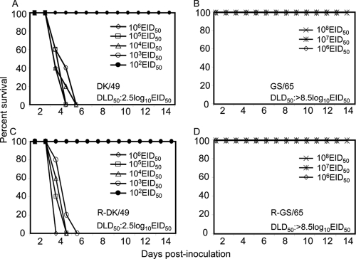
To further compare the virulence of these two viruses in ducks, we determined their DLD50. We inoculated groups of five ducks with 10-fold serial dilutions containing 102 to 106 EID50 of DK/49 or 106 to 108 EID50 of GS/65. As shown in Fig. 1, the DK/49 virus killed all of the ducks, even at doses as low as 103 EID50 (Fig. 1A), whereas GS/65 did not kill any ducks, even at the highest dose of 108 EID50 (Fig. 1B). Therefore, the two viruses differed markedly in their DLD50: 2.5 log10 EID50 for the DK/49 virus and >8.5 log10 EID50 for the GS/65 virus. These data show that DK/49 is highly virulent in ducks and that GS/65 is nonlethal, although it replicates systemically in ducks.
FIG. 1.
Virulence and death pattern of ducks infected with different H5N1 viruses. Six-week-old SPF ducks (five/group) were inoculated intranasally with different H5N1 viruses, DK/49 (A), GS/65 (B), R-DK/49 (C), or R-GS/65 (D). Doses of 102 to 106 EID50 (A, C) or 106 to 108 EID50 (B, D) were used.
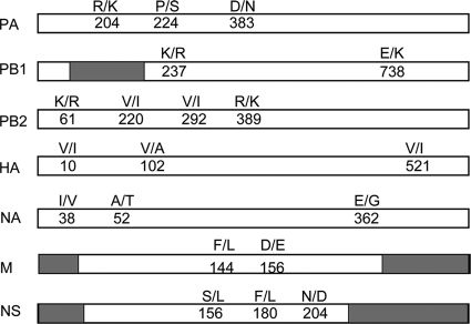
To investigate the genetic relationship between the two viruses, we sequenced and compared their genomes. We found that the two viruses are closely related and similar to the A/Anhui/1/2005(H5N1) virus (clade 2.3.4). DK/49 and GS/65 share the same products of the NP, M2, and NS2 genes at the amino acid level. We mapped a total of 20 amino acids that were different between the two viruses in the products of their PB2, PB1, PA, HA, NA, M1, and NS1 genes (Fig. 2), suggesting that single or multiple amino acid combinations among these 20 different amino acids contribute to the difference in virulence in ducks between these two viruses.
FIG. 2.
Amino acid differences between the DK/49 and GS/65 viruses. The amino acid differences between the two viruses are shown as single letters at the indicated positions. DK/49 is shown to the left of the slash, and GS/65 to the right. The PB1-F2, M2, and NS2 areas are shown in gray.
The polymerase PA gene of DK/49 dramatically increases the virulence of the GS/65 virus.
To investigate the genetic basis of the difference in virulence between the DK/49 and GS/65 viruses, we established a reverse genetics system for the two viruses, as described in Materials and Methods. The rescued viruses were designated R-DK/49 and R-GS/65, respectively. After confirming their sequences, we prepared virus stocks by the use of 10-day-old SPF eggs and tested the replication and lethality of these viruses in ducks. R-DK/49 and R-DK/65 exhibited properties similar to those of their respective parent viruses, in terms of virus titers in organs and with respect to DLD50s (Table 1 and Fig. 1C and D).
The two viruses have the same NP genes. Therefore, to investigate which gene contributes to the increased virulence of the virus in ducks, we generated seven single-gene reassortants by the use of a strategy described previously (13, 20). Each reassortant contained one gene derived from DK/49 and seven genes derived from GS/65. We inoculated groups of eight ducks with 106 EID50 of the reassortants and tested their replication and pathogenicity. The virus titers in the lungs and brains of euthanized ducks were measured on day 3 p.i. The titers of six reassortants were comparable to that of the rescued R-GS/65 virus, but in the brain, the titer of the reassortant GS/65-DK/49PA virus was significantly higher than that of the R-GS/65 virus (Table 2). Five of the seven reassortants caused deaths in only some birds within the group, whereas the remaining two reassortants, GS/65-DK/49PA and GS/65-DK/49PB2, killed all five ducks in the group within the observation period (Table 2).
TABLE 2.
Replication and virulence of H5N1 reassortants and mutants in ducksa
| Virus | Virus replication on day 3 p.i. (log10 EID50/g) |
No. of deaths/total | DLD50 (log10 EID50) | Change in DLD50 | |
|---|---|---|---|---|---|
| Lung | Brain | ||||
| R-GS/65 | 6.8 ± 0.5 | 2.5 ± 0.9 | 0/5 | >8.5 | NA |
| GS/65-DK/49PA | 6.0 ± 0.4 | 5.2 ± 0.9b | 5/5 | 3.5 | 5e |
| GS/65-DK/49PB2 | 6.5 ± 0.7 | 3.6 ± 2.4 | 5/5 | 5.5 | 3e |
| GS/65-DK/49PB1 | 6.4 ± 0.8 | 3.3 ± 1.5 | 2/5 | / | NA |
| GS/65-DK/49HA | 6.6 ± 0.2 | 2.5 ± 0.8 | 2/5 | / | NA |
| GS/65-DK/49NA | 6.4 ± 0.3 | 3.2 ± 0.6 | 2/5 | / | NA |
| GS/65-DK/49 M | 6.8 ± 0.7 | 3.5 ± 0.3 | 2/5 | / | NA |
| GS/65-DK/49NS | 6.1 ± 0.4 | 2.3 ± 0.3 | 1/5 | / | NA |
| GS/65-PA204R | 6.5 ± 0 | 3.0 ± 0.4 | 1/5 | / | NA |
| GS/65-PA224P | 6.1 ± 0.5 | 4.1 ± 0.4 | 1/5 | / | NA |
| GS/65-PA383D | 6.3 ± 0 | 3.7 ± 0.9 | 2/5 | / | NA |
| GS/65-PA204R + 224P | 6.5 ± 0.4 | 4.6 ± 0.2b | 2/5 | / | NA |
| GS/65-PA204R + 383D | 5.9 ± 0.4 | 4.5 ± 0b | 2/5 | / | NA |
| GS/65-PA224P + 383D | 6.4 ± 0.2 | 6.0 ± 0.5b | 5/5 | 3.5 | 5e |
| R-DK/49 | 7.2 ± 0.7 | 7.6 ± 0.2 | 5/5 | 2.5 | NA |
| DK/49-GS/65PA | 6.9 ± 0.5 | 6.5 ± 0.2c | 5/5 | 4.5 | 2f |
| DK/49-PA224S + 383N | 6.5 ± 0.3 | 5.3 ± 0.5d | 5/5 | 4.5 | 2f |
Six-week-old SPF Shaoxin ducks were used in these studies as described in Materials and Methods. /, not done; NA, not applicable.
P < 0.05 compared with the titers in the corresponding organs of R-GS/65-inoculated ducks.
P < 0.05 compared with the titers in the corresponding organs of R-DK/49-inoculated ducks.
P < 0.01 compared with the titers in the corresponding organs of R-DK/49-inoculated ducks.
Virulence increased compared with the R-GS/65 virus.
Virulence decreased compared with the R-DK/49 virus.
We then determined the DLD50 of GS/65-DK/49PA and GS/65-DK/49PB2. In the GS/65-DK/49PA-inoculated groups, the ducks that received 102 EID50 and 103 EID50 of virus survived, but birds inoculated with higher doses died within 8 days p.i. (DLD50, 3.5 log10 EID50 [Table 2]). In the GS/65-DK/49PB2-inoculated group, however, only the ducks that received 106 EID50 of virus died. All of the ducks inoculated with low dosages of virus survived during the observation period (DLD50, 5.5 log10 EID50 [Table 2]). These results indicate that multiple genes from the DK/49 virus contribute to the increased pathogenicity of the GS/65 virus in ducks but that the PA gene of the DK/49 virus had the greatest effect on virulence (DLD50, 3.5 versus >8.5 log10 EID50). We therefore focused on the PA gene for further analyses.
Two amino acids at positions 224 and 383 in the PA protein change the pathogenicity of the GS/65 and DK/49 viruses in ducks.
The PA proteins of the DK/49 and GS/65 viruses differ by three amino acids (Table 2). We therefore generated six mutants in the GS/65 virus background that contained one or two amino acid changes at these positions in the PA protein and tested their replication and pathogenicity in ducks. On day 3 p.i. with 106 EID50 of the test virus, the titers of all of the mutant viruses in the lungs of the ducks were comparable with the titers of the R-GS/65 virus in the lungs. However, the virus titers in the brains of the ducks that were inoculated with the three mutants containing double mutations were significantly higher than those of the R-GS/65 virus (Table 2). The virus containing the double mutation at positions 224 and 383 of the PA protein, GS/65-PA224P + 383D, killed all five ducks within 7 days p.i., whereas the other five mutant viruses killed only some of the inoculated birds during the 2-week observation (Table 2). We, therefore, determined the DLD50 of the GS/65-PA224P + 383D virus. We found that all of the ducks that received dosages between 104 and 106 EID50 died within 7 days p.i., whereas all of the ducks that received the 103 EID50 dose survived during the 2-week observation period (Table 2). These results indicate that any of the three single amino acid mutations could increase the virulence of the GS/65 virus to some degree but that the combination of the two amino acid mutations at positions 224 and 383 increased the virulence of the GS/65 virus more than 105-fold (DLD50, 3.5 versus >8.5 log10 EID50).
To investigate whether the amino acids at positions 224 and 383 of the GS/65 PA protein could attenuate the DK/49 virus in ducks, we generated the reassortant virus DK/49-GS/65PA, which contains the PA gene of GS/65 and a mutant DK/49-PA224S + 383N virus that bears two amino acid substitutions at PA224 and PA383 in the DK/49 background. Both viruses replicated well in the lungs and brains of ducks at day 3 after 106 EID50 intranasal inoculation, but the titers in the brains of the ducks inoculated with DK/49-GS/65PA and DK/49-PA224S + 383N were significantly lower than that of ducks inoculated with the R-DK/49 virus (Table 2). The virulence of these two viruses was attenuated 100-fold relative to the R-DK/49 virus (Table 2) (DLD50, 4.5 versus 2.5 log10 EID50).
These results demonstrate that the polymerase PA gene is an important virulence factor of H5N1 viruses in ducks and that the amino acids at positions 224 and 383 play a critical role in the difference in virulence in ducks observed with these two H5N1 viruses.
The amino acid at position 224 in the PA protein significantly affects viral replication in DEFs.
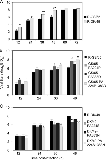
We further compared the multicycle growth of the viruses in DEFs and found that the DK/49 virus grew more rapidly than did the GS/65 virus and that the titers of DK/49 were significantly higher than those of GS/65 at 12, 36, and 48 h postinfection (Fig. 3A). We then investigated the contribution of the amino acid changes in PA to the replication of the two viruses. Titers of GS/65-PA224P and GS/65-PA224P + 383D significantly higher than those of R-GS/65 were observed at several time points postinfection, while the replication level of GS/65-PA383D was comparable to that of R-GS/65 (Fig. 3B). The P224S mutation in PA attenuated the replication of DK/49, and the titer of DK/49-PA224S was significantly lower than that of DK/49 at 48 h postinfection. The replication of DK/49-PA224S + 383N was significantly attenuated, and decreased titers were observed at 24, 36, and 48 h postinfection; however, the replication of DK/49-PA383N was similar to that of the wild-type DK/49 virus (Fig. 3C). These results indicate that the amino acid change of PA-S224P increases the replication of the H5N1 virus in DEFs, and this increase could be further enhanced by the addition of the amino acid change of PA-N383D, although N383D alone did not alter virus replication in DEFs.
FIG. 3.
Multicycle replication of H5N1 viruses in DEFs. DEF monolayers were inoculated at an MOI of 0.001 with virus, and the culture supernatants were collected at the indicated hpi and then titrated in eggs. (A) Replication of R-GS/65 and R-DK/49 viruses. *, P < 0.05 compared with that of R-GS/65-infected cells; **, P < 0.01 compared with that of R-GS/65-infected cells. (B) Replication of the mutants in the GS/65 background. *, P < 0.05 compared with that of R-GS/65-infected cells; **, P < 0.01 compared with that of R-GS/65-infected cells. (C) Replication of the mutants in the DK/49 background. *, P < 0.05 compared with that of R-DK/49-infected cells; **, P < 0.01 compared with that of R-DK/49-infected cells.
The amino acid at position 383 in the PA protein affects viral polymerase activity.
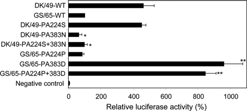
The PA protein is a component of the RNA-dependent polymerase complex and affects both transcription and replication (7, 29, 31). We investigated whether the two influenza viruses have different polymerase activities by using a minigenome assay in DEFs. As shown in Fig. 4, the polymerase activity of the DK/49 virus was more than 4-fold higher than that of the GS/65 virus. To investigate how the amino acids at positions 224 and 383 in PA affect the polymerase activity, we introduced single or double mutations into the PA proteins of both viruses. The amino acid substitution at position 224 of PA did not change the activity of the polymerase complex of the DK/49 or GS/65 virus; however, the polymerase activity of the complexes containing the single amino acid substitution at position 383 or the double mutation at positions 224 and 383 of PA changed significantly in comparison to the complex containing the wild-type PA protein (Fig. 4). These results indicate that the amino acid at position 383, but not that at position 224, contributes to the difference in the polymerase activities of the DK/49 and GS/65 viruses.
FIG. 4.
Activities of DK/49 or GS/65 ribonucleoprotein (RNP) with different PA mutations in a minigenome assay. Four protein expression plasmids (PB2, PB1, PA, and NP) for the RNP combinations were transfected into DEFs together with paviPolI-T-Luc, as described in Materials and Methods. The values shown are means ± standard deviations of results for three independent experiments and are standardized to the activity of GS/65 (100%). *, P < 0.01 compared with that of DK/49-infected cells; **, P < 0.01 compared with that of GS/65-infected cells.
The amino acid at position 383 in the PA protein affects the transport of PA and PB1 into the nucleus of DEF cells.
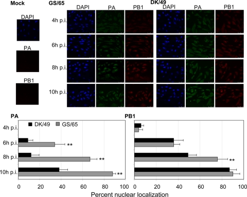
The influenza virus polymerase is active in the nucleus of the infected cell, and cytoplasmically expressed PB1 and PA are imported as a subcomplex (8). Accumulation of the polymerase PA in the nucleus correlates with the viral pathogenicity of H1N1 human influenza viruses in mice (15). We therefore tested whether the DK/49 and GS/65 viruses differ in their polymerase accumulation in the nucleus. DEFs were infected with viruses at an MOI of 2, and localization of PA and PB1 in virus-infected cells was examined at different time points postinfection. As shown in Fig. 5, in both virus-infected cells, PA and PB1 gradually shifted from the cytoplasm to the nucleus, but the accumulation of PA in the nucleus of the GS/65 virus-infected cells was faster than that of the DK/49 virus-infected cells at all time points tested. The levels of accumulation of PB1 in the nucleus of both virus-infected cells were quite similar at three of the four time points tested (4, 6, and 10 hpi); however, PB1 was detected in the nucleus of 76% of the cells infected with GS/65 at 8 hpi but in the nucleus of less than 50% of the cells infected with DK/49 (Fig. 5). The levels of accumulation of PB2 and NP in the nucleus of both virus-infected cells were comparable at all of the four time points tested (see Fig. S1 in the supplemental material).
FIG. 5.
Kinetics of the nuclear transport of PA and PB1 of H5N1 AIVs in DEFs. (Top) DEFs were infected at an MOI of 2 with DK/49 and GS/69, and the localization of PA and PB1 was determined by immunofluorescence at different time points after infection (4 hpi, 6 hpi, 8 hpi, and 10 hpi). Cell nuclei were stained with DAPI. (Bottom) PA and PB1 were quantified by counting cells (n = 100) infected with recombinant viruses under the microscope. The values shown are means ± standard deviations of results for three independent experiments. **, P < 0.01 compared with that of DK/49-infected cells.
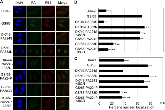
We then investigated whether the amino acids at positions 224 and 383 of PA, which contributed to the increased virulence of the H5N1 viruses in ducks, also affected viral polymerase accumulation in the nucleus. We performed immunofluorescence assays for the six mutants containing one or two amino acid changes in PA in the DK/49 or GS/65 background (Fig. 6). We found that the amino acid substitution of aspartic acid (D) to asparagine (N) at position 383 of the PA protein dramatically increased the accumulation of PA and PB1 in the nucleus of the DK/49-infected cells, whereas the amino acid substitution of N to D at position 383 of the PA protein significantly decreased the accumulation of PA and PB1 in the nucleus of the GS/65-infected cells. However, the substitution of the amino acid at position 224 of PA did not change the nuclear accumulation of PA in virus-infected cells (Fig. 6). These results indicate that the DK/49 and GS/65 viruses differ in their levels of nuclear accumulation of the polymerase and that the amino acid at position 383 of PA contributes to this difference.
FIG. 6.
Localization of PA and PB1 of different mutants in DEFs. (A) DEFs were infected at an MOI of 2 with the indicated viruses, and the localization of PA and PB1 was determined by immunofluorescence 8 h postinfection, as indicated in Materials and Methods. Cell nuclei were stained with DAPI. PA (B) and PB1 (C) were quantified by counting cells (n = 100) infected with recombinant viruses under the microscope. The values shown are means ± standard deviations of results for three independent experiments. *, P < 0.01 compared with that of DK/49-infected cells; **, P < 0.01 compared with that of GS/65-infected cells.
DISCUSSION
As H5N1 viruses have circulated, they have acquired complicated biological properties. One important property is their enhanced virulence in wild birds and ducks (5, 43), which are their natural hosts. Here, we compared two H5N1 avian influenza viruses, DK/49 and GS/65, which have similar genomes but differ in their abilities to kill ducks. The DK/49 virus replicated systemically with high viral replication in the brain and killed ducks at low doses, with a DLD50 of 2.5 log10 EID50, whereas GS/65 replicated systemically also, but with low viral replication in the brain, and was nonlethal in ducks (DLD50, >8.5 log10 EID50). We generated and tested a series of single-gene reassortants containing one gene from DK/49 in the GS/65 background and demonstrated that the PA gene is a major contributor to the increased virulence of the DK/49 virus. We identified two amino acids in the PA protein, 224P and 383D, that have cumulative effects on the pathogenicity of these H5N1 influenza viruses in ducks. We further demonstrated that amino acid 224 affects the replication of H5N1 virus in DEFs and that the amino acid at position 383 in PA affects the polymerase activity and plays an important role in the accumulation of the polymerase in the nucleus of virus-infected cells.
Based on the available sequences, the amino acids 224S and 383D in the PA protein are common in influenza A viruses, whereas 224P and 383N in PA are detected in only a few viruses isolated from ducks and humans. The 224P and 383D combination that was observed in the lethal DK/49 virus was also detected in eight other H5N1 viruses isolated after 2005 from ducks (2 strains), a wild bird (1 strain), and humans (5 strains) (accession numbers EF124756, CY034779, AB462294, CY014402, CY014410, CY014418, CY014426, and CY036236). Although the virulence of these viruses in ducks is unknown, our present study indicates that the 224P-and-383D combination in PA contributes to the increased virulence of the virus in ducks and is a recent feature of H5N1 strains.
Several H5N1 influenza viruses have been shown to be lethal in ducks in recent years (16, 38, 43). However, the viruses that were isolated from live bird markets in Hong Kong in 2002 (38), wild birds in Qinghai Lake in 2005 (43), and humans in Vietnam in 2005 (16) killed only some of the tested ducks at very high doses (106.5 to 108.75 EID50). In this study, we evaluated the virulence of the viruses in ducks by determining their DLD50s and found that the DK/49 virus is highly lethal and killed all ducks at a dose as low as 103 EID50. These findings indicate that the H5N1 influenza viruses continue to evolve and their virulence levels in ducks may be progressively increasing. The DK/49 virus could be used as a model for exploring the genetic determinants of H5N1 virulence in the natural duck host.
Hulse-Post et al. (16) previously reported that the human influenza virus isolate A/Vietnam/1203/04 (H5N1) was lethal in ducks, whereas variants containing a Y436H PB1 or a T515A PA mutation were attenuated. All of these viruses had indistinguishable replication levels in ducks. In their study, however, the DLD50 of these viruses was not determined, and the noted difference in virulence was based only on how many ducks were killed by 106 EID50 of the test virus inoculation. Therefore, the actual contribution of the amino acid at position 436 of PB1 or position 515 of PA to the virulence in ducks may be limited. Although both the DK/49 and GS/65 viruses have 436Y in PB1 and 515T in PA, GS/65 could not kill ducks, even at the highest inoculation dose of 108 EID50. We found that although both DK/49 and GS/65 replicated systemically in ducks, their titers in the brains of these birds were significantly different. Our mutagenesis studies further demonstrated that the amino acids at positions 224 and 383 of PA had a cumulative effect on the different replication levels of the two viruses in the brains of ducks, which is directly related to the virulence.
The level of polymerase accumulation in the nucleus of influenza virus-infected cells may affect the virulence of the viruses. Gabriel et al. (11) reported that the increased transport and accumulation of PB2 in the nucleus of A549 cells correlates with the adaptation and increased virulence of an H7N7 avian influenza virus in mice. In this study, we found that the accumulation of polymerase PA in the nucleus of DEF cells infected with the highly lethal DK/49 was much lower than that of PA in cells infected with the nonlethal GS/65 virus. These results suggest that the nuclear accumulation of polymerase PA may attenuate viral pathogenicity. This is in contrast to a previous study that reported that viruses bearing the PA T157A mutation are delayed in their nuclear PA accumulation and have an attenuated phenotype in mice, with respect to replication and body weight loss (15).
The polymerase complex of influenza virus is active in the nucleus of infected cells. The polymerase enters the nucleus via a host cell transport pathway that depends on the recognition of a nuclear localization sequence (NLS) of the cargo protein by an importin α/β dimer (23). NLSs have been identified in two regions of PA located between amino acids 124 and 139 and between amino acids 186 and 247 (26). Amino acid 224 is located within the second NLS of PA, but it does not affect the accumulation of PA in the nucleus of cells infected by the DK/49 and GS/65 viruses. The amino acid at position 383 of PA, however, is a major contributor to the observed difference in nuclear accumulation of the polymerase in the cells infected with the DK/49 and GS/65 viruses. These results suggest that another domain, in addition to the known NLS, may contribute to the transport and accumulation of PA in the nucleus of virus-infected cells.
The polymerase PA is a phosphoprotein with proteolytic activity. The amino-terminal third of the molecule (positions 1 to 247) is required and sufficient to activate the proteolysis function (33). Perales et al. (28) reported that mutations of the amino acids at positions 157 and 162, which result in the loss of the potential phosphorylation site, weaken the proteolysis function of PA. The amino acid 224 is located in the known second NLS of PA, and the amino acid mutation from S to P at position 224 would result in the loss of a potential casein kinase II phosphorylation site in the PA phosphoprotein. We found that the S-to-P change at position 224 in the PA protein dramatically increased the replication of GS/65 in DEFs; however, the mechanism underlying how this S224P mutation in PA could affect the virulence of the H5N1 virus in ducks remains to be determined.
In summary, we compared two naturally isolated H5N1 avian influenza viruses that differ in their lethalities in ducks and demonstrated that the polymerase PA plays an important role in the increased virulence of the H5N1 DK/49 virus in ducks. We report for the first time that the amino acids 224P and 383D in PA are critical and have cumulative effects on the highly lethal phenotype of the virus to ducks. The amino acid at position 338 in PA plays an important role in the activity of the polymerase and in the accumulation of the polymerase PA and PB1 subunits in the nucleus of virus-infected cells. Our results demonstrate that PA is a virulence factor of H5N1 avian influenza viruses.
Supplementary Material
Acknowledgments
We thank Gloria Kelly and Susan Watson for editing the manuscript.
This work was supported by the Chinese National Science Fund for Distinguished Young Scholars (30825032), by the Chinese National Key Basic Research Program (973, 2011CB505000, and 2010CB534000), by the Chinese National Key Infectious Disease Surveillance program (2009ZX10004-214), by grants from the Chinese National S&T Plan (2006BAD06A05 and 2006BAD06A04), by a contract research fund from the Ministry of Education, Culture, Sports, Science and Technology of Japan, and by the Program of Founding Research Centers for Emerging and Reemerging Infectious Diseases and is supported in part by Grants-in-Aid for Specially Promoted Research and for Scientific Research, by ERATO (Japan Science and Technology Agency), and by National Institute of Allergy and Infectious Diseases Public Health Service research grants.
Footnotes
Published ahead of print on 22 December 2010.
Supplemental material for this article may be found at http://jvi.asm.org/.
The authors have paid a fee to allow immediate free access to this article.
REFERENCES
- 1.Bussey, K. A., T. L. Bousse, E. A. Desmet, B. Kim, and T. Takimoto. 2010. PB2 residue 271 plays a key role in enhanced polymerase activity of influenza A viruses in mammalian host cells. J. Virol. 84:4395-4406. [DOI] [PMC free article] [PubMed] [Google Scholar]
- 2.Chen, H. 2009. H5N1 avian influenza in China. Sci. China C Life Sci. 52:419-427. [DOI] [PubMed] [Google Scholar]
- 3.Chen, H., et al. 2007. Polygenic virulence factors involved in pathogenesis of 1997 Hong Kong H5N1 influenza viruses in mice. Virus Res. 128:159-163. [DOI] [PubMed] [Google Scholar]
- 4.Chen, H., et al. 2004. The evolution of H5N1 influenza viruses in ducks in southern China. Proc. Natl. Acad. Sci. U. S. A. 101:10452-10457. [DOI] [PMC free article] [PubMed] [Google Scholar]
- 5.Chen, H., et al. 2006. Properties and dissemination of H5N1 viruses isolated during an influenza outbreak in migratory waterfowl in western China. J. Virol. 80:5976-5983. [DOI] [PMC free article] [PubMed] [Google Scholar]
- 6.Fan, S., et al. 2009. Two amino acid residues in the matrix protein M1 contribute to the virulence difference of H5N1 avian influenza viruses in mice. Virology 384:28-32. [DOI] [PubMed] [Google Scholar]
- 7.Fodor, E., et al. 2002. A single amino acid mutation in the PA subunit of the influenza virus RNA polymerase inhibits endonucleolytic cleavage of capped RNAs. J. Virol. 76:8989-9001. [DOI] [PMC free article] [PubMed] [Google Scholar]
- 8.Fodor, E., and M. Smith. 2004. The PA subunit is required for efficient nuclear accumulation of the PB1 subunit of the influenza A virus RNA polymerase complex. J. Virol. 78:9144-9153. [DOI] [PMC free article] [PubMed] [Google Scholar]
- 9.Gabriel, G., et al. 2007. Differential polymerase activity in avian and mammalian cells determines host range of influenza virus. J. Virol. 81:9601-9604. [DOI] [PMC free article] [PubMed] [Google Scholar]
- 10.Gabriel, G., et al. 2005. The viral polymerase mediates adaptation of an avian influenza virus to a mammalian host. Proc. Natl. Acad. Sci. U. S. A. 102:18590-18595. [DOI] [PMC free article] [PubMed] [Google Scholar]
- 11.Gabriel, G., A. Herwig, and H. D. Klenk. 2008. Interaction of polymerase subunit PB2 and NP with importin alpha1 is a determinant of host range of influenza A virus. PLoS Pathog. 4:e11. [DOI] [PMC free article] [PubMed] [Google Scholar]
- 12.Gao, Y., et al. 2009. Identification of amino acids in HA and PB2 critical for the transmission of H5N1 avian influenza viruses in a mammalian host. PLoS Pathog. 5:e1000709. [DOI] [PMC free article] [PubMed] [Google Scholar]
- 13.Hatta, M., P. Gao, P. Halfmann, and Y. Kawaoka. 2001. Molecular basis for high virulence of Hong Kong H5N1 influenza A viruses. Science 293:1840-1842. [DOI] [PubMed] [Google Scholar]
- 14.Horimoto, T., et al. 1995. Origin and molecular changes associated with emergence of a highly pathogenic H5N2 influenza virus in Mexico. Virology 213:223-230. [DOI] [PubMed] [Google Scholar]
- 15.Huarte, M., et al. 2003. Threonine 157 of influenza virus PA polymerase subunit modulates RNA replication in infectious viruses. J. Virol. 77:6007-6013. [DOI] [PMC free article] [PubMed] [Google Scholar]
- 16.Hulse-Post, D. J., et al. 2007. Molecular changes in the polymerase genes (PA and PB1) associated with high pathogenicity of H5N1 influenza virus in mallard ducks. J. Virol. 81:8515-8524. [DOI] [PMC free article] [PubMed] [Google Scholar]
- 17.Jiao, P., et al. 2008. A single-amino-acid substitution in the NS1 protein changes the pathogenicity of H5N1 avian influenza viruses in mice. J. Virol. 82:1146-1154. [DOI] [PMC free article] [PubMed] [Google Scholar]
- 18.Kawaoka, Y., and R. G. Webster. 1988. Sequence requirements for cleavage activation of influenza virus hemagglutinin expressed in mammalian cells. Proc. Natl. Acad. Sci. U. S. A. 85:324-328. [DOI] [PMC free article] [PubMed] [Google Scholar]
- 19.Li, Y., et al. 2010. Continued evolution of H5N1 influenza viruses in wild birds, domestic poultry, and humans in China from 2004 to 2009. J. Virol. 84:8389-8397. [DOI] [PMC free article] [PubMed] [Google Scholar]
- 20.Li, Z., et al. 2005. Molecular basis of replication of duck H5N1 influenza viruses in a mammalian mouse model. J. Virol. 79:12058-12064. [DOI] [PMC free article] [PubMed] [Google Scholar]
- 21.Li, Z., et al. 2006. The NS1 gene contributes to the virulence of H5N1 avian influenza viruses. J. Virol. 80:11115-11123. [DOI] [PMC free article] [PubMed] [Google Scholar]
- 22.Massin, P., P. Rodrigues, M. Marasescu, S. van der Werf, and N. Naffakh. 2005. Cloning of the chicken RNA polymerase I promoter and use for reverse genetics of influenza A viruses in avian cells. J. Virol. 79:13811-13816. [DOI] [PMC free article] [PubMed] [Google Scholar]
- 23.Mattaj, I. W., and L. Englmeier. 1998. Nucleocytoplasmic transport: the soluble phase. Annu. Rev. Biochem. 67:265-306. [DOI] [PubMed] [Google Scholar]
- 24.McAuley, J. L., et al. 2007. Expression of the 1918 influenza A virus PB1-F2 enhances the pathogenesis of viral and secondary bacterial pneumonia. Cell Host Microbe 2:240-249. [DOI] [PMC free article] [PubMed] [Google Scholar]
- 25.Mehle, A., and J. A. Doudna. 2009. Adaptive strategies of the influenza virus polymerase for replication in humans. Proc. Natl. Acad. Sci. U. S. A. 106:21312-21316. [DOI] [PMC free article] [PubMed] [Google Scholar]
- 26.Nieto, A., S. de la Luna, J. Barcena, A. Portela, and J. Ortin. 1994. Complex structure of the nuclear translocation signal of influenza virus polymerase PA subunit. J. Gen. Virol. 75(1):29-36. [DOI] [PubMed] [Google Scholar]
- 27.Office International des Epizooties. 2004. OIE manual of diagnostic tests and vaccines for terrestrial animals. Office International des Epizooties, Paris, France. [PubMed]
- 28.Perales, B., et al. 2000. The replication activity of influenza virus polymerase is linked to the capacity of the PA subunit to induce proteolysis. J. Virol. 74:1307-1312. [DOI] [PMC free article] [PubMed] [Google Scholar]
- 29.Portela, A., T. Zurcher, A. Nieto, and J. Ortin. 1999. Replication of orthomyxoviruses. Adv. Virus Res. 54:319-348. [DOI] [PubMed] [Google Scholar]
- 30.Reed, L., and H. Muench. 1938. A simple method of estimating fifty per cent endpoints. Am. J. Epidemiol. 27:493. [Google Scholar]
- 31.Regan, J. F., Y. Liang, and T. G. Parslow. 2006. Defective assembly of influenza A virus due to a mutation in the polymerase subunit PA. J. Virol. 80:252-261. [DOI] [PMC free article] [PubMed] [Google Scholar]
- 32.Salomon, R., et al. 2006. The polymerase complex genes contribute to the high virulence of the human H5N1 influenza virus isolate A/Vietnam/1203/04. J. Exp. Med. 203:689-697. [DOI] [PMC free article] [PubMed] [Google Scholar]
- 33.Sanz-Ezquerro, J. J., T. Zurcher, S. de la Luna, J. Ortin, and A. Nieto. 1996. The amino-terminal one-third of the influenza virus PA protein is responsible for the induction of proteolysis. J. Virol. 70:1905-1911. [DOI] [PMC free article] [PubMed] [Google Scholar]
- 34.Seo, S. H., E. Hoffmann, and R. G. Webster. 2002. Lethal H5N1 influenza viruses escape host anti-viral cytokine responses. Nat. Med. 8:950-954. [DOI] [PubMed] [Google Scholar]
- 35.Seo, S. H., E. Hoffmann, and R. G. Webster. 2004. The NS1 gene of H5N1 influenza viruses circumvents the host anti-viral cytokine responses. Virus Res. 103:107-113. [DOI] [PubMed] [Google Scholar]
- 36.Song, M. S., et al. 2009. The polymerase acidic protein gene of influenza A virus contributes to pathogenicity in a mouse model. J. Virol. 83:12325-12335. [DOI] [PMC free article] [PubMed] [Google Scholar]
- 37.Steel, J., A. C. Lowen, S. Mubareka, and P. Palese. 2009. Transmission of influenza virus in a mammalian host is increased by PB2 amino acids 627K or 627E/701N. PLoS Pathog. 5:e1000252. [DOI] [PMC free article] [PubMed] [Google Scholar]
- 38.Sturm-Ramirez, K. M., et al. 2004. Reemerging H5N1 influenza viruses in Hong Kong in 2002 are highly pathogenic to ducks. J. Virol. 78:4892-4901. [DOI] [PMC free article] [PubMed] [Google Scholar]
- 39.Sturm-Ramirez, K. M., et al. 2005. Are ducks contributing to the endemicity of highly pathogenic H5N1 influenza virus in Asia? J. Virol. 79:11269-11279. [DOI] [PMC free article] [PubMed] [Google Scholar]
- 40.Talon, J., et al. 2000. Influenza A and B viruses expressing altered NS1 proteins: a vaccine approach. Proc. Natl. Acad. Sci. U. S. A. 97:4309-4314. [DOI] [PMC free article] [PubMed] [Google Scholar]
- 41.Yamada, S., et al. 2010. Biological and structural characterization of a host-adapting amino acid in influenza virus. PLoS Pathog. 6:e1001034. [DOI] [PMC free article] [PubMed] [Google Scholar]
- 42.Zhang, P., et al. 2009. A novel genotype H9N2 influenza virus possessing human H5N1 internal genomes has been circulating in poultry in eastern China since 1998. J. Virol. 83:8428-8438. [DOI] [PMC free article] [PubMed] [Google Scholar]
- 43.Zhou, J. Y., et al. 2006. Characterization of a highly pathogenic H5N1 influenza virus derived from bar-headed geese in China. J. Gen. Virol. 87:1823-1833. [DOI] [PubMed] [Google Scholar]
- 44.Zhu, Q., et al. 2008. A naturally occurring deletion in its NS gene contributes to the attenuation of an H5N1 swine influenza virus in chickens. J. Virol. 82:220-228. [DOI] [PMC free article] [PubMed] [Google Scholar]
Associated Data
This section collects any data citations, data availability statements, or supplementary materials included in this article.


