Abstract
Meier-Gorlin syndrome (ear, patella, short stature syndrome) is an autosomal recessive primordial dwarfism syndrome characterised by absent/hypoplastic patellae and markedly small ears1-3. Both pre and post-natal growth are impaired in this disorder and although microcephaly is often evident, intellect is usually normal. We report here that this disorder shows marked locus heterogeneity and we identify mutations in five separate genes: ORC1, ORC4, ORC6, CDT1 and CDC6. All encode components of the pre-replication complex, implicating defects in replication licensing as the cause of a genetic syndrome with distinct developmental abnormalities.
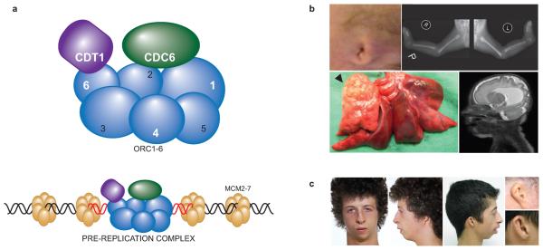
In the accompanying paper4 we identified mutations in ORC1 encoding a subunit of the origin recognition complex (ORC) in five patients with microcephalic primordial dwarfism. The ORC multiprotein complex is loaded onto chromatin at DNA origins prior to S phase to license replication. In conjunction with additional components CDT1, CDC6 and MCM helicase proteins, it then forms the pre-replication complex (described in Fig. 1). Cellular studies established that ORC1 patients had partial loss-of-function defects in replication licensing4. An orc1 zebrafish model recapitulated the dwarfism phenotype as did morpholino depletion of another pre-replication complex component, mcm5. This led us to hypothesise that further pre-replication complex genes might cause primordial dwarfism, however mutations in other ORC subunits were not found.
Figure 1. The pre-replication complex and Meier-Gorlin syndrome.
(a) Genome replication is licensed by the binding of a number of specialised proteins to origins of replication, which form the pre-replication complex9. The first step in complex formation is the loading of the heterohexameric Origin Recognition Complex (comprising ORC1-6 proteins), onto chromatin in an ATP dependent manner during M and G1 phases of the cell cycle. Further proteins, including CDC6 and CDT1 are then recruited to the pre-replicative complex, that then permit reiterative loading of the multimeric MCM helicase. At the commencement of S-phase, replication is started by the MCM helicase unwinding DNA, and the recruitment of additional replication proteins. The five proteins implicated in Meier-Gorlin syndrome are highlighted in white text (ORC1, ORC4, ORC6, CDT1, CDC6). (b) Patient 1 has a severe developmental malformation syndrome with marked microtia and extreme retroflexion and dislocation of the knees (top row). His malformations included lobar congenital emphysema (arrow head), and a severe cortical dysplasia of the brain. Parasaggital T2 weighted MRI at age 1 month demonstrating severe pachygyria, most severe frontally along with ventricular enlargement. (c) Two patients (P11, P9) with classical Meier-Gorlin syndrome. b,c Informed consent to publish the photographs was obtained from the subjects' parents.
In this study, the profound growth retardation and microcephaly in a patient with a complex lethal developmental syndrome led us to screen for ORC1 mutations. We found that he and his brother (P1, P2, Table 1 and Supplementary Table 1) were compound heterozygote for mutations in ORC1 (p.R105Q and p.V667fsX24). The combination of truncating and missense mutations was in contrast to biallelic missense mutations seen in previous ORC1 cases, suggestive of greater loss-of-function and consistent with a more severe phenotype. Unlike previous ORC1 cases, the proband (P1) had an extensive number of developmental malformations including severe cortical dysplasia, congenital emphysema, absence of the pancreatic tail and retroflexion of the knees; as well as micropenis, blepharophimosis and cranial suture stenosis (Fig. 1). Notably, he also had significant microtia and absent patellae, features classically associated with Meier-Gorlin syndrome (MIM 224690).
Table 1.
Mutations in five genes encoding pre-replication complex proteins in patients with Meier-Gorlin syndrome.
| Family | Patient | Ancestry | Gene | Nucleotide Alterations | Amino Acid Alterations | Exon(s) | Segregation | Parental Consanguinity | Gender | Current Height (SD) | Current OFC (SD) | Microtia | Absent/Small Patellae |
|---|---|---|---|---|---|---|---|---|---|---|---|---|---|
| F1 | P1 | UK | ORC1 | [c.314G>A] + [c.1999_2000delGTinsA] |
R105Q + V667fsX24 | 4; 13 | Het, M, P | No | M | −9.6 | −9.8 | + | + |
| F1 | P2 | UK | ORC1 | [c.314G>A] + [c.1999_2000delGTinsA] |
R105Q + V667fsX24 | 4; 13 | Het, M, P | No | M | NA | NA | + | u |
| F2 | P3 | USA | ORC1 | [c.314G>A] + [c.1482-2A>G] | R105Q + intron 9 splice acceptor site | 4; intron 9 |
Het, M, nps |
No | M | −6.6 | u | + | + |
| F3 | P4 | UK | ORC1 | [c.314G>A] + [c.1482-2A>G] | R105Q + intron 9 splice acceptor site | 4; intron 9 |
Het, M, P | No | F | −6.9 | −4.0 | + | + |
| F4 | P5 | USA | ORC4 | [c.521A>G] + [c.874_875insAACA] | Y174C + A292fsX19 | 8; 11 | Het, M, P | No | F | −6.4 | u | + | + |
| F5 | P6 | USA | ORC4 | c.521A>G | Y174C | 8 | Hom, M, P |
Yes | F | −4.2 | −2.1 | + | − |
| F5 | P7 | USA | ORC4 | c.521A>G | Y174C | 8 | Hom, M, P |
Yes | F | −4.1 | −3.0 | + | − |
| F6 | P8 | TR | ORC6 | [c.257_258delTT] + [c.695A>C] | F86X + Y232S | 3; 7 | Het, M, P | Yes | F | −3.3 | −1.6 | + | + |
| F6 | P9 | TR | ORC6 | [c.257_258delTT] + [c.695A>C] | F86X + Y232S | 3; 7 | Het, M, P | Yes | M | −2.4 | −2.1 | + | + |
| F6 | P10 | TR | ORC6 | [c.257_258delTT] + [c.695A>C] | F86X + Y232S | 3; 7 | Het, M, P | Yes | M | −3.2 | −2.3 | + | + |
| F7 | P11 | NZ | CDT1 | [c.1385G>A] + [c.1560C>A] | R462Q + Y520X | 9; 10 | Het, M, P | No | M | −4.7 | +0.1 | + | + |
| F8 | P12 | UK | CDT1 | [c. 196G>A ] + [c.351G>C] | A66T + Q117H(exon 2 splice donor site) |
1; 2 | Het, M, P | No | F | −5.1 | −5.0 | + | + |
| F9 | P13 | USA | CDT1 | [c.1385G>A] + [c.1560C>A] | R462Q + Y520X | 9; 10 | Het, M, P | Yes | F | −4.7 | −1.3 | + | + |
| F9 | P14 | USA | CDT1 | [c.1385G>A] + [c.1560C>A] | R462Q + Y520X | 9; 10 | Het, M, P | Yes | F | −3.9 | −1.0 | + | + |
| F9 | P15 | USA | CDT1 | [c.1385G>A] + [c.1560C>A] | R462Q + Y520X | 9; 10 | Het, M, P | Yes | M | −1.6 | +1.7 | + | + |
| F10 | P16 | UK | CDT1 | [c.351G>C] + [c.1385G>A] | Q117H(exon 2 splice donor site) + R462Q |
2; 9 | Het, nps | Yes | F | −3.3 | −0.5 | + | + |
| F11 | P17 | SA | CDT1 | [c.1081C>T] + [c.1357C>T] | Q361X + R453W | 7; 9 | Het, M, P | No | F | −0.4 | −2.1 | + | + |
| F12 | P18 | FR | CDC6 | c.968C>G | T323R | 7 | Hom, M, P |
Yes | M | −4.1 | −3.3 | + | + |
Mutations are described numbered from the first nucleotide of the initiation codon in the nucleotide sequence. For each mutation, 380 control chromosomes were screened and found to be negative for the sequence change. Abbreviations: Hom, homozygous in affected individual; Het, compound heterozygous in affected individual, M, mutation identified in mother; P, mutation identified in father; nps, parental sample(s) not available. The protein truncation Y520X in CDT1 is within the last exon and therefore the transcript is not expected to undergo nonsense-medicated mRNA decay. Additionally, in CDT1 the mutation c.351G>C confers a non-conservative amino acid substitution (p.Q117H), however the nucleotide lies within the exon 2 splice donor site and therefore this mutation is expected to result in an inframe deletion of exon 3 from the transcript and resulting protein. TR, Turkey; NZ, New Zealand; FR, France; SA, Saudi Arabia; SD, standard deviation, OFC, occipito-frontal circumference; NA, not available; u, unknown.
Consequently, we sequenced ORC1 in 33 patients with an established diagnosis of Meier-Gorlin syndrome and identified two further cases with mutations in ORC1 (Table 1, Fig. 2). Both patients from Families 2 and 3 were compound heterozygotes for the same ORC1 mutations: a splice acceptor site mutation in conjunction with the previously identified c.314G>A that encodes the R105Q amino acid substitution4, again in contrast to the biallelic missense mutations observed in the previous study. In Family 2, the c.314G>A mutation was inherited on a haplotype shared with Family 1 (Supplementary Fig. 1), consistent with common ancestry. However, the c.314G>A mutation in Family 3 and the two splice acceptor site mutations in Family 2 and 3 appear to have arisen independently. Notably, P3 in Family 2 is the original patient described by Gorlin2, unequivocally establishing that mutations in ORC1 cause Meier-Gorlin syndrome.
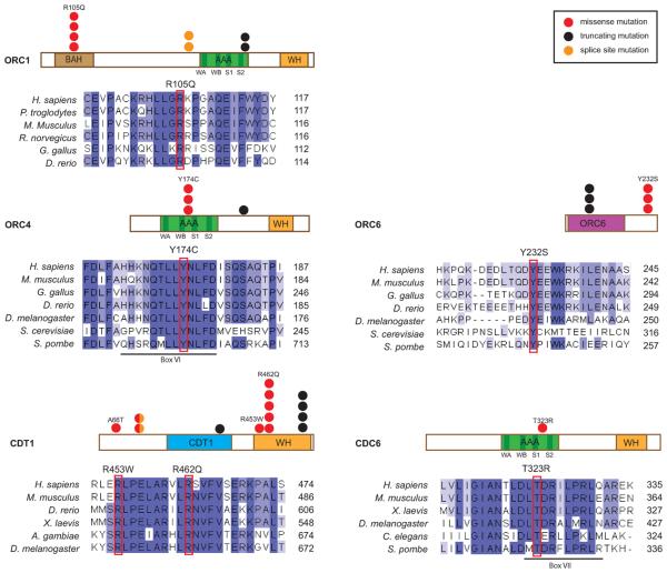
Figure 2. Pre-replication complex proteins mutated in Meier-Gorlin syndrome.
Schematics for each protein depicting known protein domains with positions of mutations shown by filled circles. Each filled circle represents one patient. Clustalw alignment of protein residues surrounding substituted amino acids (positions of substituted resides indicated by red boxes). WA, Walker A; WB Walker B, S1, Sensor 1; S2, Sensor 2; motifs. BAH, Bromo Associated Homology domain; AAA, ATPase Associated with a wide range of cellular Activites; WH, winged helix domain.
Sequencing the genes encoding other ORC subunits then identified pathogenic mutations in two further subunits of the Origin Recognition Complex (ORC4, ORC6) in three patients with Meier-Gorlin syndrome (Table 1, Supplementary Table 1). Monozygotic twins P6 and P7 were found to be homozygous for a missense mutation (c.521A>G, p.Y174C) in ORC4 (family also independently ascertained by M. Samuels and colleagues, ref. 5). The substituted tyrosine was conserved throughout evolution as distant as S. cerevisiae and S. pombe, and was located within Box VI of the AAA domain6 (Fig. 2), a domain that is essential for ATP-dependent complex formation of ORC4 with other ORC subunits7,8. A second patient, P5, is a compound heterozygote for this same Y174C substitution (present on the ancestral haplotype shared with P6 and P7; Supplementary Fig. 1), in conjunction with a four base-pair insertion mutation causing a translational frameshift and premature protein termination.
In Family 6, three affected children were compound heterozygotes for mutations in ORC6. Similar to P5, these patients inherited one loss-of-function mutation caused by a two base pair deletion and a missense mutation resulting in the substitution (p.Y232S) of an amino acid completely conserved from fungi to humans located at the C-terminus of the ORC6 protein (Fig. 2).
Given that mutations in genes encoding multiple components of the ORC complex cause Meier-Gorlin syndrome, we next examined whether additional components of the pre-replication complex could be similarly implicated. CDT1 and CDC6 are recruited to the pre-replication complex following assembly of the full ORC complex on chromatin9. As the region mutated in ORC6 is involved in CDT1 protein binding in S. cerevisiae10, we next screened our Meier-Gorlin syndrome cohort for mutations in CDT1. Five families were identified with compound heterozygous mutations in CDT1 (Table 1). In three of these families, one of the mutations was c.1385G>A, causing the substitution R462Q in the C-terminal winged helix domain of CDT1 (Fig. 2). This residue has been implicated in MCM helicase complex binding through mutagenesis of the orthologous mouse residue (p.R474Q)11, the same substitution as the human mutation we have identified here.
Finally, screening of CDC6 identified one Meier-Gorlin syndrome patient (P18) homozygous for a missense mutation (c.968C>G), resulting in the substitution T323R. This residue is absolutely conserved from fungi to humans (Fig. 2) and lies within the box VII motif of the AAA domain (Fig. 2)6, an ATP binding domain that is essential for CDC6 function in DNA replication12.
We report here that mutations in multiple components of the pre-replication complex cause Meier-Gorlin syndrome and despite finding marked genetic heterogeneity, identify an underlying functional homogeneity in this disorder. The functional studies of ORC1 in the accompanying manuscript4 establish that impairment of ORC1 function is associated with extreme growth failure in humans. Here, our molecular genetic findings implicate the entire pre-replication complex in primordial dwarfism syndromes, providing compelling genetic evidence supporting the notion that impaired replication licensing causes growth failure.
Though most of the patients described in this paper have typical features of Meier-Gorlin syndrome1-3, considerable phenotypic variation is seen from mutations in these genes, particularly with the ORC1 cases reported here and in the accompanying paper4. Growth parameters vary markedly (height ranges from −0.4 to −9.6 SD, and head circumference from +1.7 to −9.8 SD), with cognition varying from normal to severe neurological impairment associated with a severe cortical dysplasia. Thus, although microtia and small patellae are the best predictors of mutations in the pre-replication complex, the disease spectrum associated with these genes could extend from cases described as microcephalic osteodysplastic primordial dwarfism I/III to those with nonsyndromic primordial dwarfism. (Further discussion of clinical phenotypes, see Supplementary Note.)
Impaired replication might be expected to affect growth of all tissues equally; therefore it is noteworthy that specific tissues are disproportionately reduced in size, most readily apparent in the ears and patellae. Most strikingly, significant congenital malformations are also evident in several patients, affecting limb, genitourinary, lung and brain development. This finding surprisingly links mutations in a chromatin bound protein complex regulating replication with multiple congenital malformations. Such an association between chromatin bound complexes involved in essential cellular functions and development is not unprecedented. Notably in Cornelia de Lange syndrome (MIM 122470), mutations in the cohesion complex also cause severe growth retardation though with very different developmental consequences13.
The cellular mechanisms, by which preRC components cause the specific developmental phenotypes reported here, are not immediately evident. It may be that proliferation of certain cell types, such as chondrocytes in the ears and patellae are particularly sensitive to reduced preRC function. Alternatively, these and the other developmental anomalies could result from other cellular functions of the preRC components, as has been found for individual ORC proteins 14.
Previous primordial dwarfism genes have DNA damage response and centrosome functions15-17, while here we find mutations in genes involved in licensing of DNA replication origins. All are critical components required for cell cycle progression. Pathogenesis is therefore likely to be the result of impaired cellular proliferation, resulting in reduced cell number and consequently global growth failure. While in this framework genes regulating replication are rational candidates for primordial dwarfism, it is most surprising to find specific congenital malformations occurring as the consequence of mutations in the pre-replication complex. This suggests an unanticipated link between the regulation of replication and development that warrants future investigation.
Methods
Research subjects
All patients in this study had previously been published as cases of Meier-Gorlin syndrome or fulfilled diagnostic criteria for Meier-Gorlin syndrome (microtia, absent/small patellae and short stature). Genomic DNA was isolated from peripheral blood of the affected children and family members. No cytogenetic abnormalites were observed on routine karyotyping of these Meier-Gorlin syndrome cases (n=10 patients). Furthermore, no significant increase of either spontaneous, or radiation-induced chromosome breaks was observed (2 cases versus 2 control patients, ≥ 100 metaphases assessed per patient; p≥ 0.7, X2-test). Informed consent was obtained from all participating families and the studies were approved by the Scottish Multicentre Research Ethics Committee (04:MRE00/19), Regional Committee on Research Involving Human Subjects Nijmegen - Arnhem (0006-0119); or University of Texas Southwestern Medical Center at Dallas (IRB #032008-066). Informed consent was obtained for the publication of photographs.
Sequencing of candidate genes
Primers were designed using Exonprimer (Supplementary Table 2). Coding exons and exon-intron boundaries of each gene were screened by bidirectional capillary sequencing on an ABI 3730 gene sequencer. Primer sequences and PCR conditions are available upon request. Sequence analysis and mutation detection performed using Mutation Surveyor v. 2.61 (Softgenetics LLC).
Microsatellite Genotyping
Microsatellites were selected on the basis of heterozygosity and proximity to each gene. Primer sequences for microsatellites were obtained from UniSTS. Amplicons were separated by capillary electrophoresis on an ABI3730 Sequencer, and allele sizes obtained using GeneMapper (Applied Biosystems).
Protein alignments
Reference sequences for all proteins were obtained from RefSeq and alignments performed using ClustalW2. Protein domains were mapped using data obtained from Conserved Domain Database (CCD).
Supplementary Material
Acknowledgements
We thank the subjects and their families for participation in this study and all clinicians for contributing samples not included in this manuscript. Also the late Robert J. Gorlin for contributing patient P3 to the study; Elizabeth S. Gray for pathology; M. Ansari, S. McKay, S.D. van der Velde-Visser, T.H. Merkx, S. Devroy and the MRC HGU core sequencing service for advice and technical support; Eddy Maher and colleagues for cytogenetic advice and assistance; V. van Heyningen, N. Hastie, D. Fitzpatrick, I. Jackson, J. Blow for discussions and comments. This work was supported by funding from the MRC and Lister Institute of Preventative Medicine. APJ is a MRC Senior Clinical Fellow and Lister Institute Prize fellow.
Footnotes
Competing financial interests
The authors declare no competing financial interests.
Accession numbers
ORC1, NM_004153.2; ORC4, NM_181742.3; ORC6, NM_014321.2; CDT1, NM_030928.3; CDC6, NM_001254.3.
URLs
UniSTS - http://www.ncbi.nlm.nih.gov/unists
Exonprimer - http://ihg2.helmholtz-muenchen.de/ihg/ExonPrimer.html
References
- 1.Bongers EM, et al. Meier-Gorlin syndrome: report of eight additional cases and review. Am J Med Genet. 2001;102:115–24. doi: 10.1002/ajmg.1452. [DOI] [PubMed] [Google Scholar]
- 2.Gorlin RJ, Cervenka J, Moller K, Horrobin M, Witkop CJ., Jr. Malformation syndromes. A selected miscellany. Birth Defects Orig Artic Ser. 1975;11:39–50. [PubMed] [Google Scholar]
- 3.Meier Z, Poschiavo, Rothschild M. Case of arthrogryposis multiplex congenita with mandibulofacial dysostosis (Franceschetti syndrome) Helv Paediatr Acta. 1959;14:213–6. [PubMed] [Google Scholar]
- 4.Bicknell LS, et al. Mutations in ORC1, encoding the largest subunit of the Origin Recognition Complex, cause microcephalic primordial dwarfism. Nat Genet. 2011 doi: 10.1038/ng.776. [DOI] [PubMed] [Google Scholar]
- 5.Samuels ME. Nat. Genet. 2011 doi: 10.1038/nrg3011. [DOI] [PubMed] [Google Scholar]
- 6.Neuwald AF, Aravind L, Spouge JL, Koonin EV. AAA+: A class of chaperone-like ATPases associated with the assembly, operation, and disassembly of protein complexes. Genome Res. 1999;9:27–43. [PubMed] [Google Scholar]
- 7.Bowers JL, Randell JC, Chen S, Bell SP. ATP hydrolysis by ORC catalyzes reiterative Mcm2-7 assembly at a defined origin of replication. Mol Cell. 2004;16:967–78. doi: 10.1016/j.molcel.2004.11.038. [DOI] [PubMed] [Google Scholar]
- 8.Ranjan A, Gossen M. A structural role for ATP in the formation and stability of the human origin recognition complex. Proc Natl Acad Sci U S A. 2006;103:4864–9. doi: 10.1073/pnas.0510305103. [DOI] [PMC free article] [PubMed] [Google Scholar]
- 9.Mendez J, Stillman B. Perpetuating the double helix: molecular machines at eukaryotic DNA replication origins. Bioessays. 2003;25:1158–67. doi: 10.1002/bies.10370. [DOI] [PubMed] [Google Scholar]
- 10.Chen S, de Vries MA, Bell SP. Orc6 is required for dynamic recruitment of Cdt1 during repeated Mcm2-7 loading. Genes Dev. 2007;21:2897–907. doi: 10.1101/gad.1596807. [DOI] [PMC free article] [PubMed] [Google Scholar]
- 11.Jee J, et al. Structure and mutagenesis studies of the C-terminal region of licensing factor Cdt1 enable the identification of key residues for binding to replicative helicase Mcm proteins. J Biol Chem. 2010;285:15931–40. doi: 10.1074/jbc.M109.075333. [DOI] [PMC free article] [PubMed] [Google Scholar]
- 12.Herbig U, Marlar CA, Fanning E. The Cdc6 nucleotide-binding site regulates its activity in DNA replication in human cells. Mol Biol Cell. 1999;10:2631–45. doi: 10.1091/mbc.10.8.2631. [DOI] [PMC free article] [PubMed] [Google Scholar]
- 13.Liu J, Krantz ID. Cornelia de Lange syndrome, cohesin, and beyond. Clin Genet. 2009;76:303–14. doi: 10.1111/j.1399-0004.2009.01271.x. [DOI] [PMC free article] [PubMed] [Google Scholar]
- 14.Sasaki T, Gilbert DM. The many faces of the origin recognition complex. Curr Opin Cell Biol. 2007;19:337–43. doi: 10.1016/j.ceb.2007.04.007. [DOI] [PubMed] [Google Scholar]
- 15.Griffith E, et al. Mutations in pericentrin cause Seckel syndrome with defective ATR-dependent DNA damage signaling. Nat Genet. 2007 doi: 10.1038/ng.2007.80. [DOI] [PMC free article] [PubMed] [Google Scholar]
- 16.Al-Dosari MS, Shaheen R, Colak D, Alkuraya FS. Novel CENPJ mutation causes Seckel syndrome. J Med Genet. 2010;47:411–4. doi: 10.1136/jmg.2009.076646. [DOI] [PubMed] [Google Scholar]
- 17.O'Driscoll M, Ruiz-Perez VL, Woods CG, Jeggo PA, Goodship JA. A splicing mutation affecting expression of ataxia-telangiectasia and Rad3-related protein (ATR) results in Seckel syndrome. Nat Genet. 2003;33:497–501. doi: 10.1038/ng1129. [DOI] [PubMed] [Google Scholar]
Associated Data
This section collects any data citations, data availability statements, or supplementary materials included in this article.


