Abstract
An extracellular alpha-amylase (Amy1) whose gene from Cryptococcus flavus was previously expressed in Saccharomyces cerevisiae was purified to homogeneity (67 kDa) by ion-exchange and molecular exclusion chromatography. The enzyme was activated by NH4+ and inhibited by Cu+2 and Hg+2. Significant biochemical and structural discrepancies between wild-type and recombinant α-amylase with respect to Km values, enzyme specificity, and secondary structure content were found. Far-UV CD spectra analysis at pH 7.0 revealed the high thermal stability of both proteins and the difference in folding pattern of Amy1 compared with wild-type amylase from C. flavus, which reflected in decrease (10-fold) of enzymatic activity of recombinant protein. Despite the differences, the highest activity of Amy1 towards soluble starch, amylopectin, and amylase, in contrast with the lowest activity of Amy1w, points to this protein as being of paramount biotechnological importance with many applications ranging from food industry to the production of biofuels.
1. Introduction
Starch is a major storage product of many economically important crops such as wheat, rice, cassava, and potato [1]. A large variety of microorganisms employ extracellular or intracellular enzymes to hydrolyze starch thus enabling its utilization as a source of energy. One of the most important groups of enzymes that process starch is represented by the α-amylase family or family 13 glycosyl hydrolases [2, 3]. Amylases (EC 3.2.1.1, α-1,4-glucan-glucanohydrolase) are enzymes that hydrolyze starch polymers yielding diverse products including dextrins and smaller polymers of glucose. These enzymes are of great biotechnological interest with many applications ranging from food industry to the production of biofuels. Since each different application requires amylases with unique properties it is often necessary to search the biodiversity for new sources of these enzymes [4]. Several amylases isolated from yeasts such as Candida antarctica, Candida japonica [4], Lipomyces kononenkoae, Saccharomycopsis fibuligera, Schwanniomyces alluvius [5], Trichosporon pullulans, and Filobasidium capsuligenum [6] have been described. We have previously reported the characterization of an α-amylase (Amy1) from the basidiomycetous yeast Cryptococcus flavus which exhibited important biochemical properties for its industrial utilization such as high stability at pH 5.5 and optimal temperature at 50°C [7]. The gene encoding this α-amylase (AMY1) was cloned and successfully expressed in Saccharomyces cerevisiae [8]. In order to assess the use of S. cerevisiae as a host for the heterologous production of Amy1 we sought the purification and enzymatic characterization of recombinant enzyme produced in this system.
2. Material and Methods
2.1. Strains
S. cerevisiae CENPK2 (MATa/αura3-52/ura3-52 leu 2-3,112 trp1-289/trp1-289 his3-1/his3-1).
2.2. Enzyme Purification
A colony of S. cerevisiae CENPK2 harboring YEpAMY was cultured in SD medium (0,62% YNB, 2% glucose, uracil, tryptophan, and histidine), and 2 mL of this preculture was transferred to 1 L conical flask containing 200 mL of the same medium following incubation at 28°C for 60 h (200 rpm). S. cerevisiae cells were harvested by centrifugation (5,000 g/10 min), and the supernatant was dialyzed overnight against water and loaded on a Q-Sepharose fast flow column (0.9 × 18 cm) previously equilibrated in 50 mM sodium acetate (pH 5.5). The column was washed with the same buffer, and proteins were eluted by a linear gradient of 0–0.5 M NaCl. Four fractions of 3 mL were collected and monitored for the presence of proteins and amylase activity. These samples were collected, dialyzed, lyophilized, and resuspended in 1 mL of water and loaded on a Sephacryl S-200 HR (2.5 × 85 cm) column previously equilibrated with 50 mM sodium acetate containing 0.1 M NaCl. The column was washed with the same buffer at a flow rate of 12 mL·h−1. Fractions containing amylase activity were pooled, dialyzed against water, lyophilized, and stored at −20°C. In chromatography experiments, the protein content of each fraction was routinely monitored by measuring the absorbance at 280 nm. Protein concentration was measured using serum albumin as standard [9].
2.3. Electrophoresis and Zymogram Analysis
Protein integrity and the molecular mass calculation were performed by running samples on 12% sodium dodecyl sulfate-polyacrilymide gels [10]. Proteins were silver-stained as described by manufacturer [11] (1987). Molecular mass markers (Fermentas Life Sciences) were as follows: β-galactosidase (116 kDa), bovine serum albumin (66.2 kDa), ovalbumin (45 kDa), lactate dehydrogenase (35 kDa), REase Bsp98I (25 kDa), β-lactoglobulin (18.4 kDa), and lysozyme (14.4 kDa) by manufacturer. Activity gels (zymogram) were performed after running samples on 12% nondenaturing PAGE. Gels were washed with distilled water, incubated with 50 mM sodium acetate (pH 5.5) for 60 min, and then incubated at 4°C for 12 h in a solution containing 0.5% of starch (in 50 mM sodium acetate buffer [pH 5.5]). The gel was then incubated at 37°C for 2 h, and bands with amylase activity were detected after staining with iodine solution (1 mM I2 in 0.5 M KI).
2.4. Amylase Assay
α-amylase dextrinizing activity was assayed in a reaction system containing 100 μL 0.5% (w/v) soluble starch, 40 μL 0.5 M acetate buffer (pH 5.5), enzyme solution (0–60 μL), and water to a total volume of 200 μL. After 10 min at 60°C, the reaction was stopped with 200 μL 1.0 M acetic acid. Iodine reagent was then added to determine dextrinizing activity [12]. Saccharifying activity was determined by measuring the production of reducing sugars from starch by using the dinitrosalicylic acid (DNS) method [13]. One unit of dextrinizing activity was defined as the amount of enzyme necessary to hydrolyse 0.1 mg starch/min. One unit of saccharifying activity was defined as the amount of enzyme necessary to produce 1 mg glucose equivalent/min.
2.5. Enzyme Characterization
The optimum pH of the recombinant amylase was determined by varying the pH of the reaction mixtures using the following buffers (50 mM): glycine-HCl (pH 1.0–3.0), sodium acetate (pH 4.0–5.5), sodium phosphate (pH 6.0-7.0), and Tris-HCl (pH 8.0–10) at 60°C. In order to determine pH stability, the enzyme was preincubated in different buffers for 60 min at 60°C. The residual enzyme activity was assayed in 50 mM sodium acetate buffer (pH 5.5). The optimum estimated temperature of the enzyme was evaluated by measuring amylase activity at different temperatures (30°C to 70°C) in 50 mM sodium acetate (pH 5.5). The effect of temperature on enzyme stability was determined by measuring the residual activity after 15, 30, 45, and 60 min of preincubation in 50 mM sodium acetate (pH 5.5) at 55 and 60°C. In order to determine the effect of metal ions, the assay was performed after preincubation of the enzyme with various metal ions at a final concentration of 4 mM. The effect of 10 mM CaCl2 and DTT (5–10 mM) was also evaluated. Km values for the purified enzyme were determined by incubating the enzyme with 0–0.8 mg·mL−1 soluble starch in 50 mM sodium acetate (pH 5.5) at 60°C. The data obtained were fitted to a standard Lineweaver-Burk model using linear least squares regression.
2.6. Thin Layer Chromatography Analysis
Briefly, purified amylase was incubated with 0.5% starch, glycogen, pullulan, amylase, and amylopectin, in 50 mM sodium acetate (pH 5.5) at 40°C. Aliquots (10 μL) were removed after incubation for 6 and 12 h. The chromatogram was developed with n-butanol/methanol/H2O (4 : 2 : 1). Sugars were detected by thin-layer chromatography [14].
2.7. Circular Dichroism Spectroscopy
Circular Dichroism (CD) assays were carried out using Jasco J-815 spectropolarimeter (Jasco, Tokyo, Japan) equipped with a Peltier type temperature cuvette holder. Far-UV spectra were recorded using 0.1 cm path length quartz cuvette. Proteins (0.085 to 0.100 mg/mL) were analyzed in 2 mM glycine pH 3.0 and 4.0, 2 mM sodium acetate pH 5.5, 2 mM Tris-HCl pH 7.0 and 8.0. We used CD data over the wavelength range 260–200 nm to ensure a satisfactory CD signal and to prevent the high signal-noise ratio. Four consecutive measurements were accumulated, and the mean spectra were corrected for the baseline contribution of the buffer. Thermal denaturation assays were performed raising the temperature from 25 to 95°C. The observed ellipticities were converted into molar ellipticity ([θ]) based on molecular mass per residue of 115 Da. Protein structure and thermal unfolding curves were tracked by changes in [θ] at 222 or 206 nm.
The temperature dependence of the secondary structure was estimated from the Far-UV CD curves adjustments using the CDNN deconvolution software (Version 2.1, Bioinformatik.biochemtech.uni-halle.de/cdnn) [15].
3. Results and Discussion
3.1. Purification of Amy1
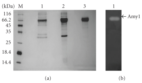
Amy1 was purified from the supernatant of S. cerevisiae cultures in a two-step chromatographic procedure. Elution profiles of both Q-Sepharose and Sephacryl-S200HR chromatography showed one peak with amylase activity (data not shown). This fraction was collected, dialyzed, and concentrated by lyophilization. The enzyme was purified to homogeneity with 3.79-fold increase in specific activity with a yield of ~10.3% as compared to the supernatant (Table 1). Comparing with the recombinant enzyme, wild-type Amy1 was purified from C. flavus cultures in only a single purification step [7]. SDS-PAGE analysis of the purified recombinant amylase showed a single protein band corresponding to ~67 kDa (Figure 1(a)) which showed α-amylase activity after zymogram analyses (Figure 1(b)). Among the amylase-producing yeasts and filamentous fungi, the α-amylases from Cryptococcus sp S-2 [16], Aspergillus fumigatus [17], Lipomyces starkeyi [18], and Lipomyces kononenkoae [19] showed similar molecular masses as Amy1 produced in S. cerevisiae.
Table 1.
Summary of the purification steps of Amy1.
| Step | Total activity (U) | Total protein (mg × 10−3) | Specific activity (U/mg × 10−3) | Purification (fold) | Yield (%) |
|---|---|---|---|---|---|
| Supernatant | 1922.2 | 1.750 | 1098.5 | 1.00 | 100.0 |
| Q-Sepharose | 490.8 | 0.120 | 4090.0 | 3.72 | 25.4 |
| Sephacryl S-200HR | 199.9 | 0.048 | 4164.5 | 3.79 | 10.3 |
Figure 1.
(a) Analysis of purified recombinant Amy1 on 12% SDS-PAGE; M, standard marker proteins; lane 1, lane 2, protein profile after ion exchange on Q-Sepharose column; lane 3, purified recombinant amylase after gel filtration on Sephacryl S-200HR column. (b) Detection of amylase activity on non-denaturing PAGE.
3.2. Enzyme Characterization
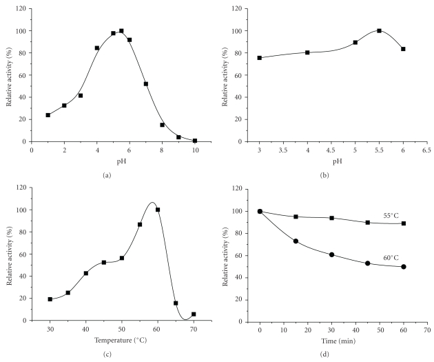
The optimum pH for the recombinant enzyme was determined as pH 5.5 (Figure 2(a)). Amy1 was tested for pH stability by preincubating the purified enzyme in appropriate buffers for 1 hour. The data suggest that Amy1 is tolerant to wide pH range (Figure 2(b)). This result was similar to that found for α-amylases from a variety of yeast strains [20, 21]. The optimum pH for yeast α-amylase activity is usually in the range of 4.0 and 6.0 [5, 7, 16, 21].
Figure 2.
Effect of pH (a-b) and temperature (c-d) on Amy1 activity and stability, respectively. The pH optimum and pH stability were determined by varying the pH of the reaction mixtures and preincubating the enzyme in different buffers for 60 min at 55°C (square) and 60°C (circle). The temperature optimum was evaluated by measuring amylase activity at different temperatures, and the effect of temperature on enzyme stability was determined by measuring the residual activity after 15, 30, 45, and 60 min of preincubation in 50 mM sodium acetate (pH 5.5) at 55 and 60°C.
Amylase activity measured as a function of temperature from 30 to 70°C shows that the activity was the highest at 60°C (Figure 2(c)). This optimum temperature is in agreement with the α-amylases from Cryptococcus sp S-2 treated with 1 mM CaCl2 [16] and Aureobasidium pullulans N13d [20]. Thermostability is considered an important and useful criterion for industrial application of α-amylase from microorganisms. As shown in Figure 2(d), the residual amylase activity is practically constant at 55°C after 1 h of incubation. However, at 60°C 50% of the residual activity was lost. Iefuji et al. [16] proposed that the C-terminal raw starch binding motifs present in Cryptococcus sp S-2 amylase (AMY-CS2) might be related to the thermostability presented by this amylase. Because Amy1 has 97% sequence identity with AMY-CS2 [8] it is possible that the same explanation applies to Amy1.
The results presented on Table 2 indicate that recombinant Amy1 is poorly affected by most of the 4 mM ions tested (Mg2+, Mn2+, and Ca2+) since the relative activity was higher than 70%. In addition, it was also found that in the presence of 10 mM CaCl2, Amy1 still maintained its original activity (data not shown) which shows that this enzyme is not affected by Ca+2. Similar behavior was found for wild-type Amy1. However, Takeuchi et al. [22] related that the Pichia burtoniiα-amylase was activated by Ca+2 (135%). These authors proposed that the activation of enzyme by calcium ions probably happened during the purification procedure when the enzyme lost activity. The Bacillus halodurans 38C-21 α-amylase also had its activity increased in presence of Ca2+ [23]. The presence of NH4+ ions had the most prominent activating effect (111%) on recombinant Amy1. However, Zn2+, Cu2+, and Hg2+ acted as inhibitors of amylase activity, with Cu+2 and Hg2+ showing a complete inhibition (Table 2). The inhibition by Hg2+ may indicate the importance of indole group of amino acid residues in enzyme function [4, 24]. The wild-type amylase is inhibited by Hg2+, Ag+, Cu2+, and Mg2+ [16] while that from L. kononenkoae CBS 5608 is inhibited by Ag+ and Cu2+ [21]. Interestingly, the amylase from yeast Aureobasidium pullulans N13d is not inhibited by Cu2+ which had an activating effect on the purified enzyme [20]. DTT is frequently used to reduce and prevent intramolecular and intermolecular disulfide bonds. The enzyme's activity was practically constant when DTT (4–25 mM) was used (data not shown). This result was different from that observed with the Thermotoga maritima MSB8 amylase expressed in E. coli which showed increased activity in presence of DDT ranging from 5 to 10 mM [25].
Table 2.
Effect of ions on purified Amy1.
| Compound (4 mM) | Relative activity (%) |
|---|---|
| Control | 100 |
| MgSO4 | 88.6 |
| MnSO4 | 77.9 |
| CaCl2 | 71.6 |
| (NH4)2SO4 | 111 |
| ZnCl2 | 20.1 |
| CuSO4 | 0 |
| HgSO4 | 0 |
Wanderley et al. [7] reported that the Km for wild-type Amy1 was 0.056 mg·mL−1 which is considerably smaller than the Km observed for the recombinant amylase (0.37 mg mL−1) which showed Michaelis-Menten type kinetics with soluble starch as substrate. The Km of the recombinant enzyme is similar to other yeast amylases such as Schwanniomyces alluvius [5] and Thermomonospora curvata [26].
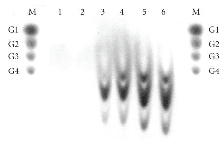
In order to establish the specificity of recombinant Amy1, the enzyme was incubated with starch, amylopectin, amylose, glycogen, and pullulan. These substrates consist of different glucose polymers containing α-1,4 linkages or a mixture of α-1,4- and α-1,6-glucosidic linkages. Amy1 showed the highest activity towards soluble starch (100%) amylopectin (97.9%), amylose (50.6%) but no activity was observed on pullulan. This result is similar to that observed for the Bacillus subtilis truncated α-amylase produced in E. coli [27], but different from that observed for wild-type Amy1 which showed the highest activity towards starch (100%), glycogen (18.9%), amylopectin (16.7%), and amylose (7.1%). The recombinant enzyme displayed typical α-amylase properties based on the evidence that oligosaccharides from several sizes were the main products from the digestion of glycogen, amylose, amylopectin, and starch (Figure 3).
Figure 3.
Thin-layer chromatogram of products released from starch after treatment with recombinant Amy1. The standards used were glucose (G1), maltose (G2), maltotriose (G3), and maltotetraose (G4). Lane 1, control (starch without enzyme); lane 2, pullulan; lane 3, glycogen; lane 4, amylose; lane 5, amylopectin, and lane 6, soluble starch.
3.3. Conformational Stability of Amy
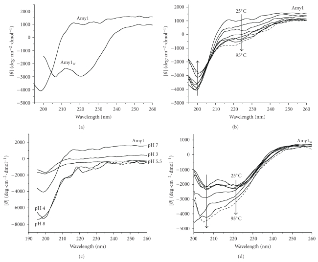
Structural characterization of recombinant (Amy1) and wild-type (Amyw) amylase from C. flavus was carried out by circular dichroism spectroscopy for comparison. Far-UV CD spectra of Amy1 and Amy1w at pH 7.0 and 25°C are quite different showing typical features of beta/unordered and beta/α-helix secondary structure pattern, respectively (Figure 4(a)). While the 222 nm dichroic band is predominantly associated with α-helical structure, the dichroic band at 198 and 206 nm may arise from changes in other secondary structure elements in the protein, as unordered or beta structure. The secondary structure content of both proteins slightly differs on helical (Amy1, 5.7% and Amy1w, 9.5%) and antiparallel β-sheet (Amy1, 42.1% and Amy1w, 32.4%) and is similar for other secondary structures.
Figure 4.
Far-UV circular dichroism analyses. Residual molar ellipticity [θ] was measured from 200 to 260 nm at 25°C or temperature arising from 25 to 95°C. (a) The distinct spectra of wild-type (Amy1w) and recombinant (Amy1) amylase are shown, indicating that they had different amount of secondary structure (see Section 3). (b) Effect of temperature on secondary structure of Amy1. Changes in molar ellipticity at 222 and 200 nm are shown as a function of temperature. (c) Effect of pH on secondary structure of Amy1. Note the slight differences of CD spectra at pH 7.0, 3.0, and 5.5 and similarity of CD spectra at pH 4.0 and 8.0. (d) Effect of temperature on secondary structure of Amy1w. Changes in molar ellipticity at 222 and 206 nm are shown as a function of temperature.
Thermal stability of the proteins at pH 7.0 was assessed upon raising the temperature from 20 to 95°C (Figures 4(b) and 4(d)). The thermal denaturation curves analyzed from Far-UV CD measurements at 206 and 222 nm at pH 7.0 strongly indicated the Amy1w and Amy1 as thermally stable proteins since no pattern of protein denaturation could be verified (data not shown). Indeed, although the CD spectra of the Amy1w in this condition show a gradual increase (downward) of the dichroic bands at 206 nm and slight at 222 nm (Figure 4(d)), no secondary structure changes were detected suggesting only conformational changes of protein or secondary structure rearrangement as a function of temperature. In contrast, CD spectra of the Amy1 show a gradual increase and decrease of the dichroic bands at 222 and 200 nm, respectively, suggesting the alteration of helical and β-sheet structures content (Figure 4(b)). A slight increase was observed in terms of α-helix (from 5.7% to 6.8%) and decrease of β-sheet (from 42.1% to 38.8%) when raising the temperature from 25 to 95°C. Altogether, these results revealed the high thermal stability of both proteins and the difference in folding pattern of Amy1 compared with wild-type amylase from C. flavus reflecting in decrease (10-fold) of enzymatic activity (Km values) of recombinant protein using a soluble starch as substrate, as discussed above.
Besides significant distinction in pH region where the maximum enzymatic activity of Amy1 occurred (Figure 2(a)), the protein achieved similar secondary structure content (helical 5.7%–6.5% and β-sheet ~42%) in a broad range of pH (pH 3.0, 5.5, and 7.0), as indicated by the CD spectra (Figure 4(c)) and CDNN estimative. At pH 8.0 as well as pH 4.0, it was verified the mainly increase in the CD signal at 200 nm (Figure 4(c)), compatible with minor β-sheet structures content (~37%) compared with pH 3.0, 5.5, and 7.0 (~42%). Noteworthy, although no secondary structure changes have been observed, analogous or different conformation of Amy1 side chains could occur as a function of those pHs, as judged by the relative activity of the enzyme in these condition (Figures 2(a) and 2(b)). In these cases, some important structural changes related with side chains ionization of aspartate, glutamate, and histidine residues at pH 4.0 and 5.5 could occur and are fundamental to enzyme activity.
In this work we have observed some significant biochemical and slight structural discrepancies between wild-type and recombinant Amy1 especially with respect to Km values, enzyme specificity, and helical and β-sheet structure content. These could be explained by small folding alterations in the structure of recombinant amylase during its expression. Different patterns of protein glycosylation between C. flavus and S. cerevisiae could also account for these differences. In fact, three putative N-glycosylation sites have been identified in Amy1 [8]. Work is under way to assess the role of glycosylation on the activity of Amy1. Because recombinant Amy1 showed considerable thermostability, its use in biotechnological processes should be considered.
4. Conclusion
In conclusion, the enzymatic and structural features of Amy1, the highest activity towards soluble starch, amylopectin, and amylase, in contrast with the lowest activity of Amy1w, points out this protein as being of paramount biotechnological importance with many applications ranging from food industry to the production of biofuels.
Acknowledgments
This work was supported by a biotechnology research grant to C. J. Ulhoa and F. A. G. Torres (CNPq, FINEP, and FUNAPE/UFG). A. S. Galdino was supported by CAPES/Brazil.
Acronyms
- SD:
Synthetic dextrose minimal media
- YNB:
Yeast nitrogen base
- REase:
Ribonuclease
- PAGE:
Polyacrilymide gel electrophoresis
- DTT:
Dithiothreitol
- CDNN:
Neural network circular dichroism.
References
- 1.Van Der Maarel MJEC, Van Der Veen B, Uitdehaag JCM, Leemhuis H, Dijkhuizen L. Properties and applications of starch-converting enzymes of the α-amylase family. Journal of Biotechnology. 2002;94(2):137–155. doi: 10.1016/s0168-1656(01)00407-2. [DOI] [PubMed] [Google Scholar]
- 2.Henrissat B. A classification of glycosyl hydrolases based on amino acid sequence similarities. Biochemical Journal. 1991;280(2):309–316. doi: 10.1042/bj2800309. [DOI] [PMC free article] [PubMed] [Google Scholar]
- 3.Henrissat B, Bairoch A. Updating the sequence-based classification of glycosyl hydrolases. Biochemical Journal. 1996;316(2):695–696. doi: 10.1042/bj3160695. [DOI] [PMC free article] [PubMed] [Google Scholar]
- 4.Gupta R, Gigras P, Mohapatra H, Goswami VK, Chauhan B. Microbial α-amylases: a biotechnological perspective. Process Biochemistry. 2003;38(11):1599–1616. [Google Scholar]
- 5.Wilson JJ, Ingledew WM. Isolation and characterization of Schwanniomyces alluvius amylolytic enzymes. Applied and Environmental Microbiology. 1982;44(2):301–307. doi: 10.1128/aem.44.2.301-307.1982. [DOI] [PMC free article] [PubMed] [Google Scholar]
- 6.De Mot R, Verachtert H. Secretion of α-amylase and multiple forms of glucoamylase by the yeast Trichosporon pullulans. Canadian Journal of Microbiology. 1986;32(1):47–51. doi: 10.1139/m86-009. [DOI] [PubMed] [Google Scholar]
- 7.Wanderley KJ, Torres FAG, Moraes LMP, Ulhoa CJ. Biochemical characterization of α-amylase from the yeast Cryptococcus flavus. FEMS Microbiology Letters. 2004;231(2):165–169. doi: 10.1016/S0378-1097(03)00955-8. [DOI] [PubMed] [Google Scholar]
- 8.Galdino AS, Ulhoa CJ, Moraes LMP, Prates MV, Bloch C, Torres FAG. Cloning, molecular characterization and heterologous expression of AMY1, an α-amylase gene from Cryptococcus flavus. FEMS Microbiology Letters. 2008;280(2):189–194. doi: 10.1111/j.1574-6968.2007.01059.x. [DOI] [PubMed] [Google Scholar]
- 9.Bradford MM. A rapid and sensitive method for the quantitation of microgram quantities of protein utilizing the principle of protein dye binding. Analytical Biochemistry. 1976;72(1-2):248–254. doi: 10.1016/0003-2697(76)90527-3. [DOI] [PubMed] [Google Scholar]
- 10.Laemmli UK. Cleavage of structural proteins during the assembly of the head of bacteriophage T4. Nature. 1970;227(5259):680–685. doi: 10.1038/227680a0. [DOI] [PubMed] [Google Scholar]
- 11.Blum H, Bier H, Gross H. Improved silver staining of plants proteins, RNA and DNA in polyacrylamide gels. Electrophoresis. 1987;8:93–99. [Google Scholar]
- 12.Fuwa H. A new method for microdetermination of amylase activity by the use of amylose as the substrate. Journal of Biochemistry. 1954;41(5):583–603. [Google Scholar]
- 13.Miller GL. Use of dinitrosalicylic acid reagent for determination of reducing sugar. Analytical Chemistry. 1959;31(3):426–428. [Google Scholar]
- 14.Lato M, Brunelli B, Ciuffini G, Mezzetti T. Thin-layer chromatography of carbohydrates on silica gel impregnated with sodium acetate, monosodium phosphate and disodium phosphate. Journal of Chromatography A. 1969;39:407–417. doi: 10.1016/s0021-9673(01)98029-x. [DOI] [PubMed] [Google Scholar]
- 15.Bohm G, Muhr R, Jaenicke R. Quantitative analysis of protein far UV circular dichroism spectra by neural networks. Protein Engineering. 1992;5(3):191–195. doi: 10.1093/protein/5.3.191. [DOI] [PubMed] [Google Scholar]
- 16.Iefuji H, Chino M, Kato M, Iimura Y. Raw-starch-digesting and thermostable α-amylase from the yeast Cryptococcus sp. S-2: purification, characterization, cloning and sequencing. Biochemical Journal. 1996;318(3):989–996. doi: 10.1042/bj3180989. [DOI] [PMC free article] [PubMed] [Google Scholar]
- 17.Goto CE, Barbosa EP, Kistner LCL, Moreira FG, Lenartovicz V, Peralta RM. Production of amylase by Aspergillus fumigatus utilizing α-methyl-D-glycoside, a synthetic analogue of maltose, as substrate. FEMS Microbiology Letters. 1998;167(2):139–143. doi: 10.1111/j.1574-6968.1998.tb13219.x. [DOI] [PubMed] [Google Scholar]
- 18.Kang HK, Lee JH, Kim D, et al. Cloning and expression of Lipomyces starkeyiα-amylase in Escherichia coli and determination of some of its properties. FEMS Microbiology Letters. 2004;233(1):53–64. doi: 10.1016/j.femsle.2004.01.036. [DOI] [PubMed] [Google Scholar]
- 19.Steyn AJC, Marmur J, Pretorius IS. Cloning, mapping and characterization of a genomic copy of the Lipomyces kononenkoaeα-amylase-encoding gene (LKA1) Yeast. 1996;12(10):925–937. doi: 10.1002/(SICI)1097-0061(199608)12:10%3C925::AID-YEA987%3E3.0.CO;2-0. [DOI] [PubMed] [Google Scholar]
- 20.Li H, Chi Z, Wang X, Duan X, Ma L, Gao L. Purification and characterization of extracellular amylase from the marine yeast Aureobasidium pullulans N13d and its raw potato starch digestion. Enzyme and Microbial Technology. 2007;40(5):1006–1012. [Google Scholar]
- 21.Prieto JA, Bort BR, Martínez J, Randez-Gil F, Buesa C, Sanz P. Purification and characterization of a new alpha-amylase of intermediate thermal stability from the yeast Lipomyces kononenkoae. Biochemistry and Cell Biology. 1995;73(1-2):41–49. doi: 10.1139/o95-005. [DOI] [PubMed] [Google Scholar]
- 22.Takeuchi A, Shimizu-Ibuka A, Nishiyama Y, et al. Purification and characterization of an α-amylase of Pichia burtonii isolated from the traditional starter "murcha" in Nepal. Bioscience, Biotechnology and Biochemistry. 2006;70(12):3019–3024. doi: 10.1271/bbb.60430. [DOI] [PubMed] [Google Scholar]
- 23.Murakami S, Nishimoto H, Toyama Y, et al. Purification and characterization of two alkaline, thermotolerant α-amylases from Bacillus halodurans 38C-2-1 and expression of the cloned gene in Escherichia coli. Bioscience, Biotechnology and Biochemistry. 2007;71(10):2393–2401. doi: 10.1271/bbb.60666. [DOI] [PubMed] [Google Scholar]
- 24.Moranelli F, Yaguchi M, Calleja GB, Nasim A. Purification and characterization of the extracellular alpha-amylase activity of the yeast Schwanniomyces alluvius. Biochemistry and Cell Biology. 1987;65(10):899–908. doi: 10.1139/o87-116. [DOI] [PubMed] [Google Scholar]
- 25.Ballschmiter M, Fütterer O, Liebl W. Identification and characterization of a novel intracellular alkaline α-amylase from the hyperthermophilic bacterium Thermotoga maritima MSB8. Applied and Environmental Microbiology. 2006;72(3):2206–2211. doi: 10.1128/AEM.72.3.2206-2211.2006. [DOI] [PMC free article] [PubMed] [Google Scholar]
- 26.Glymph JL, Stutzenberger FJ. Production, purification, and characterization of α-amylase from Thermomonospora curvata. Applied and Environmental Microbiology. 1977;34(4):391–397. doi: 10.1128/aem.34.4.391-397.1977. [DOI] [PMC free article] [PubMed] [Google Scholar]
- 27.Marco JL, Bataus LA, Valencia FF, Ulhoa CJ, Astolfi-Filho S, Felix CR. Purification and characterization of a truncated Bacillus subtilisα-amylase produced by Escherichia coli. Applied Microbiology and Biotechnology. 1996;44(6):746–752. doi: 10.1007/BF00178613. [DOI] [PubMed] [Google Scholar]


