Abstract
The prognosis of untreated advanced hepatocellular carcinoma (HCC) is grim with a median survival of less than 6 months. Spontaneous regression of HCC has been defined as the disappearance of the hepatic lesions in the absence of any specific therapy. The spontaneous regression of a very large HCC is very rare and limited data is available in the English literature. We describe spontaneous regression of hepatocellular carcinoma in a 65-year-old male who presented to our clinic with vague abdominal pain and weight loss of two months duration. He was found to have multiple hepatic lesions with elevation of serum alpha-fetoprotein (AFP) level to 6,500 µg/L (normal <20 µg/L). Computed tomography revealed advanced HCC replacing almost 80% of the right hepatic lobe. Without any intervention the patient showed gradual improvement over a period of few months. Follow-up CT scan revealed disappearance of hepatic lesions with progressive decline of AFP levels to normal. Various mechanisms have been postulated to explain this rare phenomenon, but the exact mechanism remains a mystery.
Abstract
Die Prognose eines unbehandelten, fortgeschrittenen hepatozellulären Carcinoms (HCC) ist gewöhnlich ungünstig mit einer mittleren Überlebensrate von weniger als 6 Monaten. Die spontane Rückbildung eines HCC ist definiert als Verschwinden von Leberschäden ohne eine spezifische Therapie. Die spontane Rückbildung eines sehr großen HCC ist sehr selten, und es gibt nur begrenzt Daten dazu in der englischen Forschungsliteratur. Wir beschreiben die spontane Rückbildung eines HCC bei einem 65-jährigen Mann, der sich in unserer Klinik mit vagen Bauchschmerzen und einem seit 2 Monaten andauernden Gewichtsverlust vorstellte. Man fand bei ihm multiple Leberschäden mit einem Anstieg der Alpha-Fetoprotein-Konzentration im Serum auf 6.500 µg/L (normal <20 μg/L). Durch Computertomographie (CT) des Abdomens wurde ein HCC in fortgeschrittenem Stadium diagnostiziert, das fast 80% des rechten Leberlappens einnahm. Ohne jeden Eingriff zeigte der Patient eine allmähliche Besserung über einen Zeitraum von mehreren Monaten. Ein Follw-up-CT ließ ein vollständiges Verschwinden der Leberschäden erkennen, das mit einem fortschreitenden Absinken der AFP-Werte bis auf Normwerte einherging. Verschiedene Wirkungsmechanismen wurden für dieses seltene Phänomen vorgeschlagen, aber der genaue Mechanismus bleibt unklar.
Case presentation
A 65-year-old male presented with one-week history of right upper quadrant abdominal pain preceded by anorexia and weight loss of twelve-pound over a period of two months. He denied any history of jaundice, nausea, vomiting, abnormal bowel habits or gastrointestinal bleeding. The patient had a history of hypertension, diabetes, hypercholesterolemia and obesity. There was no history of alcohol abuse or other risk factors for chronic liver disease.
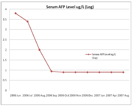
Physical examination revealed an alert, oriented patient with blood pressure 120/60 mmHg, and absence of lymphadenopathy, scleral icterus, and lower extremities showed no edema. There were no stigmata of chronic liver disease. Abdominal examination showed no organomegaly, ascitis or bruits. His cardiac, respiratory and neurological examinations were unremarkable. Initial laboratory studies showed normal hemogram. He had elevated liver enzymes with alanine aminotransferase (ALT) and aspartate aminotransferase (AST) twice the upper limits of normal, and serum bilirubin and international normalized ratio for prothrombin time were normal. Serologic tests for hepatitis B, hepatitis C, anti-mitochondrial antibody and anti-smooth muscle antibody were negative, while anti-nuclear antibody was weakly positive. Serum alpha1-antitrypsin level was normal. Serum alpha-fetoprotein (AFP) was 6,500 µg/L (normal <20 µg/L) at initial presentation and showed a steady decline on follow-up to 2,700 µg/L at 4 weeks after presentation, 8.8 µg/L 3–6 months later, and thereafter remained below 8 µg/L (Figure 1 (Fig. 1)).
Figure 1. A marked decline in serum AFP levels over time (normal <20 µg/L).
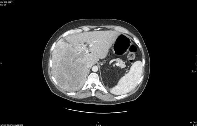
A contrast-enhanced CT scan of the abdomen at presentation showed a large heterogeneous dense mass with enhancement involving the right hepatic lobe (Figure 2 (Fig. 2)), and at least one small lesion was seen in the medial segment of the left hepatic lobe. An occlusive thrombus in the right portal vein extending into the main portal vein was noted, and a number of enlarged paraortic lymph nodes were also present. The patient was seen in consultation by the Oncology Service, and no treatment was offered. Over a period of few months his symptoms improved and the tumor showed radiological evidence of spontaneous involution coupled with a decrease in AFP levels meeting the criteria for a spontaneous resolution. A follow-up triphasic CT of the abdomen 14 weeks later revealed significant interval reduction in the size of the mass with associated atrophy of the right hepatic lobe. Persistent occlusion of the right portal vein was still present, but the main portal vein thrombus and the periaortic lymphadenopathy had resolved.
Figure 2. Axial image from a contrast-enhanced CT shows a large heterogeneous hypodense right hepatic lobe mass with thrombus extending from the right portal vein into the main portal vein (arrow).
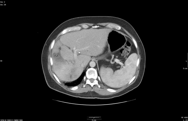
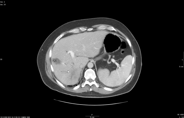
Repeat abdominal CT scan 28 weeks from the time of initial presentation showed persistent right portal vein occlusion and a small (1.2 x 2.8 cm) hypodensity in the posterior segment of the right hepatic lobe (Figure 3 (Fig. 3)). An abdominal CT almost 14 months from initial presentation showed a small irregular hypodensity in the posterior segment of the right hepatic lobe with no area of abnormality anywhere else (Figure 4 (Fig. 4)). An ultrasound 2 years after initial presentation did not show any liver lesions and the AFP level remained persistently below 10 µg/L, which provided confirmation of complete resolution of the lesion.
Figure 3. An ill-defined right hepatic lobe mass has markedly decreased in size with resolution of the main portal vein thrombus (arrow). GB = gallbladder.
Figure 4. A small irregular hypodensity in the posterior segment of the right hepatic lobe remains (arrow). GB = gallbladder.
Discussion
Advanced HCC has limited treatment options and patients succumb within few months to this disease. Spontaneous regression of HCC is a rare event, with an incidence rate of one in 140,000 cases of HCC [1]. Interestingly, there have been few case reports of spontaneous (complete) regression of HCC [2]. Nearly 62 case reports of spontaneous regression of HCC have been published from 1982 to January 2007 [2]. Most of the cases showed partial regression, while a few cases demonstrated complete regression, as in the index case. There does not appear to be a correlation between the underlying etiology of liver disease and spontaneous resolution of HCC. The index case had no cirrhosis and laboratory findings, radiological studies did not show evidence of chronic hepatitis or cirrhosis. Although several factors, such as reduction in blood supply, inflammation, and immunological factors, have been suggested as the mechanism of spontaneous regression of HCC, the precise mechanism is not understood [2], [3]. In one report, treatment by activated T-lymphocytes has been shown to reduce recurrence rates of HCC after surgery [4]. This finding suggests that related regression may be associated with host immune response, involving cytokines such as tumor necrosis factor-alpha [5]. Several other factors have been proposed to cause spontaneous regression, such as abstinence from alcohol, consumption of herbal medicines, high fevers, gastrointestinal bleeding, rapid tumor growth and the use of vitamin K [6].
Spontaneous resolution of HCC with distant metastasis to the lungs [7], chest wall [8], skull [9], peritoneum and spleen [10] has been reported. A case similar to ours was reported with a large right liver lobe and portal vein thrombosis, followed by subsequent tumor regression [11]. Tumor regression of different sizes has been described in the literature and outcome in most of the cases has been favorable with few recurrences. In cases in which surgical resections were implicated, survival was longer. It is quite possible that arterioportal shunt near the tumor may change the dynamics of blood flow to the tumor as blood supply is essential for tumor growth. Similar to transarterial chemoembolization, the tumor infarction due to disruption of the feeding artery or portal vein caused by subintimal injury, thrombus, and tumor invasion could result in tumor necrosis and regression of HCC. Another explanation considered is coagulative necrosis produced by interruption of the blood supply due to the spontaneous formation of an arterial thrombus [12], [13], [14]. In the index case considering the acute onset of symptoms, we speculated that local ischemia due to rapid tumor growth resulted in intra tumoral bleeding and/or hemorrhagic necrosis. Such an explanation was proposed for some cases in the literature which showed histologically complete necrosis in the resected mass.
We conclude that complete spontaneous resolution of HCC, though rare, provides an avenue for further research. The accumulation and careful analysis of clinical data obtained from patients with spontaneous resolution of HCC will contribute to the understanding of this enigmatic phenomenon, and potentially to the future development of new treatment strategies for HCC.
Notes
Competing interests
The authors declare that they have no competing interests.
References
- 1.Chang WY. Complete spontaneous regression of cancer: four case reports, review of literature, and discussion of possible mechanisms involved. Hawaii Med J. 2000;59(10):379–387. [PubMed] [Google Scholar]
- 2.Meza-Junco J, Montaño-Loza AJ, Martinez-Benítez B, Cabrera-Aleksandrova T. Spontaneous partial regression of hepatocellular carcinoma in a cirrhotic patient. Ann Hepatol. 2007;6(1):66–69. [PubMed] [Google Scholar]
- 3.Blondon H, Fritsch L, Cherqui D. Two cases of spontaneous regression of multicentric hepatocellular carcinoma after intraperitoneal rupture: possible role of immune mechanisms. Eur J Gastroenterol Hepatol. 2004;16(12):1355–1359. doi: 10.1097/00042737-200412000-00020. Available from: http://dx.doi.org/10.1097/00042737-200412000-00020. [DOI] [PubMed] [Google Scholar]
- 4.Anthony PP. Hepatocellular carcinoma: an overview. Histopathology. 2001;39(2):109–118. doi: 10.1046/j.1365-2559.2001.01188.x. Available from: http://dx.doi.org/10.1046/j.1365-2559.2001.01188.x. [DOI] [PubMed] [Google Scholar]
- 5.Ohba K, Omagari K, Nakamura T, Ikuno N, Saeki S, Matsuo I, Kinoshita H, Masuda J, Hazama H, Sakamoto I, Kohno S. Abscopal regression of hepatocellular carcinoma after radiotherapy for bone metastasis. Gut. 1998;43(4):575–577. doi: 10.1136/gut.43.4.575. Available from: http://dx.doi.org/10.1136/gut.43.4.575. [DOI] [PMC free article] [PubMed] [Google Scholar]
- 6.Nouso K, Uematsu S, Shiraga K, Okamoto R, Harada R, Takayama S, Kawai W, Kimura S, Ueki T, Okano N, Nakagawa M, Mizuno M, Araki Y, Shiratori Y. Regression of hepatocellular carcinoma during vitamin K administration. World J Gastroenterol. 2005;11(42):6722–6724. doi: 10.3748/wjg.v11.i42.6722. [DOI] [PMC free article] [PubMed] [Google Scholar]
- 7.Toyoda H, Sugimura S, Fukuda K, Mabuchi T. Hepatocellular carcinoma with spontaneous regression of multiple lung metastases. Pathol Int. 1999;49(10):893–897. doi: 10.1046/j.1440-1827.1999.00956.x. Available from: http://dx.doi.org/10.1046/j.1440-1827.1999.00956.x. [DOI] [PubMed] [Google Scholar]
- 8.Jeon SW, Lee MK, Lee YD, Seo HE, Cho CM, Tak WY, Kweon YO. Clear cell hepatocellular carcinoma with spontaneous regression of primary and metastatic lesions. Korean J Intern Med. 2005;20(3):268–273. doi: 10.3904/kjim.2005.20.3.268. [DOI] [PMC free article] [PubMed] [Google Scholar]
- 9.Nam SW, Han JY, Kim JI, Park SH, Cho SH, Han NI, Yang JM, Kim JK, Choi SW, Lee YS, Chung KW, Sun HS. Spontaneous regression of a large hepatocellular carcinoma with skull metastasis. J Gastroenterol Hepatol. 2005;20(3):488–492. doi: 10.1111/j.1440-1746.2005.03243.x. Available from: http://dx.doi.org/10.1111/j.1440-1746.2005.03243.x. [DOI] [PubMed] [Google Scholar]
- 10.Terasaki T, Hanazaki K, Shiohara E, Matsunaga Y, Koide N, Amano J. Complete disappearance of recurrent hepatocellular carcinoma with peritoneal dissemination and splenic metastasis: a unique clinical course after surgery. J Gastroenterol Hepatol. 2000;15(3):327–330. doi: 10.1046/j.1440-1746.2000.02092.x. Available from: http://dx.doi.org/10.1046/j.1440-1746.2000.02092.x. [DOI] [PubMed] [Google Scholar]
- 11.Iiai T, Sato Y, Nabatame N, Yamamoto S, Makino S, Hatakeyama K. Spontaneous complete regression of hepatocellular carcinoma with portal vein tumor thrombus. Hepatogastroenterology. 2003;50(53):1628–1630. [PubMed] [Google Scholar]
- 12.Yano Y, Yamashita F, Kuwaki K, Fukumori K, Kato O, Kiyomatsu K, Sakai T, Yamamoto H, Yamasaki F, Ando E, Sata M. Partial spontaneous regression of hepatocellular carcinoma: a case with high concentrations of serum lens culinaris agglutinin-reactive alpha fetoprotein. Kurume Med J. 2005;52(3):97–103. doi: 10.2739/kurumemedj.52.97. Available from: http://dx.doi.org/10.2739/kurumemedj.52.97. [DOI] [PubMed] [Google Scholar]
- 13.Ohtani H, Yamazaki O, Matsuyama M, Horii K, Shimizu S, Oka H, Nebiki H, Kioka K, Kurai O, Kawasaki Y, Manabe T, Murata K, Matsuo R, Inoue T. Spontaneous regression of hepatocellular carcinoma: report of a case. Surg Today. 2005;35(12):1081–1086. doi: 10.1007/s00595-005-3066-8. Available from: http://dx.doi.org/10.1007/s00595-005-3066-8. [DOI] [PubMed] [Google Scholar]
- 14.Imaoka S, Sasaki Y, Masutani S, Ishikawa O, Furukawa H, Kabuto T, Kameyama M, Ishiguro S, Hasegawa Y, Koyama H, et al. Necrosis of hepatocellular carcinoma caused by spontaneously arising arterial thrombus. Hepatogastroenterology. 1994;41(4):359–362. [PubMed] [Google Scholar]


