Abstract
Context:
Valid and reliable measurements of ankle-complex motion have been reported using the Hollis Ankle Arthrometer. No published normative data of ankle-complex motion obtained from ankle arthrometry are available for use as a reference for clinical decision making.
Objective:
To describe the distribution variables of ankle-complex motion in uninjured ankles and to establish normative reference values for use in research and to assist in clinical decision making.
Design:
Descriptive laboratory study.
Setting:
University research laboratory.
Patients or Other Participants:
Both ankles of 50 men and 50 women (age = 21.78 ± 2.0 years [range, 19–25 years]) were tested.
Intervention(s):
Each ankle underwent anteroposterior (AP) and inversion-eversion (I-E) loading using an ankle arthrometer.
Main Outcome Measure(s):
Recorded anterior, posterior, and total AP displacement (millimeters) at 125 N and inversion, eversion, and total I-E rotation (degrees) at 4 Nm.
Results:
Women had greater ankle-complex motion for all variables except for posterior displacement. Total AP displacement of the ankle complex was 18.79 ± 4.1 mm for women and 16.70 ± 4.8 mm for men (U = 3742.5, P < .01). Total I-E rotation of the ankle complex was 42.10° ± 9.0° for women and 34.13° ± 10.1° for men (U = 2807, P < .001). All variables were normally distributed except for anterior displacement, inversion rotation, eversion rotation, and total I-E rotation in the women's ankles and eversion rotation in the men's ankles; these variables were skewed positively.
Conclusions:
Our study increases the available database on ankle-complex motion, and it forms the basis of norm-referenced clinical comparisons and the basis on which quantitative definitions of ankle pathologic conditions can be developed.
Keywords: normal distribution, flexibility
Key Points
This study increases the available database on ankle-complex motion and forms the basis of norm-referenced clinical comparisons.
Women had greater ankle range of motion than men, and all of the range-of-motion variables measured were normally distributed except for anterior displacement, inversion rotation, eversion rotation, and total inversion-eversion rotation, which showed a higher incidence toward hypermobility.
Our findings are clinically important because they will assist in the clinical decision-making process, enabling comparisons to be made with individual patient data and enabling quantitative definitions of ankle conditions to be developed.
Instrumented ankle arthrometry allows the examiner to quantify ligamentous laxity in lieu of manual examination.1–3 Valid and reliable measurements of the combined motions within the talocrural and subtalar joints (ankle complex) have been investigated fully and reported using the Hollis Ankle Arthrometer (Blue Bay Research, Inc, Navarre, FL).3–6 Consisting of a 6-degrees-of-freedom spatial kinematic linkage, this device is described as a suitable evaluation tool that quantifies the anteroposterior (AP) load displacement and inversion-eversion (I-E) rotational characteristics of the ankle complex.3,4,7
The Hollis Ankle Arthrometer has been used in a variety of clinical and research settings involving college-aged athletes and participants less than 25 years of age. Researchers have applied this type of arthrometric assessment in studies to biomechanically assess ankle-complex laxity in vivo and in vitro,3,4,8 identify ankle instability after injury,9–13 investigate the effects of sex and athletic status on ankle-complex laxity,14 identify the relationship between ankle and knee ligamentous laxity and generalized joint laxity,15 investigate the effects of balance training on gait in patients with chronic ankle instability,13 investigate the effects of limb dominance on ankle laxity,4 and assess the effectiveness of ankle taping.16
One limitation of using the Hollis Ankle Arthrometer and of using instrumented ankle arthrometry in general is that relatively small sample sizes have been reported and no normative data are available for comparison and reference.9–14 Kovaleski et al4 investigated total AP displacement and I-E rotation between the dominant and nondominant ankles in a group of 41 male and female participants (age = 23.8 ± 4.4 years). Bilateral ankle comparisons showed no differences in ankle-complex laxity, and they reported mean total AP displacement of 18.47 ± 5.1 mm for the dominant ankle and 17.51 ± 5.4 mm for the nondominant ankle. They also reported mean total I-E rotation of 46.19° ± 12.2° for the dominant ankle and 47.38° ± 14.3° for the nondominant ankle. The relatively large SDs indicated sizable variations in AP displacement and I-E rotation measurements in the uninjured ankle. To establish normative data for ankle-complex motion, adequate sample size is important to describe the resulting distribution and to ensure confidence that the theoretical distribution fitted to the data has minimal error associated with it.
Given the importance of having normative values against which clinical findings can be compared, the purpose of our study was to describe the distribution variables of ankle-complex motion in uninjured ankles and to establish normative reference values for use in research and to assist in clinical decision making.
METHODS
Participants
Participants included 50 men (age = 21.9 ± 2.1 years, height = 178.2 ± 7.4 cm, mass = 86.9 ± 21.1 kg) and 50 women (age = 21.7 ± 2.0 years, height = 165.1 ± 7.9 cm, mass = 65.7 ± 11.1 kg) from 19 to 25 years of age (21.78 ± 2.0 years). Ninety-three participants were right-leg dominant, and 7 were left-leg dominant. The dominant leg was defined operationally as the leg used to kick a ball. None of the participants had a history of lower extremity injury, including ankle sprain. Before testing, all participants provided written informed consent, and the university's institutional review board approved the study.
Participants completed the Foot and Ankle Outcome Score (FAOS) questionnaire to gauge self-reported ankle function.17 The FAOS is a subjective self-report of ankle function in daily activities, sports, and recreation that is divided into 5 subscales. A normalized score (100 indicating no problems and 0 indicating extreme problems) was calculated for each subscale. The results of the FAOS survey showed the FAOS subscale mean scores ranged from 95.2 ± 10.5 to 99.1 ± 3.0, which implied that the ankles included in our study were free of problems associated with ankle dysfunction.
Instrumentation
Instrumented measurement of ankle-complex motion was conducted using the Hollis Ankle Arthrometer.7 The arthrometer consists of a spatial kinematic linkage, an adjustable plate fixed to the foot, a load-measuring handle attached to the footplate through which the load is applied, and a reference pad attached to the tibia.3,4 Ankle arthrometry is a method for assessing either translatory displacement or angular motion of the foot in relation to the leg that results from the combined motions within the talocrural and subtalar joints. The spatial kinematic linkage is a 6-degrees-of-freedom electrogoniometer that measures applied forces and moments and the resultant translations and rotations of the ankle complex.2,7 The arthrometer spatial linkage connected the tibial pad to the footplate and measured the motion of the footplate relative to the tibial pad. Ankle-flexion angle was measured from the plantar surface of the foot relative to the anterior tibia and was determined by the 6-degrees-of-freedom electrogoniometer within the instrumented linkage. An Inspiron 1525 computer (Dell Inc, Round Rock, TX) with an analog-to-digital converter (National Instruments Corp, Austin, TX) was used to simultaneously record and calculate the data. The resulting AP displacement (millimeters) and I-E rotation (degrees of range of motion) along with the corresponding AP load and I-E torque were recorded. We used a custom software program written in LabVIEW (National Instruments) for collection and reduction of the data.
Procedures
Testing and participant positioning replicated previously reported methods.4,5,9 Individuals participated in 1 testing session and both ankles underwent 3 trials each of AP and I-E loading. To minimize variation, the arthrometer was positioned on all participants in a similar manner for all tests, and the same examiner (N.A.S.) performed all tests.
Each participant was positioned supine on a firm table with the knee in 10° to 20° of knee flexion and the foot extended over the edge of the table. A restraining strap attached to support bars under the table was secured around the distal lower leg approximately 1 cm above the malleoli and then tightened to prevent lower leg movement during testing. The examiner placed the bottom of the foot onto the footplate and secured the foot using heel and dorsal clamps. The heel clamp prevented the device from rotating on the calcaneus, and the dorsal clamp secured the foot to the footplate. The tibial reference pad then was positioned approximately 5 cm above the malleoli and secured to the lower leg with an elastic strap.
The ankle was positioned at zero AP load and zero I-E moment at a neutral (0°) flexion angle, which was defined as the measurement reference position.2,4 The other degrees of freedom (internal-external, medial-lateral, and proximal-distal) also were maintained at their zero-load neutral position. Thus, the measurement reference position represented zero moment and force loads. This angle was measured from the plantar surface of the foot relative to the anterior tibia and determined by the 6-degrees-of-freedom electrogoniometer within the instrumented linkage. Anteroposterior loading, I-E torque, and the flexion angle were applied through the load handle in line with the footplate. Each trial involved reciprocal movements from the zero load to the maximum load. For the AP trial, the ankles were loaded to ±125 N with both anterior and posterior forces. Starting at the reference position, anterior loading was applied first; posterior loading, second. Total AP displacement of the ankle complex (millimeters) was recorded along with the loads. Anterior motion was defined as the displacement produced in response to the load changing from 0 to 125 N. Posterior motion was defined as the displacement produced in response to a load changing from 0 to −125 N. Total AP displacement was defined as the change produced in response to a load varying from −125 to 125 N. For I-E rotation, the ankles were loaded to ±4 Nm with both inversion and eversion torque. Starting at the neutral reference position, inversion loading was applied first; eversion loading, second. Rotation of the ankle complex was recorded along with the torque. Inversion rotation was defined as the angular displacement produced in response to a torque changing from 0 to 4 Nm. Eversion rotation was defined as the angular displacement produced in response to a torque changing from 0 to −4 Nm. Total I-E rotation was defined as the angular-displacement change produced in response to a torque varying from −4 to 4 Nm. By observing the computer monitor, the examiner visualized the applied load to obtain maximum AP displacement and I-E rotation.
Test order was assigned randomly between right and left ankles. After the ankle measurements were obtained, the device was removed, and the testing procedure was repeated on the contralateral ankle.
Statistical Analysis
Anterior, posterior, and total AP displacement at 125 N and inversion, eversion, and total I-E rotation at 4 Nm were used as outcome measures. Descriptive data for all variables were expressed as the mean (±SD) score, median score, SE score, range of scores, and 95% confidence interval. The dominant and nondominant ankle-complex motion data from each of the 100 participants were first tested for normality. Normality of distribution was investigated using the Kolmogorov-Smirnov test with Lilliefors correction. If a finding of the Kolmogorov-Smirnov test was significant, normality of distribution was further investigated for that finding by examining the z scores for skewness and kurtosis. If the z scores for skewness and kurtosis of the variable were from +2.00 to −2.00, the finding was considered to be normally distributed. If the z score for skewness of a finding was outside the +2.00 to −2.00 range, the finding was described as positively or negatively skewed. If the z score for kurtosis of a finding was outside the +2.00 to −2.00 range, the finding was described as leptokurtic or platykurtic.
Limb-dominance data determined to be normally distributed were analyzed using the paired-samples t test to identify any side-related difference between observations. Data determined not to be normally distributed were analyzed using the Wilcoxon test. Sex data determined to be normally distributed were analyzed using the independent-samples t test to examine differences between men and women. Data determined not to be normally distributed were analyzed using the Mann-Whitney U test. Effect size was determined using the Cohen d.18 The α level was set a priori at .05. All statistical analyses were performed with SPSS software (version 17.0; SPSS Inc, Chicago, IL).
RESULTS
Limb Dominance
The findings of the Kolmogorov-Smirnov test were significant for total AP displacement of the dominant ankle (D = 0.096, P = .02), total AP displacement of the nondominant ankle (D = 0.093, P = .03), and inversion rotation of the dominant ankle (D = 0.093, P = .03). Examination of the z scores for skewness and kurtosis showed normal distribution for total AP displacement of the dominant ankle (skewness = 0.33, SE = 0.24; kurtosis = −0.57, SE = 0.48) and total AP displacement of the nondominant ankle (skewness = 0.14, SE = 0.24; kurtosis = −0.40, SE = 0.48). The z scores for inversion rotation of the dominant ankle (skewness = 0.68, SE = 0.24; kurtosis = 0.43, SE = 0.48) showed that this variable was positively skewed. Results indicated that all variables were normally distributed except for inversion rotation of the dominant ankle.
Bilateral comparisons revealed greater dominant than nondominant ankle-complex motion for total AP displacement (dominant = 19.23 ± 4.37 mm, nondominant = 16.26 ± 4.28 mm; t99 = 6.43, P < .01, Cohen d = .68), posterior displacement (dominant = 9.64 ± 2.50 mm, nondominant = 7.30 ± 2.43 mm; t99 = 6.78, P < .01, Cohen d = .95), and eversion rotation (dominant = 15.64° ± 4.92°, nondominant = 14.57° ± 4.83°; t99 = 3.44, P < .01, Cohen d = .22). Greater inversion rotation was found for nondominant than for dominant ankle-complex motion (dominant = 22.16° ± 6.75°, nondominant = 23.85° ± 6.60°; Z = −3.5, P = .01, Cohen d = .25).
No differences were found between the dominant and nondominant ankles for anterior displacement (dominant = 9.58 ± 2.99 mm, nondominant = 8.96 ± 3.15 mm; t99 = 1.9, P = .06, Cohen d = .20) and total I-E rotation (dominant = 37.80° ± 10.44°, nondominant = 38.44° ± 10.26°; t99 = −1.22, P < .22, Cohen d = .06). Small effect sizes were found for 4 of the 6 variables and were not considered clinically important; thus, the data from both ankles were pooled for all subsequent analyses.
Sex Differences
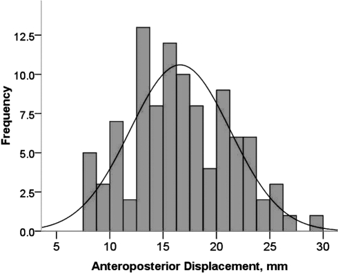
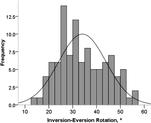
Ankle-complex motion values in the male population of 100 ankles are shown in Table 1. The findings of the Kolmogorov-Smirnov test were significant for total I-E (D = 0.097, P = .02) and eversion (D = 0.145, P = .01) rotation. Examination of the z scores showed normal distribution for total I-E rotation (skewness = 0.28, SE = 0.24; kurtosis = −0.78, SE = 0.48) and positive skewness for eversion rotation (skewness = 0.64, SE = 0.24; kurtosis = −0.30, SE = 0.48). Examination of frequency distributions for total AP displacement and total I-E rotation and their corresponding histograms graphically showed the distribution around the mean (Figures 1 and 2).
Table 1.
Descriptive Normative Data for Measurements of Ankle-Complex Motion in Men (N = 100 Ankles)
Figure 1.
Range of frequency distribution for anteroposterior displacement of the men's ankles (N = 100 ankles). The mean was 16.70 ± 4.8 mm.
Figure 2.
Range of frequency distribution for inversion-eversion rotation of the men's ankles (N = 100 ankles). The mean was 34.13° ± 10.1°.
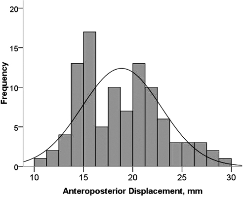
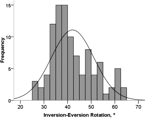
Ankle-complex motion values in the female population of 100 ankles are displayed in Table 2. The findings of the Kolmogorov-Smirnov test were significant for all the variables except posterior displacement. Examination of z scores showed normal distribution for total AP displacement (D = 0.113, P = .01; skewness = 0.47, SE = 0.24; kurtosis = −0.36, SE = 0.48); positive skewness for anterior displacement (D = 0.090, P = .04; skewness = 0.75, SE = 0.24; kurtosis = 0.85, SE = 0.48), inversion (D = 0.120, P = .01; skewness = 0.67, SE = 0.24; kurtosis = −0.05, SE = 0.48), and total I-E rotation (D = 0.121, P = .01; skewness = 0.64, SE = 0.24; kurtosis = −0.29, SE = 0.48); and positive skewness and leptokurtosis for eversion rotation (D = 0.109, P = .01; skewness = 1.07, SE = 0.24; kurtosis = 1.47, SE = 0.48). Examination of frequency distributions for total AP displacement and total I-E rotation and their corresponding histograms graphically showed the distribution around the mean (Figures 3 and 4).
Table 2.
Descriptive Normative Data for Measurements of Ankle-Complex Motion in Women (N = 100 Ankles)
Figure 3.
Range of frequency distribution for anteroposterior displacement of the women's ankles (N = 100 ankles). The mean was 18.79 ± 4.1 mm.
Figure 4.
Range of frequency distribution for inversion-eversion rotation of the women's ankles (N = 100 ankles). The mean was 42.1° ± 9.0°.
The women's ankles had greater motion than the men's ankles for all variables except posterior displacement (women's ankle motion = 8.82 ± 2.5 mm, men's ankle motion = 8.12 ± 2.9 mm; t198 = −1.83, P = .07). Mean total AP displacement of the women's ankles was 18.79 ± 4.1 mm and of the men's ankles was 16.70 ± 4.8 mm (t198 = −3.306, P < .01). Anterior displacement was 9.95 ± 2.9 mm for the women's ankles and 8.59 ± 3.1 mm for the men's ankles (U = 3708.5, P = .002). The mean total I-E rotation of the women's ankles was 42.10° ± 9.0° and of the men's ankles was 34.13° ± 10.1° (U = 2807, P < .001). Inversion rotation was 25.49° ± 6.2° for the women's ankles and 20.52° ± 6.3° for the men's ankles (U = 2800.5, P < .001). Eversion rotation was 16.60° ± 4.6° for the women's ankles and 13.61° ± 4.7° for the men's ankles (U = 3117, P < .001).
DISCUSSION
Instrumented ankle arthrometry was introduced in 1999 as an assessment tool to provide objective and quantifiable assessment of ankle-complex motion.4 Other authors3,5,6,8–16 have reported the advantages of this procedure for detecting ankle ligamentous laxity after injury. We performed this study to characterize the normal magnitude of physiologic ankle-complex motion in a population of uninjured ankles measured with the Hollis Ankle Arthrometer.
Limb Dominance
From a clinical perspective, assessment of ankle-complex motion should be made bilaterally and, when possible, against established normative data.19 This is especially important when testing an individual's functional status after ligamentous and capsular injury. Thus, knowing if ankle-complex motion between ankles in the same individual differs is imperative for accurate diagnosis. To date, few investigators have quantified differences in the uninjured ankle complex for right and left or dominant and nondominant motion,4,20,21 primarily because a reliable and repeatable method for quantifying ankle-complex motion has been unavailable.22–24 Our data are far more comprehensive than the data that normally are used to evaluate ankle-complex motion, except when researchers use a device similar to the Hollis Ankle Arthrometer as an evaluation tool.
Examination of the effect sizes for the variables quantifying ankle-complex motion confirmed that within-subjects differences between the dominant and nondominant ankles were, on average, small and, therefore, not clinically important.18,25,26 These findings of symmetry were consistent with previous reports of the mechanical laxity characteristics of the ankle complex between legs.4,20,21 Based on data obtained using 3-dimensional kinematics, Stefanyshyn and Engsberg20 determined that ranges of motion for inversion, eversion, and total I-E were not different between the right and left legs in participants with no history of ankle injury. Siegler et al21 noted no differences for inversion, eversion, or total I-E rotation comparisons of left and right ankles. They reported the average range of motion from paired-ankles data as 21.7° ± 3.8° for eversion, 20.0° ± 4.8° for inversion, and 42.0° ± 4.2° for total I-E rotation. In uninjured ankles, Kovaleski et al4 found no differences between dominant and nondominant ankles for total I-E rotation (dominant = 46.19° ± 12.2°, nondominant = 47.38° ± 14.3°) and total AP displacement (dominant = 18.47 ± 5.1 mm, nondominant = 17.51 ± 5.4 mm). This study is the only investigation that we found in which researchers examined the effect of ankle dominance on ankle-complex motion using the Hollis Ankle Arthrometer. Limb-dominance comparisons of ankle-complex motion between our study and other studies in which the authors reported using the Hollis Ankle Arthrometer were confounded because limb dominance was not identified in those studies.5,8–10,13–16 Of the other studies in which differences between left and right ankles were examined, injured ankles were included in the data analysis.11,12
Sex Differences
Joint hypermobility describes the often asymptomatic increased range of joint movement and is about 3 times more common in females than males.27 Beighton et al28 and others29,30 reported that women possess higher generalized joint hypermobility scores than men. This finding also corresponds with data presented in studies of joint-specific laxity that revealed women have greater knee and ankle laxity values than men.15,31,32 In addition, data consistently have shown differences in ankle motion and in ankle-injury patterns between men and women.31,33–35 Results from a recent study based on stress radiography measurements showed a greater mean inversion talar tilt for women's ankles (3.2° ± 3.3°) than men's ankles (1.1° ± 1.5°).33 In a prospective study of 4940 female and 6840 male collegiate basketball players, female players had a 25% greater risk of sustaining a grade I ankle sprain.35 These data indicated that female athletes might have a higher risk of sustaining an acute ankle sprain when participating in the same sport as male athletes. Knowing a sex bias can exist for the normal distribution of ankle-complex motion is imperative for accurate bilateral comparison between ankles after injury.
Our main finding regarding the effects of sex on ankle-complex motion was that women's ankles were more lax than men's ankles. The mean range of motion was 7.97° greater for total I-E rotation (42.10° ± 9.00° versus 34.13° ± 10.06°), 4.97° greater for inversion rotation (25.49° ± 6.21° versus 20.52° ± 6.29°), and 2.99° greater for eversion rotation (16.60° ± 4.59° versus 13.61° ± 4.74°) for the women's than the men's ankles. For AP displacement, mean total AP displacement was 2.09 mm (18.79 ± 4.12 versus 16.70 ± 4.76 mm), anterior displacement was 1.36 mm (9.95 ± 2.91 versus 8.59 ± 3.10 mm), and posterior displacement was 0.7 mm (8.82 ± 2.48 versus 8.12 ± 2.92 mm) greater in the women's than in the men's ankles. The relatively large SDs indicated sizable variations in both men's and women's ankle-complex motion, which implies that a relatively large range of possible motion exists within the uninjured ankle.4,5,14 Researchers using the Hollis Ankle Arthrometer have shown low standard error of measurement.3–5 This is important because high precision of measurement using the Hollis Ankle Arthrometer indicates that any measurement inconsistency occurs in an acceptably small range of values.
Authors of only 2 published studies have reported comparisons of ankle-complex motion between men's and women's ankles using the Hollis Ankle Arthrometer. Kovaleski et al14 examined the effects of sex and competitive status between collegiate athletes and nonathletes on total I-E range of motion and reported that ankle-complex rotational range of motion was greater in women than men and that it did not differ between athletes and nonathletes. They reported an average I-E rotation for the male (42.1° ± 12.8°) and female (48.3° ± 10.6°) participants' ankles that was greater than the values reported in our study (men's I-E rotation = 34.13° ± 10.1°, women's I-E rotation = 42.10° ± 9.0°). In another study, Pearsall et al15 examined the relationship between instrumented measurements of ankle and knee ligamentous laxity and generalized joint laxity. Although statistical comparisons of ankle-complex motion were not performed, the authors reported a greater average I-E rotation in the female athletes' ankles (46.6° ± 11.2°) than in the male athletes' ankles (38.11° ± 10.0°). These values were only slightly higher than the I-E rotation values observed in our study. For AP displacement, Pearsall et al15 reported a mean difference of only 0.61 mm between the 29 men (18.5 ± 5.3 mm) and 28 women (17.9 ± 4.8 mm) whom they studied. In our study, a slightly greater mean difference of 2.09 mm for AP displacement was observed between the men's and women's ankles (Table 2).
Distribution of the Data
Seven of the 12 variables quantifying ankle-complex motion were normally distributed. The remaining variables were positively skewed, with 4 of these variables (anterior displacement, total I-E rotation, inversion rotation, and eversion rotation) observed in women. For each of these variables, the means were slightly greater than the medians (Table 2). The largest difference was observed for total I-E rotation in women, for whom a mean of 42.10° and a median value of 39.90° were observed.
The bell-shaped distribution for each variable of ankle-complex motion can be described using the sample mean and SD and, because of the sample size used, these data can become good estimates of the population mean and SD for the ankles of young men and women. Therefore, these data form the basis of norm-referenced clinical tests, with the number of SDs greater or less than the mean used for classification into categories according to ankle-complex motion.25,36
Reference ranges for ankle complex motion were defined using the cut points used previously for similar studies, namely, normal (values lying in the range of mean ± 1 SD), hypomobility (−1 to −2 SDs from the mean), excessive hypomobility (>−2 SDs from the mean), hypermobility (+1 SD to +2 SDs from the mean), and excessive hypermobility (>+2 SDs from the mean).25,36,37 Using this schema, the presence and severity of ankle hypomobility and hypermobility in individuals and in patient populations can be determined (Table 3 for men and Table 4 for women). Most values fell within ±2 SDs of the mean. The tendency for outliers to be greater than +2 SDs from the mean caused the positive skewness observed for these variables. In addition, outlier scores were more frequent and larger in the women's ankles. For women's total I-E rotation, values for 7 ankles were greater than +2 SDs, whereas no values were greater than −2 SDs. For men's total I-E rotation, values for only 2 ankles were greater than +2 SDs. Total I-E rotation for the women's ankles ranged from 33.10° to 51.10°, whereas total I-E rotation ranged from 24.07° to 44.19° for the men's ankles.
Table 3.
Reference Ranges (Number of Ankles in Each Range) Illustrating the Potential Association Among Ankle-Complex Motions in Men
Table 4.
Reference Ranges (Number of Ankles in Each Range) Illustrating the Potential Association Among Ankle-Complex Motions in Women
CONCLUSIONS
Our findings illustrated that normal between-subjects ankle motion existed in a large range. Understanding normalcy is important for determining excessive motion (laxity) after injury, the need for treatment interventions, and the efficacy of treatment. Important distinctions in ankle-complex motion were noted by sex. Because ankle-complex motion was, on average, greater in women than in men, the range of motion in the normal reference range, along with hypermobility and excessive hypermobility, needs to be considered and set at a higher reference value in women than in men when clinicians contemplate intervention. In the future, researchers should focus on identifying differences in ankle-complex motion among athletes and individuals of different ages. Researchers also should study whether ankle-complex motion greater or less than the normal reference range affects incidence of ankle injury so that appropriate injury-prevention initiatives can be developed.
REFERENCES
- 1.Siegler S., Lapointe S., Nobilini R., Berman A. T. A six-degrees-of-freedom instrumented linkage for measuring the flexibility characteristics of the ankle joint complex. J Biomech. 1996;29(7):943–947. doi: 10.1016/0021-9290(95)00165-4. [DOI] [PubMed] [Google Scholar]
- 2.Hollis J. M., Blasier R. D., Flahiff C. M. Simulated lateral ankle ligamentous injury: change in ankle stability. Am J Sports Med. 1995;23(6):672–677. doi: 10.1177/036354659502300606. [DOI] [PubMed] [Google Scholar]
- 3.Kovaleski J. E., Hollis J., Heitman R. J., Gurchiek L. R., Pearsall A. W., 4th Assessment of ankle-subtalar-joint-complex laxity using an instrumented ankle arthrometer: an experimental cadaveric investigation. J Athl Train. 2002;37(4):467–474. [PMC free article] [PubMed] [Google Scholar]
- 4.Kovaleski J. E., Gurchiek L. R., Heitman R. J., Hollis J. M., Pearsall A. W., 4th Instrumented measurement of anteroposterior and inversion-eversion laxity of the normal ankle joint complex. Foot Ankle Int. 1999;20(12):808–814. doi: 10.1177/107110079902001210. [DOI] [PubMed] [Google Scholar]
- 5.Hubbard T. J., Kovaleski J. E., Kaminski T. W. Reliability of intratester and intertester measurements derived from an instrumented ankle arthrometer. J Sport Rehabil. 2003;12(3):208–220. [Google Scholar]
- 6.Heitman R. J., Kovaleski J. E., Gilley W. F., Gurchiek L. R. Application of generalizability theory in estimating the reliability of ankle laxity measurements [abstract] J Athl Train. 2006;41(suppl 2):S17. doi: 10.4085/1062-6050-44.1.48. [DOI] [PMC free article] [PubMed] [Google Scholar]
- 7.Hollis J. M. Ankle laxity measurement system. inventor. US patent 5,402,800. April 4, 1995.
- 8.Kovaleski J. E., Norrell P. M., Heitman R. J., Hollis J. M., Pearsall A. W. Knee and ankle position, anterior drawer laxity, and stiffness of the ankle complex. J Athl Train. 2008;43(3):242–248. doi: 10.4085/1062-6050-43.3.242. [DOI] [PMC free article] [PubMed] [Google Scholar]
- 9.Hubbard T. J., Kaminski T. W., Vander Griend R. A., Kovaleski J. E. Quantitative assessment of mechanical laxity in the functionally unstable ankle. Med Sci Sports Exerc. 2004;36(5):760–766. doi: 10.1249/01.mss.0000126604.85429.29. [DOI] [PubMed] [Google Scholar]
- 10.Hubbard T. J., Kramer L. C., Denegar C. R., Hertel J. Correlations among multiple measures of functional and mechanical instability in subjects with chronic ankle instability. J Athl Train. 2007;42(3):361–366. [PMC free article] [PubMed] [Google Scholar]
- 11.Hubbard T. J., Kramer L. C., Denegar C. R., Hertel J. Contributing factors to chronic ankle instability. Foot Ankle Int. 2007;28(3):343–354. doi: 10.3113/FAI.2007.0343. [DOI] [PubMed] [Google Scholar]
- 12.Hubbard T. J. Ligament laxity following inversion injury with and without chronic ankle instability. Foot Ankle Int. 2008;29(3):305–311. doi: 10.3113/FAI.2008.0305. [DOI] [PubMed] [Google Scholar]
- 13.McKeon P. O., Paolini G., Ingersoll C. D., et al. Effects of balance training on gait parameters in patients with chronic ankle instability: a randomized controlled trial. Clin Rehabil. 2009;23(7):609–621. doi: 10.1177/0269215509102954. [DOI] [PubMed] [Google Scholar]
- 14.Kovaleski J. E., Hollis M. J., Norrell P. M., Vicory J. R., Heitman R. J. Sex and competitive status in ankle inversion-eversion range of motion of college students. Percept Mot Skills. 2004;99(3, pt 2):1257–1262. doi: 10.2466/pms.99.3f.1257-1262. [DOI] [PubMed] [Google Scholar]
- 15.Pearsall A. W., Kovaleski J. E., Heitman R. J., Gurchiek L. R., Hollis J. M. The relationships between instrumented measurements of ankle and knee ligamentous laxity and generalized joint laxity. J Sports Med Phys Fitness. 2006;46(1):104–110. [PubMed] [Google Scholar]
- 16.Wilkerson G. B., Kovaleski J. E., Meyer M., Stawiz C. Effects of the subtalar sling ankle taping technique on combined talocrural-subtalar joint motions. Foot Ankle Int. 2005;26(3):239–246. doi: 10.1177/107110070502600310. [DOI] [PubMed] [Google Scholar]
- 17.Roos E. M., Brandsson M. D., Karlsson J. Validation of the Foot and Ankle Outcome Score for ankle ligament reconstruction. Foot Ankle Int. 2001;22(10):788–794. doi: 10.1177/107110070102201004. [DOI] [PubMed] [Google Scholar]
- 18.Cohen J. Statistical Power Analysis for Behavioral Sciences. 2nd ed. Hillsdale, NJ:: Lawrence Erlbaum Assoc; 1988. pp. 145–178. [Google Scholar]
- 19.Starkey C., Brown S. D., Ryan J. L. Examination of Orthopedic and Athletic Injuries. 3rd ed. Philadelphia, PA:: FA Davis Co; 2010. [Google Scholar]
- 20.Stefanyshyn D., Engsberg J. R. Right to left differences in the ankle joint complex range of motion. Med Sci Sports Exerc. 1994;26(5):551–555. [PubMed] [Google Scholar]
- 21.Siegler S., Wang D., Plasha E., Berman A. T. Technique for in vivo measurement of the three-dimensional kinematics and laxity characteristics of the ankle joint complex. J Orthop Res. 1994;12(3):421–431. doi: 10.1002/jor.1100120315. [DOI] [PubMed] [Google Scholar]
- 22.Frost S. C., Amendola A. Is stress radiography necessary in the diagnosis of acute or chronic ankle instability? Clin J Sport Med. 1999;9(1):40–45. doi: 10.1097/00042752-199901000-00008. [DOI] [PubMed] [Google Scholar]
- 23.Fujii T., Luo Z. P., Kitaoka H. B., An K. N. The manual stress test may not be sufficient to differentiate ankle ligament injuries. Clin Biomech (Bristol, Avon) 2000;15(8):619–623. doi: 10.1016/s0268-0033(00)00020-6. [DOI] [PubMed] [Google Scholar]
- 24.Martin D. E., Kaplan P. A., Kahler D. M., Dussault R., Randolph B. J. Retrospective evaluation of graded stress examination of the ankle. Clin Orthop Relat Res. 1996;328:165–170. doi: 10.1097/00003086-199607000-00026. [DOI] [PubMed] [Google Scholar]
- 25.Moseley A. M., Crosbie J., Adams R. Normative data for passive ankle plantarflexion-dorsiflexion flexibility. Clin Biomech (Bristol, Avon) 2001;16(6):514–521. doi: 10.1016/s0268-0033(01)00030-4. [DOI] [PubMed] [Google Scholar]
- 26.Welkowitz J., Ewen R. B., Cohen J. Introductory Statistics for the Behavioral Sciences. 5th ed. Orlando, FL:: Harcourt Brace College Publishers; 2000. [Google Scholar]
- 27.Hakim A., Grahame R. Joint hypermobility. Best Pract Res Clin Rheumatol. 2003;17(6):989–1004. doi: 10.1016/j.berh.2003.08.001. [DOI] [PubMed] [Google Scholar]
- 28.Beighton P., Solomon L., Soskolne C. Articular mobility in an African population. Ann Rheum Dis. 1973;32(5):413–418. doi: 10.1136/ard.32.5.413. [DOI] [PMC free article] [PubMed] [Google Scholar]
- 29.Remvig L., Jensen D. V., Ward R. C. Epidemiology of general joint hypermobility and basis for the proposed criteria for benign joint hypermobility syndrome: review of the literature. J Rheumatol. 2007;34(4):804–809. [PubMed] [Google Scholar]
- 30.Jansson A., Saartok T., Werner S., Renström P. General joint laxity in 1845 Swedish school children of different ages: age- and gender-specific distributions. Acta Paediatr. 2004;93(9):1202–1206. doi: 10.1080/08035250410023971. [DOI] [PubMed] [Google Scholar]
- 31.Beynnon B. D., Bernstein I. M., Belisle A., et al. The effect of estradiol and progesterone on knee and ankle joint laxity. Am J Sports Med. 2005;33(9):1298–1304. doi: 10.1177/0363546505275149. [DOI] [PubMed] [Google Scholar]
- 32.Rozzi S. L., Lephart S. M., Gear W. S., Fu F. H. Knee joint laxity and neuromuscular characteristics of male and female soccer and basketball players. Am J Sports Med. 1999;27(3):312–319. doi: 10.1177/03635465990270030801. [DOI] [PubMed] [Google Scholar]
- 33.Wilkerson R. D., Mason M. A. Differences in men's and women's mean ankle ligamentous laxity. Iowa Orthop J. 2000;20:46–48. [PMC free article] [PubMed] [Google Scholar]
- 34.Hootman J. M., Dick R., Agel J. Epidemiology of collegiate injuries for 15 sports: summary and recommendations for injury prevention initiatives. J Athl Train. 2007;42(2):311–319. [PMC free article] [PubMed] [Google Scholar]
- 35.Hosea T. M., Carey C. C., Harrer M. F. The gender issue: epidemiology of ankle injuries in athletes who participate in basketball. Clin Orthop Relat Res. 2000;372:45–49. doi: 10.1097/00003086-200003000-00006. [DOI] [PubMed] [Google Scholar]
- 36.Redmond A. C., Crane Y. Z., Menz H. B. Normative values for the Foot Posture Index. J Foot Ankle Res. 2008;1(1):6. doi: 10.1186/1757-1146-1-6. [DOI] [PMC free article] [PubMed] [Google Scholar]
- 37.Ekstrand J., Gillquist J. The frequency of muscle tightness and injuries in soccer players. Am J Sports Med. 1982;10(2):75–78. doi: 10.1177/036354658201000202. [DOI] [PubMed] [Google Scholar]


