Fig. 1.
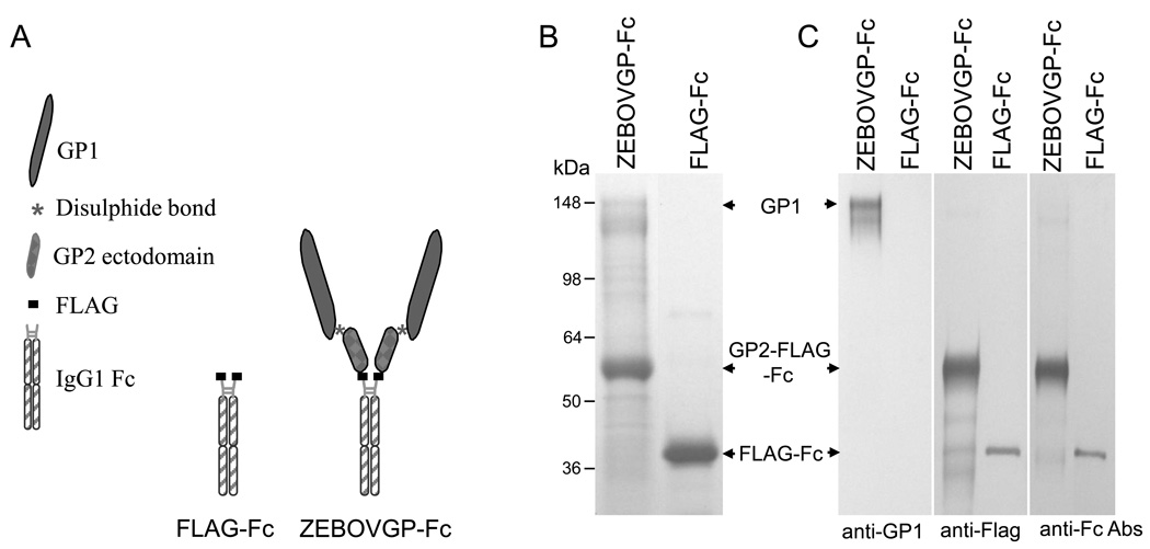
Schematic representation and purification of the ZEBOVGP-Fc and FLAG-Fc proteins. A) Schematic representation of the fusion proteins. The ZEBOVGP-Fc fusion protein contains the ectodomain of ZEBOV GP tagged at its C terminus with a FLAG peptide and fused to the hinge and Fc regions of human IgG1. The FLAG-Fc fusion protein contains a FLAG tag fused to the hinge and Fc regions of IgG1.
B) SDS-PAGE analysis of fusion proteins. Protein A-purified ZEBOVGP-Fc and FLAG-Fc preparations were analyzed by denaturing SDS-PAGE in a 4–12% gradient gel and stained with Coomassie blue. C) Western blot analysis of ZEBOVGP-Fc and FLAG-Fc. Proteins were resolved by SDS-PAGE under denaturing conditions, transferred to PVDF membranes, and probed with ZEBOV-specific anti-GP1 mAb 13F6-1-2, anti-Flag M2 mAb, or goat anti-human Fc Ab. ZEBOV GP1, GP2-FLAG-Fc, and FLAG-Fc bands are indicated with arrows. Positions and size of molecular weight markers are indicated in kDa.

