Abstract
The receptor Notch interacts with the Abl tyrosine kinase signaling pathway to control axon growth and guidance in Drosophila motor neurons. In part, this is mediated by binding to Trio, a guanine nucleotide exchange factor (GEF) for Rho GTPases. We show here that one of the two GEF domains of Trio, the Rac-specific GEF1, is essential for Trio-dependent motor axon guidance and for the genetic suppression of Notch function in motor axon patterning, but the Rho-specific GEF2 domain is not. Consistent with this, we show that Rac, and not Rho1 or Cdc42, interacts genetically with Notch in a manner indistinguishable from that of bona fide Abl signaling components. We therefore infer that Rac is a key component of Abl signaling in Drosophila motor axons, and specifically that it is the crucial Rho GTPase in “non-canonical” Notch/Abl signaling.
Keywords: Notch, Trio, Rac, Abl, axon guidance, Drosophila
Introduction
As an axon navigates through its environment during nervous system development, the growth cone at its tip responds to signals from many guidance cues by executing dynamic rearrangements of the actin cytoskeleton. The Rho subfamily of small GTPases - Rho, Rac and Cdc42 – is critical for this modulation of the actin cytoskeleton (Hall, 1992; Tapon and Hall, 1997; Hall, 1998). The different Rho GTPases are thought to act upon different kinds of actin structures. For example, Rho stimulates formation of focal adhesions and stress fibers (Ridley and Hall, 1992), Rac promotes lamellipodial structures (Ridley et al., 1992) and Cdc42 activates filopodia (Nobes and Hall, 1995).
Among the Rho GTPases, Rac has been the most enigmatic. Activation of Rac displays many effects on cell morphology, cell polarity and cell migration (Etienne-Manneville and Hall, 2002; Jaffe and Hall, 2005). Specifically in the growth cone, Rac plays pivotal roles in outgrowth, branching and guidance of axons (Luo, 2000b; Guan and Rao, 2003). In Drosophila, Rac mutant embryos display severe axon growth defects both in CNS and PNS (Hakeda-Suzuki et al., 2002). A very large number of molecules have been identified as Rac binding partners, and it is not clear which among them are the key effectors for actin rearrangement in particular contexts (Bishop and Hall, 2000; Luo, 2000a).
A variety of axon guidance signals seem to act through Rac GTPases (Tapon and Hall, 1997; Luo, 2000b). For example, in C. elegans, Rac acts downstream of UNC-40, a netrin receptor (Gitai et al., 2003). Axonal repulsion by Slit-Robo signaling is mediated by Rac and restricted Rac function also limits Slit-Robo signaling (Hakeda-Suzuki et al., 2002; Fan et al., 2003; Yang and Bashaw, 2006). The intracellular domain of Plexin B, a semaphorin receptor, binds to Rac-GTP and reduces active Rac by sequestering it from its target Pak, resulting in repulsion of axons (Vikis et al., 2000; Hu et al., 2001). Rac1 deficient cerebellar granule neurons in primary culture display reduced PAK1 phosphorylation and mislocalization of WAVE complex from the growth cone membrane (Tahirovic et al., 2010)
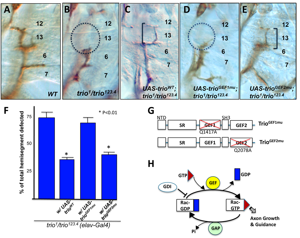
Like other GTPases, Rac GTPases cycle between an active GTP-bound state and an inactive GDP-bound state (Fig 1H). Rac activity is controlled by three main classes of regulatory proteins, Guanine nucleotide Exchange Factors (GEFs), GTPase –Activating Proteins (GAPs), and Guanine nucleotide Dissociation Inhibitors (GDIs). GEFs initiate the release of GDP resulting in accumulation of GTP-bound active GTPases (Rossman et al., 2005). GAPs convert the active state to the inactive GDP-bound form (Moon et al., 2003). GDIs bind to the GDP-bound form, preventing the release of GDP and keeping Rac in the inactive state (Dovas and Couchman, 2005). The overwhelming number of GEFs (~60) and GAPs (~70) in mammals (Etienne-Manneville and Hall, 2002) suggests that each of these proteins likely has its own specific roles in different contexts.
Figure 1. Trio-GEF1 activity is required for motor axon guidance.
Embryos of the indicated genotypes were stained with anti fasciclin II (Fas II; 1D4) at late stage 16 and dissected. (A) Wild type. (B) Embryos of loss of function trio mutant (trio1/trio123.4) exhibit highly penetrant ISNb stall phenotype where axons cannot reach their distal target, the muscle 12/13 cleft (dotted circle). (C), (E) ISNb growth is partially rescued (brackets) by transgenes of UAS-trioWT (C) and UAS-trioGEF2mu (E). (D) UAS-trioGEF1mu fails to rescue ISNb growth (dotted circle). (F) Expressivities of trio phenotypes. For control (left bar), we scored embryos of elav-Gal4; trio1/trio123.4, which displayed similar expressivity to trio1/trio123.4 alone (74%, n=248). Asterisk (*) indicates that difference compared to the control is statistically significant (P<0.01). More than 200 hemisegments per each genotype were analyzed. (G) Schematic diagram of mutant trio transgenes. trioGEF1mu construct has Q1417A mutation abolishing GEF activities in GEF1 and trioGEF2mu has Q2078A mutation in GEF2 (Newsome et al., 2000). NTD; N-terminal Domain. SR; Spectrin Repeats. (H) Cycle of Rac GTPase in axonal growth and guidance. GEF: Guanine nucleotide Exchange Factor; GAP: GTPase Activating Protein; GDI: Guanine nucleotide Dissociation Inhibitor.
Among the many Rac-specific GEFs and GAPs, the GEF Trio is perhaps one of the best characterized Rac regulators in axon patterning. Trio has two tandem Dbl-homology (DH-PH) GEF domains. GEF1 is a specific activator of Rac GTPase. This was shown by genetic epistasis in Drosophila, as a Rac mutation blocks the dominant effects of expressing the GEF1 domain in isolation. It was also verified by direct biochemical experiments, as Trio GEF1 activates Rac in vitro, but not Rho or Cdc 42, both for Drosophila Trio (Newsome et al., 2000) and mammalian Trio (Bellanger et al., 1998). GEF1 was shown genetically to be critically required for Trio function in photoreceptor axon guidance in Drosophila, in part to regulate the activity of PAK kinase (Newsome et al., 2000). The GEF2 domain of Trio, in contrast, is a specific activator of Rho (Bellanger et al., 1998; Spencer et al., 2001) and is not required for photoreceptor axon guidance in the fly eye (Newsome et al., 2000; Vanderzalm et al., 2009). A specific axonal function of GEF1 activity was also found in C. elegans trio (UNC73) (Vanderzalm et al., 2009), while GEF2 regulates pharynx and vulva musculature, synaptic neurotransmission (Steven et al., 2005) and P cell migration (Spencer et al., 2001).
The role of Trio in axon guidance has been linked to Abl tyrosine kinase (Liebl et al., 2000). Abl and Trio mutants have similar axon guidance phenotypes by themselves, and in combination they interact synergistically. Abl is one of the key molecules required in axon pathfinding (Wills et al., 2002; Forsthoefel et al., 2005; Song et al., 2008). The notion that Rac activity is required for Abl function has also been suggested in other contexts (reviewed in Hern·ndez 2004). Rac promotes the activities of oncogenic constitutively-activated forms of Abl such as p210Bcr-Abl and v-Abl in mammalian cultured cells (Renshaw et al., 1996; Bassermann et al., 2002). Abl activates Rac in conjunction with receptor tyrosine kinase signaling in part by phosphorylation of the Ras GEF, Sos-1 (Sini et al., 2004) and is also required for Rac activation following stimulation of cadherin-mediated cell-cell adhesion (Zandy et al., 2007).
We have shown previously that Abl and Trio participate in a non-canonical function of the receptor Notch in axon patterning in Drosophila (Giniger, 1998; Crowner et al., 2003). In contrast to the usual Notch signaling mechanism, the function of Notch during axon guidance does not require the canonical molecular events of nuclear translocation of the intracellular domain to control target gene expression mediated by the transcription factor Su(H). Instead, Notch is present in vivo in a multiprotein complex together with Trio, and also with Disabled, another core component of Abl signaling, as shown by co-immunoprecipitation of Notch with Trio and Disabled proteins from wild type Drosophila extracts (Le Gall et al., 2008; Song et al., 2010). This physical association of Notch with Trio and Disabled is essential for Notch-dependent control of axon growth and guidance (Le Gall et al., 2008).
Motivated by these observations, we investigated the potential involvement of small Rho GTPases in non-canonical Notch signaling during axon guidance in Drosophila embryos. Here, we first show that the Rac-specific GEF1 activity of Trio is selectively required for Trio-dependent axon patterning in embryonic motor nerves, and specifically for the interaction with Notch. Furthermore, we show a selective genetic interaction of Rac, and not Rho1 or Cdc42, with Notch, modifying its axonal function. These data support the hypothesis that Rac is a critical player in the Abl- and Trio-dependent mechanism by which Notch controls axon growth and guidance.
Results
Trio GEF1 activity is essential for motor axon guidance
Motor nerve guidance in the fly embryonic nervous system provides a powerful system for quantitatively assaying the contribution of signaling proteins to axon growth and guidance in vivo. In late stage 16 embryos, subsets of motor neurons display simple and distinguishable axonal projections. Inter-Segmental Nerve b (ISNb) has 7 motor axons that exit from the ISN root at a specific choice point to innervate ventrolateral muscles (VLM) (Fig 1A). Abl and its accessory signaling components such as Neurotactin (Nrt), Disabled (Dab), Failed Axon Connections (Fax) and Trio, are essential for proper growth and guidance of this motor nerve (Gertler et al., 1989; Hill et al., 1995; Song et al., 2010)
Like other mutations in Abl signaling, trio mutants display a specific axonal defect in ISNb, ‘stalling’ in the middle of the target field (Fig.1B and Table 1)(Awasaki et al., 2000; Bateman et al., 2000). We found that expression of a trio transgene with a mutation inactivating the GEF1 domain (UAS-trioGEF1mu)(Newsome et al., 2000) was unable to rescue the ISNb axonal phenotypes of a trio mutant. In contrast, expression of a transgene bearing the equivalent lesion in GEF2 (UAS-trioGEF2mu) rescued the axonal defects of trio as effectively as a wild type transgene (UAS-trioWT)(Fig 1C, D and E and Table 1). We verified by immunostaining that all three Trio derivatives accumulated to similar levels and trafficked properly to axons (data not shown). Therefore, the activity of GEF1 is essential for the function of Trio in motor axon guidance whereas GEF2 is not. This is consistent with previous data showing that the GEF1 domain of Trio is preferentially required for sensory axon guidance in photoreceptor cells, while GEF2 is dispensable in this context (Newsome et al., 2000).
Table 1.
Genetic rescue of trio mutant and modification of Notch-trio interaction by trio transgenes in ISNb motor axon guidance.
| Genotype | n | ISNb defects |
|---|---|---|
| Rescue of trio mutant by trio transgenes | ||
| trio1/trio123.4 | 190 | 74% |
| UAS-trioWT; trio1/trio123.4 | 240 | 35%* |
| UAS-trioGEF1mu; trio1/trio123.4 | 242 | 70%ns |
| UAS-trioGEF2mu; trio1/trio123.4 | 132 | 40%* |
| Modification of N-trio interaction by trio transgenes | ||
| Nts1 | 562 | 36% |
| Nts1; trio123.4/+ | 300 | 27% |
| Nts1; UAS-trioWT; trio123.4/+ | 286 | 35%** |
| Nts1; UAS-trioGEF1mu; trio123.4/+ | 259 | 29%ns |
| Nts1; UAS-trioGEF2mu; trio123.4/+ | 284 | 35%** |
| trio123.4/+ | 120 | <2% |
Abdominal segments A2 – A7 were examined in late stage 16 embryos for quantification of ISNb phenotypes. n, total hemisegments counted. All trio transgenes were expressed by pan-neuronal elav-Gal4 driver.
For rescue of trio mutant, single asterisks (*) denote statistically significant rescue by transgenes relative to trio1/trio123.4 (P<0.005).
For rescue of Notch-trio interaction, double asterisks (**) denote statistically significant modification by transgenes relative to Nts1; trio123.4/+ (P<0.005). P-values were determined by χ2 test.
Not statistically significant. P-value relative to czontrol was more than 0.1 in each comparison.
Function of Trio in non-canonical Notch signaling during axonal guidance is mediated by GEF1 function
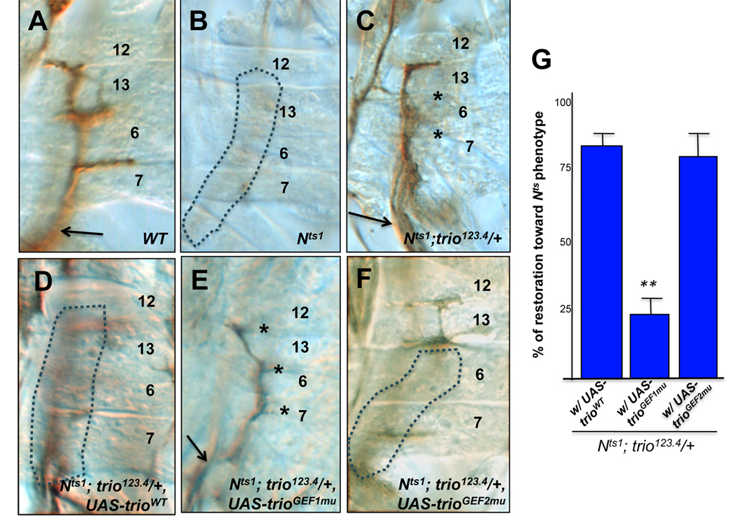
We next examined the interaction of Trio with the receptor Notch in axon growth and guidance. Inactivation of Notch at the time of axon growth selectively misroutes some motor axons, causing specific guidance defects. For example, in ISNb, Notch mutant (Nts1) axons often grow past the “choice point” at which they should exit the main ISN and enter the ventrolateral muscle domain (VLM). This results in an abnormal “bypass” innervation pattern, with few or no axon projections into the VLM (36% of total hemisegments defective, dotted area in Fig. 2B and Supplementary Fig 1) under appropriate temperature shifting conditions (details in Experimental Procedures). Guidance errors also occur in Segmental Nerve a (SNa) in Nts1 mutants (Supplementary Fig 2). The axonal action of Notch is mediated by a non-canonical signaling mechanism by which Notch locally inhibits the activity of Abl and associated cofactors(Crowner et al., 2003). Consistent with this, reduction of Abl signaling components, including Trio, suppresses the axon patterning phenotypes of Nts1. For example, heterozygosity for a loss of function trio mutation significantly restores the ability of ISNb axons to enter the VLM field in a Notch mutant genetic background (Nts1; trio123.4/+, 28%, n=190, P<0.05 (Fig 2); Nts; trio8/+, 27%, n=236, P<0.001). The effect of trio heterozygosity on the Notch phenotype is quantitatively modest, but it is observed with unrelated trio alleles, and is similar to the degree of suppression produced by heterozygosity for other components of the Abl signaling pathway, such as Abl and Nrt (Crowner et al., 2003). In the Discussion (below) we expand on the motivation and significance of using hypomorphic manipulations, such as heterozygous mutations, in analyzing the Notch/trio interaction.
Figure 2. Modulation of ISNb axon patterning by expression of Trio derivatives in Notchts1.
(A) Wild type. Anti-FasII stained ISNb axons normally exit from the ISN at a specific choice point (arrow). (B) Notchts1 (Nts1). ISNb axons fail to diverge from the ISN pathway and do not enter the ventrolateral muscle layer (VLM) and therefore are not visible in this plane of focus (dotted line). (C) Nts1; trio123.4/+. Reduction of trio gene dosage rescues the ISNb bypass phenotype of the Notch mutant. Note re- entrance of ISNb axons to VLM field at choice point (arrow), though in a fraction of the rescued hemisegments some of the normal muscle innervations fail to form properly (asterisks). (D – F) Nts1;elav-GAL4; trio123.4/+ embryos bearing the indicated trio transgenes. Pan-neuronal expression of wildtype trio (D) or trioGEF2mu (F) reverts the suppression produced by a heterozygous trio mutation, restoring the expressivity of the original Nts1 phenotype (quantified in (G)). In contrast, expression of trioGEF1mu (E) has no effect on the Nts; trio−/+ genetic interaction (though in this genotype ISNb axons seem not always to fully invade the clefts between adjacent muscles (asterisks)). (G) Quantification of the effect of trio transgenes on the Notch-trio interaction. Effect of expressing the indicated trio transgene is graphed as percent restoration of the Nts phenotype, relative to the phenoytpe of Nts1; trio123.4/+. Double asterisk (**) indicates that the difference between Nts1; UAS-trioWT; trio123.4/+ and Nts1; UAS-trioGEF1mu; trio123.4/+ is statistically significant (P<0.005).
We used the Notch/Trio interaction to further dissect the mechanism of Trio action in Notch-dependent axon guidance. We found that the suppression of Notch phenotype by a trio mutation was reverted by pan-neuronal expression of UAS-trioWT (elav-Gal4 driven), once again causing ISNb to bypass the VLM as in Nts1 (Fig. 2D and G, 86% restoration of the VLM bypass phenotype [P<0.005], and Table 1). This suggests that neuronal trio is largely responsible for the genetic interaction of Notch with trio. Next, we examined which domains of Trio, in particular which GEF domains, contribute to the Notch-Trio interaction. Trio constructs bearing mutations that selectively inactivate either GEF1 or GEF2 domain, UAS-trioGEF1mu and UAS-trioGEF2mu, were pan-neuronally expressed in the Nts1; trio123.4/+ background. We did not detect any significant alteration of the Notch-Trio interaction with elav-GAL4-driven trioGEF1mu expression (Fig. 2E and G, 23% suppression [Not significant, P=0.14] and Table 1), while trioGEF2mu expression, in contrast, restored the bypass phenotype nearly as effectively as did UAS-trioWT (Fig. 2F and G, restore 81% of Nts1 phenotype [P<0.005] and Table 1). Pan-neuronal overexpression of wild type trio or GEF-inactive trio transgenes did not produce any dominant axon patterning defects in a wild type background (data not shown). These results suggest that GEF1 activity of Trio is selectively required for the interaction with Notch in axon patterning. Consistent with this hypothesis, expression of the constitutively active Trio GEF1 domain (Newsome et al., 2000; Ferraro et al., 2007) alone mimics the Notch ISNb phenotype (48% bypass, n=456 driven by elav-Gal4), whereas expression of Trio (GEF2) alone does not perturb ISNb patterning (less than 2% bypass, n=228, driven by elav-Gal4).
Rac small GTPase is selectively required in Notch mediated motor axon guidance
The observation that activity of Trio GEF1 is necessary and sufficient for the functional interaction of Notch with Trio in axon patterning led us to investigate whether Rac, the specific target for activation by GEF1, acts in this context. First, we investigated whether genetic reduction of Rac levels modifies motor axon phenotypes of Nts1, and found that heterozygosity for a Rac triple mutant, Rac1J10Rac2ΔMtlΔ/+ suppressed the axonal defects of Nts1 (Table 2, 23% of hemisegments defective, N=132, vs 36% for Nts1, P<0.05). In contrast, heterozygosity for mutations of other small Rho GTPases (i.e. Cdc424 and Rho1rev220) did not alter the expressivity of ISNb bypass in Nts1 (Table 2). This result suggests that among Rho GTPases, Rac is preferentially required in the genetic pathway of Notch signaling for patterning ISNb motor axons.
Table 2.
Quantification of genetic interactions between Notch and Rho small GTPases in ISNb motor axon guidance.
| Genotype | na | %b | Control | na | %b |
|---|---|---|---|---|---|
| Modification by mutation | |||||
| Nts1 | 562 | 36 | |||
| Nts1; Rac1J10Rac2ΔMtlΔ/+ | 132 | 23* | Rac1J10Rac2ΔMtlΔ/+ | 120 | <2 |
| Nts1;Rho1rev220/+ | 192 | 36 ns | Rho1rev220/+ | 120 | <2 |
| Nts1,Cdc424/Nts1,+ | 144 | 35 ns | Cdc424/+ | 120 | <2 |
| Modification by transgenes | |||||
| Nts1; Gal4-60/+ | 230 | 35 | |||
| Nts1; elav-Gal4/+ | 192 | 37 | |||
| Nts1; UAS-RacV12/Gal4-60 | 180 | 55* | UAS-RacV12/Gal4-60 | 168 | 6 |
| Nts1; UAS-RacN17/ Gal4-60 | 156 | 27*c | UAS-RacN17/ Gal4-60 | 180 | 3 |
| Nts1; UAS-Rac1WT/ Gal4-60 | 168 | 41 ns | UAS-Rac1 WT / Gal4-60 | 132 | 5 |
| Nts1; UAS-Rho1V14/Gal4-60 | 192 | 39 ns | UAS-Rho1V14/Gal4-60 | 120 | <2 |
| Nts1; UAS-Rho1N19/ elav-Gal4 | 120 | 38ns | UAS-Rho1N19/elav-Gal4 | 202 | <2 |
| Nts1; UAS-Cdc42V12/Gal4-60 | 300 | 39 ns | UAS-Cdc42V12/Gal4-60 | 120 | <2 |
| Nts1; UAS-Cdc42N17/elav-Gal4 | 168 | 36 ns | UAS-Cdc42N17/elav-Gal4 | 240 | <2 |
Abdominal segments A2 – A7 were examined in late stage 16 embryos for quantification of ISNb bypass phenotypes. Asterisks denote statistically significant modification by mutants and transgenes (P<0.05). Comparison is to Nts1 for effect of mutations; comparison is to Nts1 with the appropriate GAL4 driver for transgenes. P-values were determined by χ2 test. UAS-Cdc42N17 and UAS-Rho1N19 were pan-neuronally expressed by elav-Gal4 driver. Since elav-driven expression of some dominant forms of GTPases (RacN17, RacV12, Rho1V14 and Cdc42V12) and UAS-Rac1 WT show massive axonal defects in most hemisegments (also described in(Kaufmann et al., 1998; Fritz and VanBerkum, 2002), for these transgenes instead we used Gal4-60, a less active GAL4 driver (Luo et al., 1994). In a completely independent set of experiments, the critical genotypes were assayed in a somewhat different temperature shift protocol and gave the same result, supporting the robustness of the genetic interaction (D.Crowner and E.G unpublished observation)
Number of hemisegments of the designated genotype that were scored.
The percentage of the hemisegments examined that exhibited ISNb bypass phenotype (described in Supplementary 1).
Besides the bypass phenotype, we observed a different guidance defect, 11% of trio-like stall phenotypes.
Not statistically significant. P-values relative to control were > 0.4 in these comparisons.
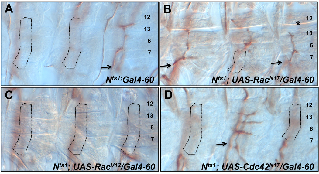
To test this hypothesis further, we employed dominant negative or constitutively active forms of Rac. Expression of a constitutively active form of Rac, RacV12 significantly increased the occurrence of ISNb bypass in Nts1 embryos (Fig. 3C and Table 2), while expression of the dominant negative RacN17 suppressed the ISNb bypass phenotype (Fig. 3B and Table 2). In contrast, the ISNb phenotype of Nts1 was not significantly modulated by expression of dominant negative or constitutively active forms of Cdc42 or Rho1 (Fig. 3D and Table 2). None of the GTPase transgenes produces dominant bypass phenotypes on their own under the conditions that we used, though RacN17 produces a low frequency of trio-like stall phenotypes (11%). Moreover, the effect of UAS-Rac1WT expression in Nts1 is not statistically significant (Table 2), suggesting that Rac activity, rather than amount, is critical for the genetic interaction with Notch. Taken together, our results suggest that Rac selectively modulates Notch function in motor axon guidance, while Rho1 and Cdc42 do not have strong effects in this context.
Figure 3. Modification of Notch ISNb axon patterning by dominant mutations of small GTPases.
(A) Nts1;Gal4-60. Like Nts1, ISNb axons often fail to diverge from the ISN pathway and do not enter the ventrolateral muscle layer (VLM) and therefore are not visible in this plane of focus (dotted line). (B) Nts1; UAS-RacN17/Gal4-60. Gal4-60 driven RacN17, a dominant negative form of Rac, significantly restores the Notch ISNb bypass phenotype, causing ISNb axons to enter the VLM field at the choice point (arrows), though in a fraction of the rescued hemisegments some of the normal muscle innervations are incomplete (asterisks). (C) Nts1; UAS-RacV12/Gal4-60. Gal4-60 driven RacV12, a constitutively active form of Rac, strongly enhanced Notch ISNb phenotype. However, dominant mutations of other small GTPases such as Cdc42 (D) or RhoA (not shown) did not change significantly the expressivity of Notch ISNb bypass phenotype.
Discussion
The guanine exchange factor Trio physically associates with Notch in vivo (Le Gall et al., 2008) and is a genetic component of Notch signaling in motor axon guidance (Crowner et al., 2003; Le Gall et al., 2008). In this paper, we found that one of the tandem GEF domains of Trio, the Rac-specific GEF1, is essential for this genetic interaction. Consistent with this observation, the axonal phenotypes of a Notch mutant are suppressed by reducing the level of the three Rac paralogs, to a degree similar to that caused by reduction of trio. This interaction appears to be specific, because mutations of other closely related small GTPases, Rho1 and Cdc42, did not modify the axonal phenotypes of Nts1. This was further confirmed by testing the effect of dominant Rac transgenes. Expression of a dominant negative RacN17 suppressed the ISNb phenotype of a Notch mutant, while a constitutively active Racv12 enhanced it. In contrast, introduction of other dominant transgenes such as Cdc42N17 did not alter the Notch mutant phenotypes. Thus, Rac appears to be the key Rho GTPase for control of motor axon guidance by Notch.
Our data support the hypothesis that Rac is a key component of Abl signaling in Drosophila motor axons. Notch-dependent axon patterning is executed by an alternate, “non-canonical” Notch signaling pathway defined by the Abl tyrosine kinase (Giniger, 1998). Notch protein associates in vivo with the Abl cofactors, Disabled and Trio, and genetic experiments suggest that Notch antagonizes the activity of the Abl signaling pathway (Crowner et al., 2003; Le Gall et al., 2008). We find here that Rac interacts functionally with Notch in the same way as do the core Abl signaling proteins. Reduction of Rac activity suppresses Notch axonal phenotypes, just as do reduction of Abl pathway components or expression of the Abl antagonist, Enabled. Enhancement of Rac activity exacerbates Notch axonal phenotypes, just as does activation of Abl signaling or mutation of Enabled. These data are therefore consistent with the hypothesis we proposed previously that the key role of Notch in ISNb guidance is to limit Abl-dependent adhesion of ISNb growth cones to the ISN nerve pathway (Crowner et al., 2003). By this model, excessive Abl, Rac-dependent substratum adhesion in a Notch mutant prevents ISNb growth cones from defasiculating from the ISN to enter the target muscle field. Modulation of Rac activity directly modifies this adhesion, aiding or hindering the Notch-dependent release of the growth cone from the ISN pathway at the choice point. These observations are also consistent with results from vertebrate cell culture models that suggested a crucial role for Rac in Abl-dependent signaling and cell adhesion (Zandy et al., 2007; Zandy and Pendergast, 2008).
The biological function of Rac has been difficult to investigate in vivo. First, Rac performs a wide range of functions in many cells, so experimental modulation of Rac often produces complex combinations of effects. Second, the phenotypes caused by overexpression of constitutively active Rac are often similar to those produced by overexpression of a dominant negative form, rather than being opposite, making interpretation of experimental manipulations extremely challenging (Luo et al., 1994; Luo, 2000a). For example, both increase and decrease of Rac activity can cause growth cone stalling in Drosophila neurons. At the molecular level, however, this occurs for opposite reasons: extreme activation of Rac causes excessive stabilization of actin filaments, while inactivation of Rac excessively destabilizes them (Luo et al., 1994). In either event, however, the result is to block growth cone advance. These properties motivated us to introduce two technical modifications to our experiments that have been essential to obtaining clear conclusions. First, we used a sensitized genetic background in which a single molecular process was limiting for a specific axon guidance decision. This minimized the confusions normally introduced by the pleiotropic functions of Rac. We achieved this by employing a precise temperature shift of a temperature-sensitive allele of Notch in a synchronized population of embryos, and assaying the turning of a single nerve comprising seven closely related axons at a precise point in their trajectory. Second, rather than using severe manipulation of Rac level or activity we used the mildest manipulations we could achieve and averaged over a large number of trials to detect quantitative modulation of an intermediate (hypomorphic) Notch phenotype by Rac. Axon growth and guidance rely on a cycle of actin dynamics. Extreme manipulations run the risk of halting that cycle altogether. We reasoned that more modest manipulations would allow us to interrogate sensitively the effect of a particular signaling molecule on the dynamics of the actin cycle. We therefore employed heterozygous Rac mutations rather than homozygotes for investigating genetic interactions with Notch. Moreover, when expressing dominant Rac transgenes, we searched for GAL4 drivers that expressed at low, rather than high level, and that did not produce phenotypes on their own in a wild type genetic background. These perturbations nonetheless sufficed to produce significant quantitative effects on axon phenotype in the sensitized Nts1 background. While the subtle manipulations used here necessarily produced effects that were relatively modest quantitatively, they were consistent across genotypes, such as different mutant alleles, or reduction of different genes within the Abl pathway, and they were internally consistent when comparing different kinds of perturbations, such as gain- vs loss-of-function experiments. In contrast, use of more extreme manipulations, such as interaction with homozygous mutations in the Abl pathway, were often uninterpretable due to defects in other, unrelated developmental processes.
The data reported here suggest that Rac, acting downstream of Trio, is a major player in the non-canonical signaling pathway by which Notch controls axon growth and guidance. The key challenges now are to uncover the molecular mechanism by which Notch antagonizes Abl signaling, and to understand how and why suppression of Abl signaling by Notch promotes proper growth and guidance of Notch-dependent axons.
Experimental Procedures
Fly stocks
Fly stocks were raised on standard Drosophila media at room temperature (23–25°C) except for Notch mutant, Nts1 in 18°C. We obtained fly stocks as follows: trio123.4-E. Leibl (Dennison University, OH, USA); Gal4-60 -G. Technau (University of Mainz, Mainz, Germany); Rho1rev220, S. Parker (Fred Hutchinson Cancer Research Center, WA, USA); elav-Gal4, Y.N. Jan (UCSF, CA, USA); Rac1J10Rac2ΔMtl, UAS-RacN17, UAS-RacV12, UAS- Rac1, UAS-Cdc42V12, UAS-Cdc42N17 (L. Luo, Stanford University, CA, USA); Cdc424 (R. Fehon, Duke University, NC, USA); trio1,UAS-trioWT, UAS-Rho1N19, UAS-Rho1v14 -the Bloomington Drosophila Stock Center (Bloomington, IN, USA). To examine the specificity of GEF activity of Trio, we generate transgenic lines of GEF1-inactive (UAS-trioGEF1mu) and GEF2-inactive (UAS-trioGEF2mu)(mutant trio constructs were kindly provided by B.J. Dickson). Chromosome balancer containing β-galactosidase (TM6B-T8-LacZ and CyOact-LacZ) were used in all genetic experiments.
Sample collection and preparation
Egg collection and fixation was carried out as previously described (Crowner et al., 2003) with a slight modification of temperature shift condition; 0–3 hour Nts1 embryos were placed at 18°C for 13 hours and then 32°C for 6 hours. In this late-shifting condition, a significant portion of motor axons display abnormal patterning including ISNb phenotype (Supplementary 1) without affecting relevant cell fates, including number and location of neurons and glia (confirmed by marker staining, data not shown) and morphogenesis of muscles. For scoring SNa phenotypes, embryos were placed at 18°C for 14 hours and then 32°C for 5.5 hours.
Immunohistochemistry
Collected embryos were stained with antibodies by standard methods (Bodmer 1987, Bier 1989). Anti-Sxl (M114, Developmental Studies Hybridoma Bank, Iowa, USA) was used at 1:50 for segregating hemizygote for X chromosome, negative for Sxl expression. Anti-Fasciclin II (1D4, Developmental Studies Hybridoma Bank) antibody was used at 1:150 for labeling motor nerves. Rabbit anti β-gal (1:1000, Cappel, pre-absorbed prior to use) was used for sorting negative embryos out for further analysis.
Statistical analysis
ISNb was scored in hemisegments A2–A7 by staining with anti-Fasciclin II, as described previously (Song et al., 2010). Data for a given genotype was pooled and significance vs control was assessed by χ2 test. Average variation between duplicate trials was less than 15% of the mean value for a given condition (Table 2).
Supplementary Material
Examples of ISNb bypass phenotypes in Nts1 (A) Wild type (B, C and D) Nts1 (anti-FasII staining, 1D4). Under our temperature shift condition (described in Experimental Procedures), Nts1 embryos display a range of axonal defects in ISNb All defects share the common feature that ISNb fails to exit at the choice point (dotted arrows in B,C, D and diagrams), resulting in bypass of the target muscle area (dotted lines in B, C, D and diagrams). (B) Turn back; After ISNb axons bypass the VLM, they exit the ISN and enter the dorsal part of the VLM (arrows), muscle 12 and 13. (C) Mid insertion; Bypassing axons exit the ISN at the mid VLM field, causing abnormal axon projection between muscle 13 and 6 (arrows). (D) Complete bypass; All ISNb axons run together or parallel with ISN root and never enter the VLM field. Note that the micrographs show lateral views, whereas the schematics below represent cross sections taken at the position of the nerve trajectory. Vertical, dashed line in each cross section indicates the position of the focal plane shown in the micrograph above it.
SNa axonal phenotype in Nts1(anti-FasII staining, 1D4). (A) In wild type, SNa axons diverge to make two major projections, dorsal (D) and lateral (L). (B) Many hemisegments in Nts1 mutant (~70%, n=360) lack the lateral projection (dotted circle). Lateral SNa axons fail to exit at the choice point (arrows) and proceed along the dorsal projection, (C) In the presence of a heterozygous trio mutation, SNa axon defect is significantly rescued (asterisk). Note: The temperature shift condition for SNa assay is different from that for ISNb; 0–3 hour old eggs were incubated at 18° for 14 hours and then at 32°C for 5.5 hours.
Acknowledgements
We thank K. Zinn, L. Belluscio and the members of the Giniger Lab for comments on the manuscript, D. Crowner and Z. Liu for technical assistance, the Bloomington Drosophila Stock Center for fly lines, and Developmental Studies Hybridoma Bank for antibody reagents. This work was supported in part by the Intramural Research Program of the NIH (NINDS: Z01NS003013 to E. G.).
Grant Information : NIH grant (NINDS: Z01NS003013 to E. Giniger)
References
- Awasaki T, Saito M, Sone M, Suzuki E, Sakai R, Ito K, Hama C. The Drosophila trio plays an essential role in patterning of axons by regulating their directional extension. Neuron. 2000;26:119–131. doi: 10.1016/s0896-6273(00)81143-5. [DOI] [PubMed] [Google Scholar]
- Bassermann F, Jahn T, Miething C, Seipel P, Bai R, Coutinho S, Tybulewicz V, Peschel C, Duyster J. Association of Bcr-Abl with the proto-oncogene Vav is implicated in activation of the Rac-1 pathway. J Biol Chem. 2002;277:12437–12445. doi: 10.1074/jbc.M112397200. [DOI] [PubMed] [Google Scholar]
- Bateman J, Shu H, Van Vactor D. The guanine nucleotide exchange factor trio mediates axonal development in the Drosophila embryo. Neuron. 2000;26:93–106. doi: 10.1016/s0896-6273(00)81141-1. [DOI] [PubMed] [Google Scholar]
- Bellanger JM, Lazaro JB, Diriong S, Fernandez A, Lamb N, Debant A. The two guanine nucleotide exchange factor domains of Trio link the Rac1 and the RhoA pathways in vivo. Oncogene. 1998;16:147–152. doi: 10.1038/sj.onc.1201532. [DOI] [PubMed] [Google Scholar]
- Bishop AL, Hall A. Rho GTPases and their effector proteins. Biochem J. 2000;348(Pt 2):241–255. [PMC free article] [PubMed] [Google Scholar]
- Crowner D, Le Gall M, Gates MA, Giniger E. Notch steers Drosophila ISNb motor axons by regulating the Abl signaling pathway. Curr Biol. 2003;13:967–972. doi: 10.1016/s0960-9822(03)00325-7. [DOI] [PubMed] [Google Scholar]
- Dovas A, Couchman JR. RhoGDI: multiple functions in the regulation of Rho family GTPase activities. Biochem J. 2005;390:1–9. doi: 10.1042/BJ20050104. [DOI] [PMC free article] [PubMed] [Google Scholar]
- Etienne-Manneville S, Hall A. Rho GTPases in cell biology. Nature. 2002;420:629–635. doi: 10.1038/nature01148. [DOI] [PubMed] [Google Scholar]
- Fan X, Labrador JP, Hing H, Bashaw GJ. Slit stimulation recruits Dock and Pak to the roundabout receptor and increases Rac activity to regulate axon repulsion at the CNS midline. Neuron. 2003;40:113–127. doi: 10.1016/s0896-6273(03)00591-9. [DOI] [PubMed] [Google Scholar]
- Ferraro F, Ma XM, Sobota JA, Eipper BA, Mains RE. Kalirin/Trio Rho guanine nucleotide exchange factors regulate a novel step in secretory granule maturation. Mol Biol Cell. 2007;18:4813–4825. doi: 10.1091/mbc.E07-05-0503. [DOI] [PMC free article] [PubMed] [Google Scholar]
- Forsthoefel D, Liebl E, Kolodziej P, Seeger M. The Abelson tyrosine kinase, the Trio GEF and Enabled interact with the Netrin receptor Frazzled in Drosophila. Development. 2005;132:1983–1994. doi: 10.1242/dev.01736. [DOI] [PubMed] [Google Scholar]
- Fritz JL, VanBerkum MF. Regulation of rho family GTPases is required to prevent axons from crossing the midline. Dev Biol. 2002;252:46–58. doi: 10.1006/dbio.2002.0842. [DOI] [PubMed] [Google Scholar]
- Gertler FB, Bennett RL, Clark MJ, Hoffmann FM. Drosophila abl tyrosine kinase in embryonic CNS axons: a role in axonogenesis is revealed through dosage-sensitive interactions with disabled. Cell. 1989;58:103–113. doi: 10.1016/0092-8674(89)90407-8. [DOI] [PubMed] [Google Scholar]
- Giniger E. A role for Abl in Notch signaling. Neuron. 1998;20:667–681. doi: 10.1016/s0896-6273(00)81007-7. [DOI] [PubMed] [Google Scholar]
- Gitai Z, Yu T, Lundquist E, Tessier-Lavigne M, Bargmann C. The netrin receptor UNC-40/DCC stimulates axon attraction and outgrowth through enabled and, in parallel, Rac and UNC-115/AbLIM. Neuron. 2003;37:53–65. doi: 10.1016/s0896-6273(02)01149-2. [DOI] [PubMed] [Google Scholar]
- Guan K, Rao Y. Signalling mechanisms mediating neuronal responses to guidance cues. Nat Rev Neurosci. 2003;4:941–956. doi: 10.1038/nrn1254. [DOI] [PubMed] [Google Scholar]
- Hakeda-Suzuki S, Ng J, Tzu J, Dietzl G, Sun Y, Harms M, Nardine T, Luo L, Dickson BJ. Rac function and regulation during Drosophila development. Nature. 2002;416:438–442. doi: 10.1038/416438a. [DOI] [PubMed] [Google Scholar]
- Hall A. Ras-related GTPases and the cytoskeleton. Mol Biol Cell. 1992;3:475–479. doi: 10.1091/mbc.3.5.475. [DOI] [PMC free article] [PubMed] [Google Scholar]
- Hall A. Rho GTPases and the actin cytoskeleton. Science. 1998;279:509–514. doi: 10.1126/science.279.5350.509. [DOI] [PubMed] [Google Scholar]
- Hill KK, Bedian V, Juang JL, Hoffmann FM. Genetic interactions between the Drosophila Abelson (Abl) tyrosine kinase and failed axon connections (fax), a novel protein in axon bundles. Genetics. 1995;141:595–606. doi: 10.1093/genetics/141.2.595. [DOI] [PMC free article] [PubMed] [Google Scholar]
- Hu H, Marton T, Goodman C. Plexin B mediates axon guidance in Drosophila by simultaneously inhibiting active Rac and enhancing RhoA signaling. Neuron. 2001;32:39–51. doi: 10.1016/s0896-6273(01)00453-6. [DOI] [PubMed] [Google Scholar]
- Jaffe A, Hall A. Rho GTPases: biochemistry and biology. Annu Rev Cell Dev Biol. 2005;21:247–269. doi: 10.1146/annurev.cellbio.21.020604.150721. [DOI] [PubMed] [Google Scholar]
- Kaufmann N, Wills ZP, Van Vactor D. Drosophila Rac1 controls motor axon guidance. Development. 1998;125:453–461. doi: 10.1242/dev.125.3.453. [DOI] [PubMed] [Google Scholar]
- Le Gall M, De Mattei C, Giniger E. Molecular separation of two signaling pathways for the receptor, Notch. Dev Biol. 2008;313:556–567. doi: 10.1016/j.ydbio.2007.10.030. [DOI] [PMC free article] [PubMed] [Google Scholar]
- Liebl E, Forsthoefel D, Franco L, Sample S, Hess J, Cowger J, Chandler M, Shupert A, Seeger M. Dosage-sensitive, reciprocal genetic interactions between the Abl tyrosine kinase and the putative GEF trio reveal trio's role in axon pathfinding. Neuron. 2000;26:107–118. doi: 10.1016/s0896-6273(00)81142-3. [DOI] [PubMed] [Google Scholar]
- Luo L. Rho GTPases in neuronal morphogenesis. Nat Rev Neurosci. 2000a;1:173–180. doi: 10.1038/35044547. [DOI] [PubMed] [Google Scholar]
- Luo L. Trio quartet in D. (melanogaster) Neuron. 2000b;26:1–2. doi: 10.1016/s0896-6273(00)81129-0. [DOI] [PubMed] [Google Scholar]
- Luo L, Liao YJ, Jan LY, Jan YN. Distinct morphogenetic functions of similar small GTPases: Drosophila Drac1 is involved in axonal outgrowth and myoblast fusion. Genes Dev. 1994;8:1787–1802. doi: 10.1101/gad.8.15.1787. [DOI] [PubMed] [Google Scholar]
- Moon SY, Zang H, Zheng Y. Characterization of a brain-specific Rho GTPase-activating protein, p200RhoGAP. J Biol Chem. 2003;278:4151–4159. doi: 10.1074/jbc.M207789200. [DOI] [PubMed] [Google Scholar]
- Newsome T, Schmidt S, Dietzl G, Keleman K, Asling B, Debant A, Dickson B. Trio combines with dock to regulate Pak activity during photoreceptor axon pathfinding in Drosophila. Cell. 2000;101:283–294. doi: 10.1016/s0092-8674(00)80838-7. [DOI] [PubMed] [Google Scholar]
- Nobes CD, Hall A. Rho, rac, and cdc42 GTPases regulate the assembly of multimolecular focal complexes associated with actin stress fibers, lamellipodia, and filopodia. Cell. 1995;81:53–62. doi: 10.1016/0092-8674(95)90370-4. [DOI] [PubMed] [Google Scholar]
- Renshaw M, Lea-Chou E, Wang J. Rac is required for v-Abl tyrosine kinase to activate mitogenesis. Curr Biol. 1996;6:76–83. doi: 10.1016/s0960-9822(02)00424-4. [DOI] [PubMed] [Google Scholar]
- Ridley AJ, Hall A. The small GTP-binding protein rho regulates the assembly of focal adhesions and actin stress fibers in response to growth factors. Cell. 1992;70:389–399. doi: 10.1016/0092-8674(92)90163-7. [DOI] [PubMed] [Google Scholar]
- Ridley AJ, Paterson HF, Johnston CL, Diekmann D, Hall A. The small GTP-binding protein rac regulates growth factor-induced membrane ruffling. Cell. 1992;70:401–410. doi: 10.1016/0092-8674(92)90164-8. [DOI] [PubMed] [Google Scholar]
- Rossman KL, Der CJ, Sondek J. GEF means go: turning on RHO GTPases with guanine nucleotide-exchange factors. Nat Rev Mol Cell Biol. 2005;6:167–180. doi: 10.1038/nrm1587. [DOI] [PubMed] [Google Scholar]
- Sini P, Cannas A, Koleske A, Di Fiore P, Scita G. Abl-dependent tyrosine phosphorylation of Sos-1 mediates growth-factor-induced Rac activation. Nat Cell Biol. 2004;6:268–274. doi: 10.1038/ncb1096. [DOI] [PubMed] [Google Scholar]
- Song J, Giniger E, Desai C. The receptor protein tyrosine phosphatase PTP69D antagonizes Abl tyrosine kinase to guide axons in Drosophila. Mech Dev. 2008;125:247–256. doi: 10.1016/j.mod.2007.11.005. [DOI] [PMC free article] [PubMed] [Google Scholar]
- Song J, Kannan R, Merdes GS, ingh J, M M, Giniger E. Disabled is a bona fide component of the Abl signaling network. development. 2010;137:3719–3727. doi: 10.1242/dev.050948. [DOI] [PMC free article] [PubMed] [Google Scholar]
- Spencer AG, Orita S, Malone CJ, Han M. A RHO GTPase-mediated pathway is required during P cell migration in Caenorhabditis elegans. Proc Natl Acad Sci U S A. 2001;98:13132–13137. doi: 10.1073/pnas.241504098. [DOI] [PMC free article] [PubMed] [Google Scholar]
- Steven R, Zhang L, Culotti J, Pawson T. The UNC-73/Trio RhoGEF-2 domain is required in separate isoforms for the regulation of pharynx pumping and normal neurotransmission in C. elegans. Genes Dev. 2005;19:2016–2029. doi: 10.1101/gad.1319905. [DOI] [PMC free article] [PubMed] [Google Scholar]
- Tahirovic S, Hellal F, Neukirchen D, Hindges R, Garvalov BK, Flynn KC, Stradal TE, Chrostek-Grashoff A, Brakebusch C, Bradke F. Rac1 regulates neuronal polarization through the WAVE complex. J Neurosci. 2010;30:6930–6943. doi: 10.1523/JNEUROSCI.5395-09.2010. [DOI] [PMC free article] [PubMed] [Google Scholar]
- Tapon N, Hall A. Rho, Rac and Cdc42 GTPases regulate the organization of the actin cytoskeleton. Curr Opin Cell Biol. 1997;9:86–92. doi: 10.1016/s0955-0674(97)80156-1. [DOI] [PubMed] [Google Scholar]
- Vanderzalm P, Pandey A, Hurwitz M, Bloom L, Horvitz H, Garriga G. C. elegans CARMIL negatively regulates UNC-73/Trio function during neuronal development. Development. 2009;136:1201–1210. doi: 10.1242/dev.026666. [DOI] [PMC free article] [PubMed] [Google Scholar]
- Vikis H, Li W, He Z, Guan K. The semaphorin receptor plexin-B1 specifically interacts with active Rac in a ligand-dependent manner. Proc Natl Acad Sci U S A. 2000;97:12457–12462. doi: 10.1073/pnas.220421797. [DOI] [PMC free article] [PubMed] [Google Scholar]
- Wills Z, Emerson M, Rusch J, Bikoff J, Baum B, Perrimon N, Van Vactor D. A Drosophila homolog of cyclase-associated proteins collaborates with the Abl tyrosine kinase to control midline axon pathfinding. Neuron. 2002;36:611–622. doi: 10.1016/s0896-6273(02)01022-x. [DOI] [PubMed] [Google Scholar]
- Yang L, Bashaw GJ. Son of sevenless directly links the Robo receptor to rac activation to control axon repulsion at the midline. Neuron. 2006;52:595–607. doi: 10.1016/j.neuron.2006.09.039. [DOI] [PubMed] [Google Scholar]
- Zandy NL, Pendergast AM. Abl tyrosine kinases modulate cadherin-dependent adhesion upstream and downstream of Rho family GTPases. Cell Cycle. 2008;7:444–448. doi: 10.4161/cc.7.4.5452. [DOI] [PubMed] [Google Scholar]
- Zandy NL, Playford M, Pendergast AM. Abl tyrosine kinases regulate cell-cell adhesion through Rho GTPases. Proc Natl Acad Sci U S A. 2007;104:17686–17691. doi: 10.1073/pnas.0703077104. [DOI] [PMC free article] [PubMed] [Google Scholar]
Associated Data
This section collects any data citations, data availability statements, or supplementary materials included in this article.
Supplementary Materials
Examples of ISNb bypass phenotypes in Nts1 (A) Wild type (B, C and D) Nts1 (anti-FasII staining, 1D4). Under our temperature shift condition (described in Experimental Procedures), Nts1 embryos display a range of axonal defects in ISNb All defects share the common feature that ISNb fails to exit at the choice point (dotted arrows in B,C, D and diagrams), resulting in bypass of the target muscle area (dotted lines in B, C, D and diagrams). (B) Turn back; After ISNb axons bypass the VLM, they exit the ISN and enter the dorsal part of the VLM (arrows), muscle 12 and 13. (C) Mid insertion; Bypassing axons exit the ISN at the mid VLM field, causing abnormal axon projection between muscle 13 and 6 (arrows). (D) Complete bypass; All ISNb axons run together or parallel with ISN root and never enter the VLM field. Note that the micrographs show lateral views, whereas the schematics below represent cross sections taken at the position of the nerve trajectory. Vertical, dashed line in each cross section indicates the position of the focal plane shown in the micrograph above it.
SNa axonal phenotype in Nts1(anti-FasII staining, 1D4). (A) In wild type, SNa axons diverge to make two major projections, dorsal (D) and lateral (L). (B) Many hemisegments in Nts1 mutant (~70%, n=360) lack the lateral projection (dotted circle). Lateral SNa axons fail to exit at the choice point (arrows) and proceed along the dorsal projection, (C) In the presence of a heterozygous trio mutation, SNa axon defect is significantly rescued (asterisk). Note: The temperature shift condition for SNa assay is different from that for ISNb; 0–3 hour old eggs were incubated at 18° for 14 hours and then at 32°C for 5.5 hours.


