Abstract
“Thioalkalivibrio sulfidophilus” HL-EbGr7 is an obligately chemolithoautotrophic, haloalkaliphilic sulfur-oxidizing bacterium (SOB) belonging to the Gammaproteobacteria. The strain was found to predominate a full-scale bioreactor, removing sulfide from biogas. Here we report the complete genome sequence of strain HL-EbGr7 and its annotation. The genome was sequenced within the Joint Genome Institute Community Sequencing Program, because of its relevance to the sustainable removal of sulfide from bio- and industrial waste gases.
Keywords: haloalkaliphilic, sulfide, thiosulfate, sulfur-oxidizing bacteria (SOB)
Introduction
“Thioalkalivibrio sulfidophilus” HL-EbGr7 is an obligately chemolithoautotrophic SOB using CO2 as a carbon source and reduced inorganic sulfur compounds as an energy source. It belongs to the genus Thioalkalivibrio. This genus is characterized by obligate haloalkaliphily and forms a monophyletic group within the family Ectothiorhodospiraceae. The genus currently includes nine validly described species [1] and many yet uncharacterized isolates [2,3]. The members are slow growing and well-adapted to hypersaline (up to salt saturation) and alkaline (up to pH 10.5) conditions. They can oxidize sulfide, thiosulfate, elemental sulfur, sulfite and polythionates (see Table 1 for summary). Moreover, some species can reduce nitrate, nitrite or nitrous oxide [17,18] or utilize thiocyanate (SCN-) as an energy and nitrogen source [19,20]. Genetic diversity analysis of 85 Thioalkalivibrio strains isolated from different soda lakes located in Mongolia, Kenya, California, Egypt and south Siberia, indicated a high genetic diversity and an endemic character, i.e., the majority of the genotypes (85.9%) were found to be unique to one region [15].
Table 1. Classification and general features of “Thioalkalivibrio sulfidophilus” strain HL-EbGR7 according to the MIGS recommendations [4].
| MIGS ID | Property | Term | Evidence code |
|---|---|---|---|
| Current classification | Domain Bacteria | TAS [5] | |
| Phylum Proteobacteria | TAS [6] | ||
| Class Gammaproteobacteria | TAS [7,8] | ||
| Order Chromatiales | TAS [7,9] | ||
| Family Ectothiorhodospiraceae | TAS [10] | ||
| Genus Thioalkalivibrio | TAS [11-13] | ||
| Species “Thioalkalivibrio sulfidophilus” HL-EbGR7 | NAS | ||
| Gram stain | negative | TAS [2] | |
| Cell shape | rod-shaped | TAS [2] | |
| Motility | motile | TAS [2] | |
| Sporulation | non-sporulating | TAS [2] | |
| Temperature range | Mesophile | TAS [2] | |
| Optimum temperature | 34 | TAS [2] | |
| MIGS-6.3 | Salinity range | 0.2-1.5M Na+ (opt.0.4 M) | TAS [2] |
| MIGS-22 | Oxygen requirement | microaerophilic | TAS [2] |
| Carbon source | HCO3- | TAS [2] | |
| Energy source | Sulfide/polysulfide, thiosulfate, sulfur | TAS [2] | |
| MIGS-6 | Habitat | Alkaline bioreactors; soda lakes | TAS [2] |
| MIGS-15 | Biotic relationship | free-living | TAS [2] |
| MIGS-14 | Pathogenicity | none | NAS |
| Biosafety level | 1 | TAS [14] | |
| Isolation | Thiopaq bioreactor | TAS [15] | |
| MIGS-4 | Geographic location | Eerbeek, The Netherlands | TAS [15] |
| MIGS-5 | Sample collection time | 2005 | NAS |
| MIGS-4.1 | Latitude | 52.11 | TAS [16] |
| MIGS-4.2 | Longitude | 6.07 | TAS [16] |
| MIGS-4.3 | Depth | Not applicable | |
| MIGS-4.4 | Altitude | Sea level | NAS |
Evidence codes - IDA: Inferred from Direct Assay (first time in publication); TAS: Traceable Author Statement (i.e., a direct report exists in the literature); NAS: Non-traceable Author Statement (i.e., not directly observed for the living, isolated sample, but based on a generally accepted property for the species, or anecdotal evidence). These evidence codes are from of the Gene Ontology project. If the evidence code is IDA, then the property was directly observed by one of the authors or an expert mentioned in the acknowledgements.
Apart from their role in the sulfur cycle of soda lakes, Thioalkalivibrio species also play a key role in the sustainable removal of sulfide from wastewater and gas streams. In this so-called ‘Thiopaq-process’, hydrogen sulfide is stripped from the gas phase into an alkaline solution, which is subsequently transferred to a bioreactor where Thioalkalivibrio oxidizes HS- almost exclusively to elemental sulfur at a low red-ox potential [21]. Removal of toxic sulfide is needed, not only for a clean and healthy environment, but also to protect gas turbines from corrosion. In contrast to chemical desulfurization processes, such as the ‘Claus-process’, biological removal is cheaper, cleaner and more sustainable, as the produced hydrophilic bio-sulfur is a better fertilizer and fungicide than the chemically produced crystalline hydrophobic sulfur.
To get insight into the molecular mechanism by which Thioalkalivibrio strains adapt to haloalkaline conditions (i.e., pH 10 and up to 4 M of Na+) identification of the genes that are involved in these adaptations is needed. The most important issues are sulfide specialization, carbon assimilation at high pH and bioenergetic adaptation to high salt/high pH. In addition, information on the genome might help in optimizing the sulfur removal process. Here we present a summary classification and a set of features for “T. sulfidophilus” HL-EbGr7, together with the description of the genomic sequencing and annotation.
Classification and features
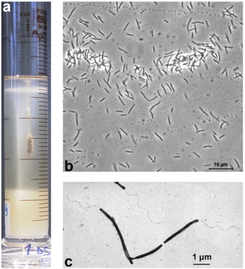
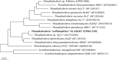
“T. sulfidophilus” HL-EbGr7 was isolated from a full-scale Thiopaq bioreactor in the Netherlands used to remove H2S from biogas [21]. The reactor biomass had a very peculiar property, which made it different from the usual SOB biomass, i.e., an almost complete sulfide specialization and no thiosulfate-oxidizing activity. This was probably the result of a very low red-ox potential at which the reactor was operated. Therefore, the dominant SOB could originally be enriched only with sulfide as substrate in cylinders with agarose-stabilized medium containing opposing gradients of oxygen and sulfide [Figure 1a, 22]. Subsequently, the strain was purified using serial dilutions in gradient cultures and finally from a colony on solid medium with sulfide at micro-oxic conditions. It has rod-shaped, elongated cells with a polar flagellum (Figure 1b and c). The strain is obligately alkaliphilic with a pH optimum of 9.5. It can tolerate a salinity of 1.5 M (optimum at 0.4 M) of total sodium, sulfide concentrations up to 5 mM and a temperature up to 40°C. It utilizes ammonium and urea, but not nitrate or nitrite, as a N-source. On the basis of 16S rRNA gene sequencing the strain belongs to the Gammaproteobacteria with Thioalkalivibrio denitrificans as the closest, described species (Figure 2). Despite this relation, strain HL-EbGr7 cannot grow anaerobically with NOx. Both phylogeny and specific physiology indicate that this strain represents a novel species within the genus Thioalkalivibrio for which a tentative species epithet “sulfidophilus” is proposed.
Figure 1.
a, Enrichment of “Thioalkalivibrio sulfidophilus” HL-EbGr7 in a gradient culture, whereby sulfide is diffusing from an agarose plug in the bottom of the cylinder and O2 from the top. The bacterial cells accumulate in a dense band at the most favorable sulfide and oxygen concentration. b, phase-contrast microphotograph; c, electron microscopy microphotograph of a total cell preparation contrasted with phosphotungstic acid.
Figure 2.
Phylogenetic tree based on 16S rRNA sequences showing the phylogenetic position of “Thioalkalivibrio sulfidophilus” HL-EbGr7. The sequence was aligned to sequences stored in the SILVA database using the SINA Webaligner [23]. Subsequently, the aligned sequences were imported into ARB [24], and a neighbor joining tree was constructed. Sequences of members from the Alphaproteobacteria were used as an outgroup, but were pruned from the tree. The scale bar indicates 1% sequence difference.
Genome sequencing information
Genome project history
Strain HL-EbGr7 was selected for sequencing in the 2007 Joint Genome Institute Community Sequencing Program, because of its relevance to bioremediation. A summary of the project information is presented in Table 2. The complete genome sequence was finished in December 2008. The GenBank accession number for the project is NC_011901. The genome project is listed in the Genome OnLine Database (GOLD) [25] as project Gc00934. Sequencing was carried out at the Joint Genome Institute (JGI). Finishing was done by JGI-Los Alamos National Laboratory (LANL) and initial automatic annotation by JGI-Oak Ridge National Laboratory (ORNL).
Table 2. Genome sequencing project information.
| MIGS ID | Characteristic | Details |
|---|---|---|
| MIGS-28 | Libraries used | 6kb Sanger and 454 standard libraries |
| MIGS-29 | Sequencing platform | ABI-3730, 454 GS FLX Titanium |
| MIGS-31.2 | Sequencing coverage | 8.19 × Sanger, 23.3 × pyrosequence |
| MIGS-31 | Finishing quality | Finished |
| Sequencing quality | Less than one error per 50kb | |
| MIGS-30 | Assembler | Newbler, PGA |
| MIGS-32 | Gene calling method | Prodigal, GenePRIMP |
| GenBank ID | NC_011901 | |
| GenBank date of release | December 29, 2008 | |
| GOLD ID | Gc00934 | |
| NCBI project ID | 29177 | |
| IMG Taxon ID | 643348585 | |
| MIGS-13 | Source material identifier | Personal culture collection, Winogradsky Institute of Microbiology, Moscow |
| Project relevance | Bioremediation |
Growth conditions and DNA isolation
After a long-term gradual adaptation on mixed substrate medium, the isolate was able to grow solely with thiosulfate at micro-oxic conditions. The medium contained 40 mM thiosulfate as an energy source and a standard sodium carbonate-bicarbonate buffer [2] (Sorokin et al., 2006) at pH 10 and 0.6 M Na+. The cells were harvested by centrifugation and stored at -80°C for DNA extraction. Genomic DNA was obtained using phenol-chloroform-isoamylalcohol (PCI) extraction. Briefly, the cell pellet was suspended in a Tris-EDTA buffer at pH 8, and lysed with a mixture of SDS and Proteinase K. The genomic DNA was extracted using PCI and precipitated with ethanol. The pellet was dried under vacuum and subsequently dissolved in water. The quality and quantity of the extracted DNA was evaluated using the DNA Mass Standard Kit provided by the JGI.
Genome sequencing and assembly
The genome of “T. sulfidophilus” HL-EbGr7 was sequenced using a combination of Sanger and 454 sequencing platforms. All general aspects of library construction and sequencing can be found at the JGI website [26]. Pyrosequencing reads were assembled using the Newbler assembler version 1.1.02.15 (Roche). Large Newbler contigs were broken into 7,722 overlapping fragments of 1,000 bp and entered into assembly as pseudo-reads. The sequences were assigned quality scores based on Newbler consensus q-scores with modifications to account for overlap redundancy and adjust inflated q-scores. A hybrid 454/Sanger assembly was made using the Parallel Genome Assembler (PGA). Possible mis-assemblies were corrected and gaps between contigs were closed by editing in Consed, or by custom primer walks of sub-clones or PCR products. A total of 518 Sanger finishing reads were produced to close gaps, to resolve repetitive regions, and to raise the quality of the finished sequence. The error rate of the completed genome sequence is less than 1 in 100,000. Together, the combination of the Sanger and 454 sequencing platforms provided a 31.49-times coverage of the genome. The final assembly contains 32,486 Sanger reads and 390,057 pyrosequencing reads.
Genome annotation
Genes were identified using Prodigal [27] as part of the Oak Ridge National Laboratory genome annotation pipeline followed by a round of manual curation using the JGI GenePRIMP pipeline [28]. The predicted CDSs were translated and used to search the National Center for Biotechnology Information (NCBI) nonredundant database, UniProt, TIGRFam, Pfam, PRIAM, KEGG, COG, and InterPro, databases. Additional gene prediction analysis and functional annotation were performed within the Integrated Microbial Genomes Expert Review (IMG-ER) platform [29].
Genome properties
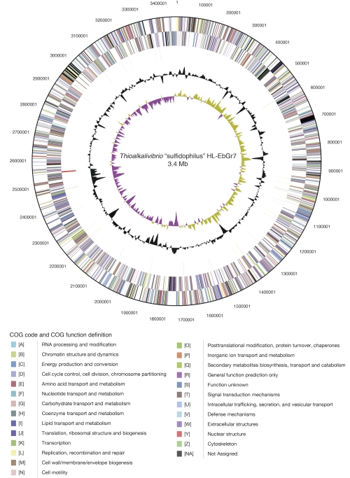
The genome of strain HL-EbGr7 consists of a single circular chromosome (Figure 3) with a size of 3.46 Mbp. The G+C percentage determined from the genome sequence is 65.06%, which is a little higher than the G+C content determined by thermal denaturation (63.5 + 0.5 mol%). There are 3,366 genes of which 3,319 are protein-coding genes and the remaining 47 are RNA genes. 36 pseudogenes were identified, constituting 1.07% of the total number of genes. The properties and statistics of the genome are summarized in Table 3, and genes belonging to COG functional categories are listed in Table 4.
Figure 3.
Graphical circular map of the chromosome of “Thioalkalivibrio sulfidophilus” HL-EbGr7. From outside to the center: Genes on the forward strand (Colored by COG categories), Genes on the reverse strand (colored by COG categories), RNA genes (tRNAs green, rRNAs red, other RNAs black), GC content, GC skew.
Table 3. Genome statistics.
| Attribute | Value | % of Total |
|---|---|---|
| Genome size (bp) | 3,464,554 | 100.00% |
| DNA coding region (bp) | 3,030,998 | 87.49% |
| DNA G+C content (bp) | 2,254,142 | 65.06% |
| Number of replicons | 1 | |
| Extrachromosomal elements | 0 | |
| Total genes | 3366 | 100.00% |
| RNA genes | 47 | 1.40% |
| rRNA operons | 3 | 0.09% |
| Protein-coding genes | 3319 | 98.06% |
| Pseudogenes | 36 | 1.07% |
| Genes in paralog clusters | 294 | 8.73% |
| Genes assigned to COGs | 2512 | 74.36% |
| Genes assigned Pfam domains | 2653 | 78.82% |
| Genes with signal peptides | 719 | 21.36% |
| CRISPR repeats | 2 |
Table 4. Number of genes associated with the general COG functional categories.
| Code | value | %age | COG category |
|---|---|---|---|
| E | 180 | 6.39 | Amino acid transport and metabolism |
| G | 94 | 3.34 | Carbohydrate transport and metabolism |
| D | 49 | 1.74 | Cell cycle control, cell, division, chromosome partitioning |
| N | 109 | 3.87 | Cell motility |
| M | 199 | 7.06 | Cell wall/membrane/envelope biogenesis |
| B | 2 | 0.07 | Chromatin structure and dynamics |
| H | 139 | 4.93 | Coenzyme transport and metabolism |
| Z | 0 | 0 | Cytoskeleton |
| V | 37 | 1.31 | Defense mechanism |
| C | 184 | 6.53 | Energy production and conversion |
| W | 0 | 0 | Extracellular structures |
| S | 270 | 9.58 | Function unknown |
| R | 310 | 11.00 | General function prediction only |
| P | 152 | 5.39 | Inorganic ion transport and metabolism |
| U | 117 | 4.15 | Intracellular trafficking, secretion, and vesicular transport |
| I | 70 | 2.48 | Lipid transport and metabolism |
| Y | 0 | 0 | Nuclear structure |
| F | 58 | 2.06 | Nucleotide transport and metabolism |
| O | 142 | 5.04 | Posttranslational modification, protein turnover, chaperones |
| A | 2 | 0.07 | RNA processing and modification |
| L | 145 | 5.15 | Replication, recombination and repair |
| Q | 47 | 1.67 | Secondary metabolites biosynthesis, transport and catabolism |
| T | 216 | 7.67 | Signal transduction mechanisms |
| K | 134 | 4.76 | Transcription |
| J | 162 | 5.75 | Translation, ribosomal structure and biogenesis |
| - | 854 | 25.37 | Not in COGs |
Insights from the genome sequence
Autotrophic growth
One of the major problems of autotrophic growth at high pH is the assimilation of inorganic carbon (Ci); carbon dioxide concentrations are very low and most inorganic carbon is present as HCO3- or even as CO32- at pH values of 10 and higher. The latter is not available to the cell, which is the main reason for growth limitation of haloalkaliphilic SOB at pH above 10.5, since their energy generation respiratory system is still active up to pH 11-11.5 [2]. Inside the cells, where Ci assimilation occurs, the pH is around 8.5, which means that HCO3- must be taken up as a substrate at an exterior pH of 10. This demands active transport by means of a Na+/HCO3- symporter, such as StbA, which has been found in the alkaliphilic cyanobacterium Synechocystis sp. strain PCC6803 [30]. However, genes encoding StbA have not been detected in strain HLEbGr7. Another means of growth at limited CO2 concentrations is the use of a carbon-concentrating mechanism (CCM), which has been described for other autotrophic microorganisms [31]. Part of the CCM is the presence of carboxysomes, in which ribulose-1,5-biphosphate carboxylase/oxygenase (RuBisCO) and carbonic anhydrase, the key enzymes in CO2 fixation, are located in close proximity for an efficient carbon fixation [32]. The genome of strain HL-EbGr7 contains the genes for the large (rbcL) and small subunit (rbcS) of RuBisCO form 1Ac, and for the synthesis of a-carboxysomes, including csoSCA (formerly know as csoS3) encoding a carboxysome shell carbonic anhydrase. The latter is necessary to convert the transported HCO3- into CO2 – the actual substrate of RuBisCO. In contrast to Thiomicrospira crunogena, the genomes of Thioalkalivibrio are lacking genes for RuBisCO form 1Aq and form II, which has been confirmed by Tourova et al. [33]. Expression studies at different CO2 concentrations in the chemolithoautotroph Hydrogenovibrio marinus indicated the preferential expression of RuBisCO form 1Ac at low CO2 concentrations and RuBisCO form 1Aq and/or form II at higher CO2 concentrations [34]. This result indicates that our strain is indeed adapted to low CO2 concentrations.
Sulfur metabolism
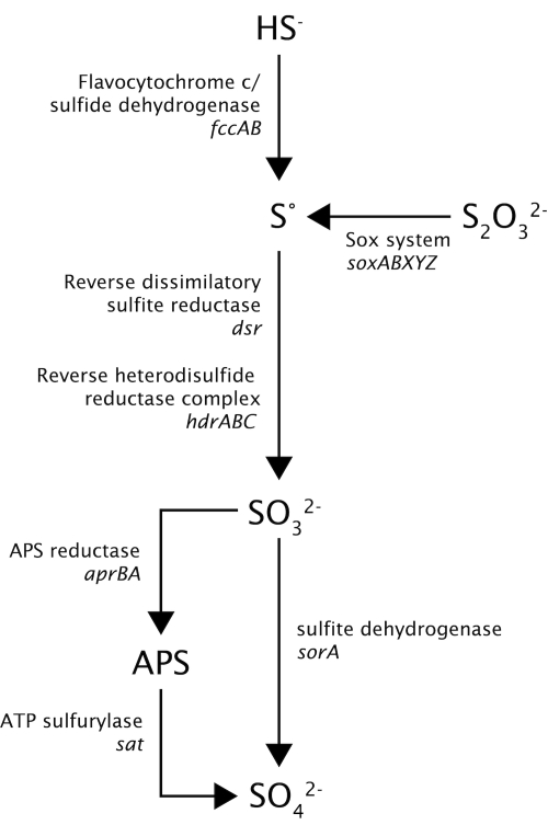
Thioalkalivibrio species can oxidize reduced sulfur compounds, such as sulfide and thiosulfate, to elemental sulfur and subsequently to sulfate. However, little is known about the enzymes that are involved in the sulfur metabolism of these organisms. Figure 4 shows the pathway of sulfur metabolism in strain HL-EbGr7 inferred from the genes found in the genome. Sulfide is oxidized by flavocytochrome c/sulfide dehydrogenase; both genes, encoding the small subunit A (fccA), and the large subunit B (fccB) were present in 3 copies. Although sulfide:quinone oxidoreductase (SQR) activity had been found in Thioalkalivibrio species, the sqr gene could not be detected. A similar result has also been found for Allochromatium vinosum [35]. The presence of a truncated Sox cluster consisting of soxXAYZB, but lacking soxCD, leads to the formation of elemental sulfur as an intermediate [35], and also gives the organism the possibility to oxidize the sulfane moiety of thiosulfate, which has been confirmed by culture studies. The soxXA genes were present in 4 copies; the sox YZ genes in 2 copies. Subsequently, elemental sulfur is oxidized to sulfite by a reversed dissimilatory sulfite reductase pathway, consisting of dsrABEFHCMKLJOPNR. In addition, we found genes (hdrABC) encoding a heterodisulfide reductase complex, which was highly similar to that found in Acidithiobacillus ferrooxidans and was hypothesized to work in reverse [36]. Sulfite can be oxidized to sulfate, either directly by sulfite dehydrogenase (sorA) or indirectly via adenosine-5′-phosphosulfate (APS [37]) by APS reductase encoded by aprBA and ATP sulfurylase encoded by sat. Obviously, the two alternative pathways may operate depending on the red-ox potential: (i) a sulfide-dependent microoxic pathway at very low red-ox conditions, such as present in the Thiopaq reactor, involving the ‘reversed sulfidogenic’ pathway, and (ii) an aerobic sulfide/thiosulfate oxidation pathway at high red-ox potential, such as in batch cultures with thiosulfate, involving the truncated Sox cluster and SorA. The presence of different copies of genes involved in the sulfur metabolism might indicate the adaptation of this organism to a highly specialized sulfide oxidation lifestyle.
Figure 4.
Hypothetical pathways for oxidation of sulfur compounds in “Thioalkalivibrio sulfidophilus” HL-EbGr7.
Energy metabolism
Although we are gaining some insight into the bioenergetics of alkaliphilic heterotrophs, such as Bacillus species [38], it is a complete mystery how haloalkaliphilic chemolithoautotrophic bacteria obtain enough energy for growth. To generate NADH for CO2 fixation, chemolithoautotrophic bacteria, using inorganic compounds (e.g. H2S or NH3) as electron donors, have to transport electrons against the thermodynamic gradient (‘reverse electron transport’), which is an energy-requiring process. In addition, those that are living at high salt concentrations, have to invest extra energy in the production of organic compatible solutes. And thirdly, bacteria that live at high pH have to invest additional energy to maintain their pH homeostasis.
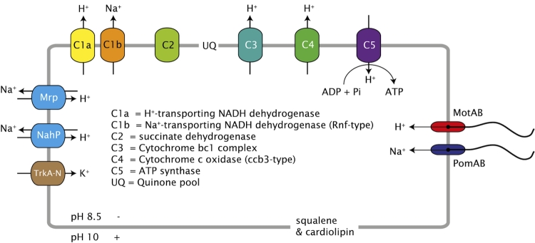
So, to obtain enough energy for growth the haloalkaliphilic chemolithoautotrophic SOB must have a special adaptation of their bioenergetics. The most obvious solution would be the presence of primary sodium pumps, such as a sodium-driven ATP synthase, but genes for this could not be detected; instead we found all the genes for a proton-driven F0F1-type ATP synthase (i.e., subunit A, B, and C of the F0 subcomplex, and subunit alpha, beta, gamma, delta, and epsilon of the F1 subcomplex). The presence of a proton-driven ATP synthase instead of a sodium-driven ATP synthase has been found in all genomes of so far studied aerobic alkaliphilic bacteria studied thus far [39]. However, we could detect several genes encoding different sodium-dependent pumps, such as the primary sodium pump Rnf and secondary pumps, such as the Na+/H+ antiporters NhaP and Mrp, a sodium:sulfate symporter (SulP), and the sodium-depending flagellar motor PomA/B. Apart from the genes encoding the proton-translocating NADH dehydrogenase (nuoABCDEFGHIJKLMN), we also found genes (rnfABCDGE) that are homologous to the nqr genes encoding the sodium-translocating NADH:quinone oxidoreductase (Na+-NQR [40], ). Na+-NQR was first discovered in the marine bacterium Vibrio alginolyticus [41]. It is coupled to the respiratory chain, and oxidizes NADH with ubiquinone as electron acceptor. The free energy released is used to generate a sodium motive force at the FAD-quinone coupling site. The presence of both a proton- and sodium-translocating NADH:quinone oxidoreductase in one organism was described previously by Takada et al. [42]. They showed that both pumps were very active in a psychrophilic bacterium, Vibrio sp. strain ABE-1, growing at low temperatures. It is, of course, not clear what the role of either pump is in our strain, but it is tempting to speculate that they are a special adaptation to generate enough energy for growth under these extreme conditions. Future transcriptomic and proteomic studies are necessary to validate this speculation. NhaP is a Na+/H+-antiporter (a secondary sodium pump), which plays a role in the regulation of the internal pH of the cell; it pumps sodium out of the cell and leaves protons and ensuing energy generated by the respiratory chain. Furthermore, we found all 7 genes (mnhA-G) for the multisubunit Na+/H+-antiporter Mrp, which may play a similar role as NhaP. Apart from genes encoding proton-driven flagellar motors (motA/B), we also found genes encoding sodium-driven flagellar motors (pomA/B). Phylogenetic analysis of the motA/B and pomA/B grouped them with sequences of other bacteria, such as Halorhodospira halophila and Alkalilimnicola ehrlichii (results are not shown).
We have also found genes for the production of cardiolipin (cardiolipin synthase) and of squalene (squalene synthase), confirming the high concentrations of these compounds in the cell membranes of another Thioalkalivibrio strain, strain ALJ15 [43]. These compounds contribute indirectly to an efficient energy metabolism, as the negatively charged cardiolipids might trap protons at the cell membrane preventing them from diffusing into the environment [44], and squalene lowers the proton permeability of the lipid bilayer [45]. From the genes that we found, we made the following conceptual model (Figure 5).
Figure 5.
Conceptual model of the different proton and sodium primary pumps, secondary transporters and flagellar motors in Thioalkalivibrio “sulfidophilus” HL-EbGr7.
Compatible solutes
Thioalkalivibrio species are characterized by their tolerance to high salt concentrations, which can be up to 4.3M total sodium [2,17]. To withstand these hypersaline conditions, these species synthesize glycine-betaine as the main compatible solute. In one of the high-salt Thioalkalivibrio strains, Banciu et al. [43] showed a positive correlation between salinity and the intracellular glycine-betaine concentration, and found that glycine-betaine constituted 9% of cell dry weight at 4M of sodium in the culture medium. In most cases, betaine is synthesized from choline by a two-step oxidation pathway [46]. However, an alternative route is the synthesis of betaine by a series of methylation reactions [47]. The genome of strain HL-EbGr7 contains genes coding for glycine sarcosine N-methyltransferase and sarcosine dimethylglycine methyltransferase, that are catalyzing betaine synthesis from glycine in a three-step methylation process, i.e., glycine -> sarcosine -> dimethylglycine -> betaine. The sequences of the 2 enzymes have high similarities to sequences found in the close relatives Halorhodospira halophila and Nitrococcus mobilis. Apart from glycine-betaine Thioalkalivibrio species also produce sucrose as a minor compatible solute (up to 2.5% of cell dry weight at 2M of sodium) [43]. The genomes of strain HL-EbGr7 contain genes coding for the enzymes sucrose synthase and sucrose phosphate synthase, which both play a role in the synthesis of sucrose. In contrast to other members of the Ectothiorhodospiraceae, i.e., Alkalilimnicola ehrlichii, and Halorhodospira halodurans, no genes were found for ectoine synthesis in the genome of HL-EbGr7.
Acknowledgments
This work was performed under the auspices of the US Department of Energy Office of Science, Biological and Environmental Research Program, and by the University of California, Lawrence Berkeley National Laboratory under contract No. DE-AC02-05CH11231, Lawrence Livermore National Laboratory under Contract No. DE-AC52-07NA27344, and Los Alamos National Laboratory under contract No. DE-AC02-06NA25396, UT-Battelle and Oak Ridge National Laboratory under contract DE-AC05-00OR22725. DS was supported financially by RFBR grant 10-04-00152.
References
- 1.Sorokin DY, Kuenen JG. Haloalkaliphilic sulfur-oxidizing bacteria from soda lakes. FEMS Microbiol Rev 2005; 29:685-702 10.1016/j.femsre.2004.10.005 [DOI] [PubMed] [Google Scholar]
- 2.Sorokin DY, Banciu H, Robertson LA, Kuenen JG. Haloalkaliphilic sulfur-oxidizing bacteria. In: The Prokaryotes. Volume 2: Ecophysiology and Biochemistry 2006; pp. 969-984. Dworkin, M., Falkow S, Rosenberg E, Schleifer KH, Stackebrandt E. (Ed's). Springer, New York. [Google Scholar]
- 3.Sorokin DY, van den Bosch PLF, Abbas B, Janssen AJH, Muyzer G. Microbiological analysis of the population of extremely haloalkaliphilic sulfur-oxidizing bacteria dominating in lab-scale sulfide-removing bioreactors. Appl Microbiol Biotechnol 2008; 80:965-975 10.1007/s00253-008-1598-8 [DOI] [PMC free article] [PubMed] [Google Scholar]
- 4.Field D, Garrity G, Gray T, Morrison N, Selengut J, Sterk P, Tatusova T, Thomson N, Allen MJ, Angiuoli SV, et al. The minimum information about a genome sequence (MIGS) specification. Nat Biotechnol 2008; 26:541-547 10.1038/nbt1360 [DOI] [PMC free article] [PubMed] [Google Scholar]
- 5.Woese CR, Kandler O, Wheelis ML. Towards a natural system of organisms: proposal for the domains Archaea, Bacteria, and Eucarya. Proc Natl Acad Sci USA 1990; 87:4576-4579 10.1073/pnas.87.12.4576 [DOI] [PMC free article] [PubMed] [Google Scholar]
- 6.Garrity GM, Holt JG. The Road Map to the Manual. In: Garrity GM, Boone DR, Castenholz RW (eds), Bergey's Manual of Systematic Bacteriology, Second Edition, Volume 1, Springer, New York, 2001, p. 119-169. [Google Scholar]
- 7.List Editor Validation of publication of new names and new combinations previously effectively published outside the IJSEM. List no. 106. Int J Syst Evol Microbiol 2005; 55:2235-2238 10.1099/ijs.0.64108-0 [DOI] [PubMed] [Google Scholar]
- 8.Garrity GM, Bell JA, Lilburn T. Class III. Gammaproteobacteria class. nov. In: Garrity GM, Brenner DJ, Krieg NR, Staley JT (eds), Bergey's Manual of Systematic Bacteriology, Second Edition, Volume 2, Part B, Springer, New York, 2005, p. 1. [Google Scholar]
- 9.Imhoff J. Order I. Chromatiales ord. nov. In: Garrity GM, Brenner DJ, Krieg NR, Staley JT (eds), Bergey's Manual of Systematic Bacteriology, Second Edition, Volume 2, Part B, Springer, New York, 2005, p. 1-3. [Google Scholar]
- 10.Imhoff JF. Reassignment of the genus Ectothiorhodospira Pelsh 1936 to a new family, Ectothiorhodospiraceae fam. nov., and emended description of the Chromatiaceae Bavendamm 1924. Int J Syst Bacteriol 1984; 34:338-339 10.1099/00207713-34-3-338 [DOI] [Google Scholar]
- 11.Sorokin DY, Lysenko AM, Mityushina LL, Tourova TP, Jones BE, Rainey FA, Robertson LA, Kuenen GJ. Thioalkalimicrobium aerophilum gen. nov., sp. nov. and Thioalkalimicrobium sibericum sp. nov., and Thioalkalivibrio versutus gen. nov., sp. nov., Thioalkalivibrio nitratis sp.nov., novel and Thioalkalivibrio denitrificancs sp. nov., novel obligately alkaliphilic and obligately chemolithoautotrophic sulfur-oxidizing bacteria from soda lakes. Int J Syst Evol Microbiol 2001; 51:565-580 [DOI] [PubMed] [Google Scholar]
- 12.Banciu H, Sorokin DY, Galinski EA, Muyzer G, Kleerebezem R, Kuenen JG. Thioalkalivibrio halophilus sp. nov., a novel obligately chemolithoautotrophic, facultatively alkaliphilic, and extremely salt-tolerant, sulfur-oxidizing bacterium from a hypersaline alkaline lake. Extremophiles 2004; 8:325-334 10.1007/s00792-004-0391-6 [DOI] [PubMed] [Google Scholar]
- 13.List Editor Notification that new names and new combinations have appeared in volume 51, part 2, of the IJSEM. Int J Syst Evol Microbiol 2001; 51:795-796 [DOI] [PubMed] [Google Scholar]
- 14.Classification of Bacteria and Archaea in risk groups. www.baua.de TRBA 466.
- 15.Foti M, Ma S, Sorokin DY, Rademaker JLW, Kuenen GJ, Muyzer G. Genetic diversity and biogeography of haloalkaliphilic sulfur-oxidizing bacteria beloning to the genus Thioalkalivibrio. FEMS Microbiol Ecol 2006; 56:95-101 10.1111/j.1574-6941.2006.00068.x [DOI] [PubMed] [Google Scholar]
- 16.Maps and Utilities for the PC, Mac, iPhone, iPod touch and Smartphones. http://www.iTouchMap.com
- 17.Sorokin DY, Kuenen JG, Jetten M. Denitrification at extremely alkaline conditions in obligately autotrophic alkaliphilic sulfur-oxidizing bacterium “Thioalkalivibrio denitrificans”. Arch Microbiol 2001; 175:94-101 10.1007/s002030000210 [DOI] [PubMed] [Google Scholar]
- 18.Sorokin DY, Antipov AN, Kuenen JG. Complete denitrification in a coculture of haloalkaliphilic sulfur-oxidizing bacteria from a soda lake. Arch Microbiol 2003; 180:127-133 10.1007/s00203-003-0567-y [DOI] [PubMed] [Google Scholar]
- 19.Sorokin DY, Tourova TP, Lysenko AM, Kuenen JG. Microbial thiocyanate utilization under high alkaline conditions. Appl Environ Microbiol 2001; 67:528-538 10.1128/AEM.67.2.528-538.2001 [DOI] [PMC free article] [PubMed] [Google Scholar]
- 20.Sorokin DY, Tourova TP, Antipov AN, Muyzer G, Kuenen JG. Anaerobic growth of the haloalkaliphilic denitrifying sulfur-oxidizing bacterium Thialkalivibrio thiocyanodenitrificans sp. nov. with thiocyanate. Microbiology 2004; 150:2435-2442 10.1099/mic.0.27015-0 [DOI] [PubMed] [Google Scholar]
- 21.Janssen AJH, Lens PNL, Stams AJM, Plugge CM, Sorokin DY, Muyzer G, Dijkmane H, Van Zessene E, Luimesf P, Buisman CJN. Application of bacteria involved in the biological sulfur cycle for paper mill effluent purification. Sci Total Environ 2009; 407:1333-1343 [DOI] [PubMed] [Google Scholar]
- 22.Nelson DC, Jannasch HW. Chemoautotrophic growth of a marine Beggiatoa in sulfide-gradient cultures. Arch Microbiol 1983; 136:262-269 10.1007/BF00425214 [DOI] [Google Scholar]
- 23.Pruesse E, Quast C, Knittel K, Fuchs B, Ludwig W, Peplies J, Glöckner FO. SILVA: a comprehensive online resource for quality checked and aligned ribosomal RNA sequence data compatible with ARB. Nucleic Acids Res 2007; 35:7188-7196 10.1093/nar/gkm864 [DOI] [PMC free article] [PubMed] [Google Scholar]
- 24.Ludwig W, Strunk O, Westram R, Richter L, Meier H, Kumar Y, Buchner A, Lai T, Steppi S, Jobb G. ARB: a software environment for sequence data. Nucleic Acids Res 2004; 32:1363-1371 10.1093/nar/gkh293 [DOI] [PMC free article] [PubMed] [Google Scholar]
- 25.Liolios K, Chen IM, Mavromatis K, Tavernarakis N, Hugenholtz P, Markowitz VM, Kyrpides NC. The Genomes OnLine Database (GOLD) in 2009: status of genomic and metagenomic projects and their associated metadata. Nucleic Acids Res 2010; 38:D346-D354 10.1093/nar/gkp848 [DOI] [PMC free article] [PubMed] [Google Scholar]
- 26.DOE Joint Genome Institute http://www.jgi.doe.gov/
- 27.Hyatt D, Chen GL, LoCascio PF, Land ML, Larimer FW, Hauser LJ. Prodigal: prokaryotic gene recognition and translation initiation site identification. BMC Bioinformatics 2010; 11:119 10.1186/1471-2105-11-119 [DOI] [PMC free article] [PubMed] [Google Scholar]
- 28.Pati A, Ivanova NN, Mikhailova N, Ovchinnikova G, Hooper SD, Lykidis A, Kyrpides NC. GenePRIMP: a gene prediction improvement pipeline for prokaryotic genomes. Nat Methods 2010; 7:455-457 10.1038/nmeth.1457 [DOI] [PubMed] [Google Scholar]
- 29.Markowitz VM, Szeto E, Palaniappan K, Grechkin Y, Chu K, Chen IM, Dubchak I, Anderson I, Lykidis A, Mavromatis K, et al. The integrated microbial genomes (IMG) system in 2007: data content and analysis tools extensions. Nucleic Acids Res 2008; 36:D528-D533 10.1093/nar/gkm846 [DOI] [PMC free article] [PubMed] [Google Scholar]
- 30.Shibata M, Katoh H, Sonoda M, Ohkawat H, Shimoyama M, Fukuzawa H, Kaplan A, Ogawa T. Genes essential to sodium-dependent bicarbonate transport in cyanobacteria: function and phylogenetic analysis. J Biol Chem 2002; 277:18658-18664 10.1074/jbc.M112468200 [DOI] [PubMed] [Google Scholar]
- 31.Dobrinski KP, Longo DL, Scott KM. The carbon-concentrating mechanism of the hydrothermal vent chemolithoautotroph Thiomicrospira crunogena. J Bacteriol 2005; 187:5761-5766 10.1128/JB.187.16.5761-5766.2005 [DOI] [PMC free article] [PubMed] [Google Scholar]
- 32.Yeates TO, Kerfeld CA, Heinhorst S, Cannon GC, Shively JM. Protein-based organelles in bacteria: carboxysomes and related microcompartments. Nat Rev Microbiol 2008; 6:681-691 10.1038/nrmicro1913 [DOI] [PubMed] [Google Scholar]
- 33.Tourova TP, Spiridonova EM, Berg IA, Slobodova NV, Boulygina ES, Sorokin DY. Phylogeny and evolution of the family Ectothiorhodospiraceae based on comparison of 16S rRNA, cbbL and nifH genes. Int J Syst Evol Microbiol 2007; 57:2387-2398 10.1099/ijs.0.65041-0 [DOI] [PubMed] [Google Scholar]
- 34.Yoshizawa Y, Toyoda K, Arai H, Ishii M, Igarashi Y. CO2-responsive expression and gene organization of three ribulose-1,5-biphosphate carboxylase/oxygenase enzymes and carboxysomes in Hydrogenovibrio marinus strain MH-110. J Bacteriol 2004; 186:5685-5691 10.1128/JB.186.17.5685-5691.2004 [DOI] [PMC free article] [PubMed] [Google Scholar]
- 35.Frigaard NU, Dahl C. Sulfur metabolism in phototrophic sulfur bacteria. Adv Microb Physiol 2009; 54:103-200 10.1016/S0065-2911(08)00002-7 [DOI] [PubMed] [Google Scholar]
- 36.Quatrini R, Appia-Ayme C, Dennis Y, Jedlicki E, Holmes DS, Bonnefoy V. Extending the models for iron and sulfur oxidation in the extreme acidophile Acidithiobacillus ferrooxidans. BMC Genomics 2009; 10: 394 10.1186/1471-2164-10-394 [DOI] [PMC free article] [PubMed] [Google Scholar]
- 37.Kappler U. Bacterial sulfite-oxidizing enzymes. Biochim Biophys Acta 2011; 1807:1-10 10.1016/j.bbabio.2010.09.004 [DOI] [PubMed] [Google Scholar]
- 38.Padan E, Bibi E, Ito M, Krulwich TA. Alkaline pH homeostasis in bacteria: New insights. Biochim Biophys Acta 2005; 1717:67-88 10.1016/j.bbamem.2005.09.010 [DOI] [PMC free article] [PubMed] [Google Scholar]
- 39.Hicks DB, Liu J, Fujisawa M, Krulwich TA. F1F0-ATP synthases of alkaliphilic bacteria: Lessons from their adaptations. Biochim Biophys Acta 2010; 1797:1362-1377 10.1016/j.bbabio.2010.02.028 [DOI] [PMC free article] [PubMed] [Google Scholar]
- 40.Verkhovsky MI, Bogachev AV. Sodium-translocating NADH:quinone oxidoreductase as a redox-driven ion pump. Biochim Biophys Acta 2010; 1797:738-746 10.1016/j.bbabio.2009.12.020 [DOI] [PubMed] [Google Scholar]
- 41.Tokuda H, Unemoto T. A respiratory-dependent primary sodium extrusion system functioning at alkaline pH in the marine bacterium Vibrio alginolyticus. Biochem Biophys Res Commun 1981; 102:265-271 10.1016/0006-291X(81)91516-3 [DOI] [PubMed] [Google Scholar]
- 42.Takada Y, Fukunaga N, Sasaki S. Respiration-dependent proton and sodium pumps in a psychrophilic bacterium, Vibrio sp. strain ABE-1. Plant Cell Physiol 1988; 29:207-214 [Google Scholar]
- 43.Banciu H, Sorokin DY, Rijpstra WI, Sinninghe Damsté JS, Galinski EA, Takaichi S, Muyzer G, Kuenen JG. Fatty acid, compatible solute and pigment composition of the obligately chemolithoautotrophic alkaliphilic sulfur-oxidizing bacteria from soda lakes. FEMS Microbiol Lett 2005; 243:181-187 10.1016/j.femsle.2004.12.004 [DOI] [PubMed] [Google Scholar]
- 44.Haines TH, Dencher NA. Cardiolipin: a proton trap for oxidative phosphorylation. FEBS Lett 2002; 528:35-39 10.1016/S0014-5793(02)03292-1 [DOI] [PubMed] [Google Scholar]
- 45.Hauß T, Dantea S, Dencher NA, Haines TH. Squalene is in the midplane of the lipid bilayer: implications for its function as a proton permeability barrier. Biochim Biophys Acta 2002; 1556:149-154 10.1016/S0005-2728(02)00346-8 [DOI] [PubMed] [Google Scholar]
- 46.Boch J, Kempf B, Schmid R, Bremer E. Synthesis of the osmoprotectant glycine betaine in Bacillus subtilis: Characterization of the gbsAB genes. J Bacteriol 1996; 178:5121-5129 [DOI] [PMC free article] [PubMed] [Google Scholar]
- 47.Lai MC, Yang DR, Chuang MJ. Regulatory factors associated with synthesis of the osmolyte glycine betaine in the halophilic methanoarchaeon Methanohalophilus portucalensis. Appl Environ Microbiol 1999; 65:828-833 [DOI] [PMC free article] [PubMed] [Google Scholar]


