Abstract
Botulinum neurotoxins cleave synaptic SNAREs and block exocytosis, demonstrating that these proteins function in neurosecretion. However, the function of the SNARE syntaxin remains less clear because no neurotoxin cleaves it selectively. Starting with a botulinum neurotoxin which cleaves both syntaxin and SNAP-25, we engineered a version that retains activity against syntaxin but spares SNAP-25. These mutants block synaptic release in neurons and norepinephrine release in neuroendocrine cells, thus establishing an essential role for syntaxin in Ca2+-triggered exocytosis. These mutants can generate syntaxin-free cells as a useful experimental system for research, and may lead to pharmaceuticals that target syntaxin selectively.
Keywords: Exocytosis, membrane trafficking, zinc protease, botulinum neurotoxin
A large body of experimental work supports the view that soluble N-ethylmaleimide sensitive factor attachment protein receptors (SNAREs) function in Ca2+-triggered exocytosis of neurotransmitters and hormones (1, 2). However, for one of the three synaptic SNAREs, the plasma membrane protein syntaxin, unequivocal support for such a role is lacking. For the two other synaptic SNAREs, SNAP-25 and synaptobrevin (also known as VAMP), experiments with botulinum neurotoxin (BoNT) and tetanus toxin, have provided some of the most convincing evidence in support of a function in exocytosis (3). Most of these toxins selectively cleave synaptobrevin or SNAP-25, and they all block exocytosis. However, none of them cleave syntaxin selectively. BoNT/C1 is the only one that cleaves syntaxin, but it also cleaves SNAP-25. Genetic studies have confirmed the roles of SNAP-25 (4) and synaptobrevin (5), and by providing cells for testing mutants on null backgrounds, these knock-outs have enabled researchers to investigate the roles of these two proteins in remarkable detail (6, 7). While loss of syntaxin from invertebrates impairs synaptic transmission (8), ablation of different exons of the syntaxin 1A gene either leaves synaptic transmission normal (9) or is embryonic lethal (10). Thus, for vertebrates the function of syntaxin has yet to be rigorously tested, and a null system for detailed mutagenesis studies is still unavailable.
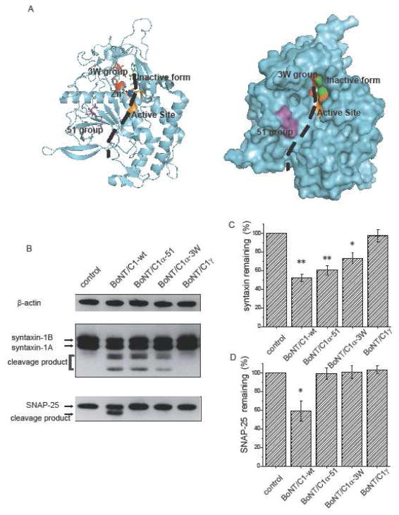
All of the BoNT serotypes have a similar overall structure, with a ~100 kD heavy chain that mediates cell surface binding and entry, and a ~50 kD light chain that acts as a selective protease. The homology between the various BoNT light chains indicated that we can use the crystal structure of the BoNT/A-substrate complex (11) to locate residues important for substrate recognition and specificity within the BoNT/C1 structure (12) (Fig. 1A). We created more than 150 mutants in these and other parts of the BoNT/C1 light chain and assayed their proteolytic activity by using a lentiviral vector to introduce the encoding DNA into neurons. Western blot analysis was then used to test for proteolysis of syntaxin and SNAP-25 (Supplementary Methods).
Fig. 1.
A. Structure of BoNT/C1 light chain (12) in ribbon (left) and space-filling (right) representations (PDB ID: 2QN0). The position of the substrate, indicated by a black dashed curve, was inferred from the BoNT/A-substrate crystal structure (11). B. Western blot analysis of extracts from cultured hippocampal neurons blotted for syntaxin and SNAP-25. Untransfected control cells (lane 1) show full length syn-taxin 1A, syntaxin 1B, and SNAP-25. Infection with wild type BoNT/C1 light chain cleaves all three proteins (lane 2). BoNT/C1α-51 (lane 3) and BoNT/C1α-3W (lane 4) cleaved syntaxin and spared SNAP-25. BoNT/C1γ showed no catalytic activity toward any substrate. C and D. Gel quantification of BoNT/C1 cleavage. The syntaxin (C) and SNAP-25 (D) band densities were reduced by cleavage in parallel with the appearance of lower molecular mass cleavage products. Syntaxin and SNAP-25 signals were normalized to the β-actin control and averaged (N=3).
Extracts from control cells (cultured rat hippocampal neurons not transfected with a BoNT/C1 light chain) contained syntaxin 1A and 1B, which appeared as two closely spaced bands, and SNAP-25 which appeared as a single band (Fig. 1B). Transfection with wild type BoNT/C1 light chain reduced the amount of full length protein and produced lower molecular weight cleavage products. Most of the mutants we created either showed dual specificity or no activity (Supplementary Material), but two were found to cleave only syntaxin and spare SNAP-25 (lanes 3 and 4 of Fig. 1B). These two highly selective mutants were designated BoNT/C1α-3W and BoNT/C1α-51. BoNT/C1α-3W was generated by mutating the three residues in the S1′ pocket (L200/M221/I226) (Fig 1A, red) to tryptophan. BoNT/C1α-51 was another triple mutant, 51T/52N/53P, in a region that has not received prior attention. Wild type BoNT/C1, BoNT/C1α-51, and BoNT/C1α-3W reduced the syntaxin bands by 52.3%, 60.5%, and 73.3% of control, respectively, while only wild type BoNT/C1 reduced the density of the SNAP-25 band (Fig. 1C). No cleavage of synaptobrevin was seen with BoNT/C1α-51 in one experiment (data not shown). None of the many other mutants we tested showed such clear selectivity for syntaxin over SNAP-25. These two mutants can thus serve as tools for testing the role of syntaxin. A third mutant, the inactive form denoted as BoNT/C1γ has mutations in two critical catalytic residues (R372A and Y375F). This mutant had no proteolytic activity (lane 5 of Fig. 1B), and was used as a control for transfection and actions unrelated to catalysis.
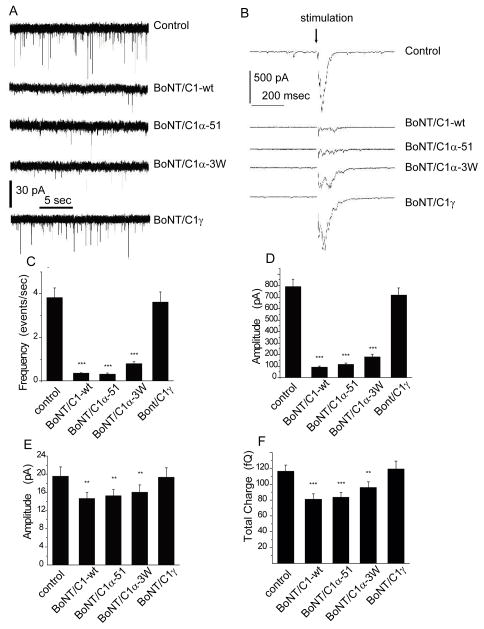
To evaluate the role of syntaxin in synaptic transmission we performed patch clamp recordings from cultured hippocampal neurons expressing wild type BoNT/C1 and BoNT/C1 mutants. Toxins effective in cleaving syntaxin reduced both miniature excitatory postsynaptic currents (mEPSCs) (Fig. 2A) and electrically evoked synaptic responses (Fig. 2B). The frequency of mEPSCs (Fig. 2C) and the amplitudes of evoked synaptic currents (Fig. 2D) were reduced approximately 10-fold below controls. BoNT/C1α-51 blocked both forms of release with similar effectiveness, while BoNT/C1α-3W was somewhat less effective, reducing spontaneous (Fig. 2C) and evoked (Fig. 2D) release by about 5-fold. BoNT/C1γ, the inactive control, produced no significant reduction in spontaneous or evoked release or quantal size (Figs. 2A–2F). The reduction in synaptic transmission was greater than the reduction in syntaxin levels (compare Figs. 2A–2D with Fig. 1C). This disparity has been noted previously for wild type neurotoxins and interpreted to mean that the exocytosis of one vesicle requires the cooperative action of multiple SNAREs (13).
Fig. 2.
Synaptic release. Patch clamp recordings from cultured hippocampal neurons 72–96 hours after infection. A. Miniature excitatory synaptic currents (mEPSCs). B. Synaptic currents evoked by extracellular stimulation (200 μA, 0.4 msec). C. Mean mEPSC frequency (N = 8–10 cells; ~20–300 events per cell). D. Mean mEPSC amplitude. E. Mean evoked synaptic current. F. Mean mEPSC charge. **p<0.01, *** p<0.001
Wild type BoNT/C1 and the two active mutants both produced a small (~20%) but significant reduction in the amplitude of miniature synaptic currents (Fig. 2E) and charge per mEPSC (Fig. 2F), indicating that with lower levels of either syntaxin, or of both syntaxin and SNAP-25, the few vesicles still able to fuse are smaller and contain less glutamate (14). This could reflect the lower energy barrier for exocytosis of smaller vesicles (15), which can be overcome by the assembly of fewer SNARE complexes (16).
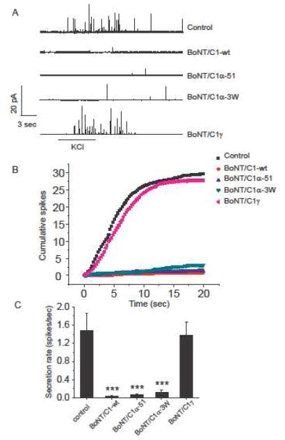
To evaluate syntaxin in the exocytosis of dense-core vesicles we transfected PC12 cells with lentiviral vector DNA. Release was monitored using amperometry, which reveals the release of individual vesicles as spikes (17) (Fig. 3A). BoNT/C1 variants that cleave syntaxin blocked depolarization-induced release of norepinephrine from PC12 cells. Depolarized PC12 cells showed much fewer spikes following transfection with wild type BoNT/C1, BoNT/C1α-51, or BoNT/C1α-3W (Fig. 3A). The cumulative spike plots (Fig. 3B) and overall secretion rates (Fig. 3C) indicated a roughly 50-fold reduction, but the action of BoNT/C1α-3W was somewhat weaker, and the control toxin BoNT/C1γ had no effect.
Fig. 3.
Norepinephrine release from PC12 cells 2–3 days after transfection. A. Amperometric traces, with exocytosis induced by application of 105 mM KCl (bar below). B. Cumulative spike counts from traces such as in A (control, N = 9–16 cells C. Mean frequency of spikes per cell. **** p<0.001
These experiments establish engineered versions BoNT/C1 as tools for the acute and selective elimination of syntaxin from cells. Using these reagents we were able to demonstrate that syntaxin is essential for Ca2+-triggered exocytosis of synaptic vesicles in neurons and dense-core vesicles in endocrine cells. Like cleavage of SNAP-25 and synaptobrevin, cleavage of syntaxin 1A and 1B dramatically reduced exocytosis. The present experiments thus place syntaxin on an equal footing with the other synaptic SNAREs as essential components of the exocytotic apparatus. Furthermore, by providing a syntaxin-free background, the engineered BoNT/C1 light chains developed for this study will serve as valuable tools for detailed studies of the mechanism by which syntaxin functions in exocytosis.
We generated two series of BoNT/C1 light chain variants that are specific syntaxin proteases. These newly engineered BoNT/C1 light chain proteins may also aid in extending the therapeutic potential of the clostridial neurotoxins (18). BoNT/C1 offers a potential alternative for BoNT/A in BoNT/A nonresponsive patients (19, 20). Furthermore, BoNT/C1α could be useful in the treatment of diseases involving syntaxin, which has a number of functions in addition to exocytosis, including regulation of K+ channels (21), Ca2+ channels (22) and K-ATP channels (23). Regulation of K-ATP channels is very important in type II diabetes treatment, many cases of which are caused by hyperactive beta-cells and are treated by K-ATP channel openers to limit electrical activity. Syntaxin 1A down regulates K-ATP channels to make beta cells more excitable (24). Cleavage of syntaxin would thus increase K-ATP channel activity to reduce beta cell firing and restore normal insulin secretion.
Supplementary Material
Acknowledgments
Funded by NIH grant NS44057 and the Howard Hughes Medical Institute.
Footnotes
BRIEFS. An engineered botulinum neurotoxin selectively cleaves syntaxin.
Supporting Information Available Detailed methodology. Western blot analysis of ~150 BoNT/C1 light chain mutants. This material is available free of charge via the Internet at http://pubs.acs.org.
References
- 1.Chen YA, Scheller RH. Nat Rev Mol Cell Biol. 2001;2:98–106. doi: 10.1038/35052017. [DOI] [PubMed] [Google Scholar]
- 2.Jahn R, Scheller RH. Nat Rev Mol Cell Biol. 2006;7:631–643. doi: 10.1038/nrm2002. [DOI] [PubMed] [Google Scholar]
- 3.Schiavo G, Matteoli M, Montecucco C. Physiol Rev. 2000;80:717–766. doi: 10.1152/physrev.2000.80.2.717. [DOI] [PubMed] [Google Scholar]
- 4.Washbourne P, Thompson PM, Carta M, Costa ET, Mathews JR, Lopez-Bendito G, Molnar Z, Becher MW, Valenzuela CF, Partridge LD, Wilson MC. Nat Neurosci. 2002;5:19–26. doi: 10.1038/nn783. [DOI] [PubMed] [Google Scholar]
- 5.Borisovska M, Zhao Y, Tsytsyura Y, Glyvuk N, Takamori S, Matti U, Rettig J, Sudhof T, Bruns D. Embo J. 2005;24:2114–2126. doi: 10.1038/sj.emboj.7600696. [DOI] [PMC free article] [PubMed] [Google Scholar]
- 6.Kesavan J, Borisovska M, Bruns D. Cell. 2007;131:351–363. doi: 10.1016/j.cell.2007.09.025. [DOI] [PubMed] [Google Scholar]
- 7.Sorensen JB, Wiederhold K, Muller EM, Milosevic I, Nagy G, de Groot BL, Grubmuller H, Fasshauer D. Embo J. 2006;25:955–966. doi: 10.1038/sj.emboj.7601003. [DOI] [PMC free article] [PubMed] [Google Scholar]
- 8.Schulze KL, Broadie K, Perin MS, Bellen HJ. Cell. 1995;80:311–320. doi: 10.1016/0092-8674(95)90414-x. [DOI] [PubMed] [Google Scholar]
- 9.Fujiwara T, Mishima T, Kofuji T, Chiba T, Tanaka K, Yamamoto A, Akagawa K. J Neurosci. 2006;26:5767–5776. doi: 10.1523/JNEUROSCI.0289-06.2006. [DOI] [PMC free article] [PubMed] [Google Scholar]
- 10.McRory JE, Rehak R, Simms B, Doering CJ, Chen L, Hermosilla T, Duke C, Dyck R, Zamponi GW. Biochem Biophys Res Commun. 2008;375:372–377. doi: 10.1016/j.bbrc.2008.08.031. [DOI] [PubMed] [Google Scholar]
- 11.Breidenbach MA, Brunger AT. Nature. 2004;432:925–929. doi: 10.1038/nature03123. [DOI] [PubMed] [Google Scholar]
- 12.Jin R, Sikorra S, Stegmann CM, Pich A, Binz T, Brunger AT. Biochemistry. 2007;46:10685–10693. doi: 10.1021/bi701162d. [DOI] [PubMed] [Google Scholar]
- 13.Montecucco C, Schiavo G, Pantano S. Trends Biochem Sci. 2005;30:367–372. doi: 10.1016/j.tibs.2005.05.002. [DOI] [PubMed] [Google Scholar]
- 14.Bekkers JM, Richerson GB, Stevens CF. Proc Natl Acad Sci (U S A) 1990;87:5359–5362. doi: 10.1073/pnas.87.14.5359. [DOI] [PMC free article] [PubMed] [Google Scholar]
- 15.Zhang Z, Jackson MB. Biophys J. 2010;98:2524–2534. doi: 10.1016/j.bpj.2010.02.043. [DOI] [PMC free article] [PubMed] [Google Scholar]
- 16.Jackson MB. J Membr Biol. 2010;235:89–100. doi: 10.1007/s00232-010-9258-1. [DOI] [PMC free article] [PubMed] [Google Scholar]
- 17.Wightman RM, Jankowski JA, Kennedy RT, Kawagoe KT, Schroeder TJ, Leszczyszyn DJ, Near JA, Diliberto DJ, Jr, Viveros OH. TProc Natl Acad Sci (USA) 1991;88:10754–10758. doi: 10.1073/pnas.88.23.10754. [DOI] [PMC free article] [PubMed] [Google Scholar]
- 18.Chen S, Barbieri JT. Proc Natl Acad Sci U S A. 2009;106:9180–9184. doi: 10.1073/pnas.0903111106. [DOI] [PMC free article] [PubMed] [Google Scholar]
- 19.Eleopra R, Tugnoli V, Rossetto O, Montecucco C, De Grandis D. Neurosci Lett. 1997;224:91–94. doi: 10.1016/s0304-3940(97)13448-6. [DOI] [PubMed] [Google Scholar]
- 20.Eleopra R, Tugnoli V, Quatrale R, Rossetto O, Montecucco C, Dressler D. Neurotox Res. 2006;9:127–131. doi: 10.1007/BF03033930. [DOI] [PubMed] [Google Scholar]
- 21.Fili O, Michaelevski I, Bledi Y, Chikvashvili D, Singer-Lahat D, Boshwitz H, Linial M, Lotan I. J Neurosci. 2001;21:1964–1974. doi: 10.1523/JNEUROSCI.21-06-01964.2001. [DOI] [PMC free article] [PubMed] [Google Scholar]
- 22.Bergsman JB, Tsien RW. J Neurosci. 2000;20:4368–4378. doi: 10.1523/JNEUROSCI.20-12-04368.2000. [DOI] [PMC free article] [PubMed] [Google Scholar]
- 23.Leung YM, Kwan EP, Ng B, Kang Y, Gaisano HY. Endocr Rev. 2007;28:653–663. doi: 10.1210/er.2007-0010. [DOI] [PubMed] [Google Scholar]
- 24.Kang Y, Leung YM, Manning-Fox JE, Xia F, Xie H, Sheu L, Tsushima RG, Light PE, Gaisano HY. J Biol Chem. 2004;279:47125–47131. doi: 10.1074/jbc.M404954200. [DOI] [PubMed] [Google Scholar]
Associated Data
This section collects any data citations, data availability statements, or supplementary materials included in this article.


