Abstract
Pancreatic cancer is an exceptionally aggressive disease in great need of more effective therapeutic options. Epithelial-mesenchymal transition (EMT) plays a key role in cancer invasion and metastasis and there is a gain of stem cell properties during EMT. Here we report increased expression of the putative pancreatic stem cell marker DCAMKL-1 in an established KRAS transgenic mouse model of pancreatic cancer and in human pancreatic adenocarcinoma. Co-localization of DCAMKL-1 with vimentin, a marker of mesenchymal lineage, along with 14-3-3 σ was observed within pre-malignant PanIN lesions that arise in the mouse model. siRNA-mediated knockdown of DCAMKL-1 in human pancreatic cancer cells induced microRNA miR-200a, an EMT inhibitor, along with down-regulation of EMT-associated transcription factors ZEB1, ZEB2, Snail, Slug and Twist. Furthermore, DCAMKL-1 knockdown resulted in downregulation of c-Myc and KRAS through a let-7a microRNA-dependent mechanism, and downregulation of Notch-1 through a miR-144 microRNA-dependent mechanism. These findings illustrate direct regulatory links between DCAMKL-1, microRNAs and EMT in pancreatic cancer. Moreover, they demonstrate a functional role for DCAMKL-1 in pancreatic cancer. Together, our results rationalize DCAMKL-1 as a therapeutic target for eradicating pancreatic cancers.
Keywords: DCAMKL-1, Dclk-1, Cancer Stem Cell Marker, 14-3-3 σ, epithelial-mesenchymal transition (EMT), vimentin, pancreatic intraepithelial neoplasia (PanIN), Let-7a, miR-144, miR-200a, Notch-1
Introduction
Pancreatic adenocarcinoma has the worst prognosis of any major malignancy with a 3% 5-year survival (1). Major obstacles in treating pancreatic cancer include extensive local tumor invasion and early metastasis. There is increasing evidence that a small subset of cells termed “cancer stem cells” (CSCs) are capable of initiating and sustaining tumor growth (2). CSCs share unique properties with normal adult stem cells, including the ability to self-renew and differentiate. CSCs are often refractory to current standard chemotherapeutic agents and radiation therapies, as those treatment strategies are designed to eradicate actively cycling cells, not slowly cycling CSCs. This results in tumor shrinkage but often fails to prevent tumor recurrence, due to the surviving CSCs ability to regenerate the tumor (3). Thus, novel therapies that specifically target the CSC population, either alone or in conjunction with current strategies may be more effective in obliterating solid tumors.
The existence of CSCs was first demonstrated in acute myelogenous leukemia (4) and subsequently verified in breast (5), pancreatic (3) and brain tumors (6–8). The CD133+ subpopulations from brain tumors could initiate clonally derived neurospheres in vitro showing self-renewal, differentiation, and proliferative characteristics similar to normal brain stem cells (6–8). In a recent study a subpopulation of CD44+CD24+ESA+ cells derived from primary human pancreatic adenocarcinomas CSCs (3) were implanted in immunocompromised mice and identified a subpopulation of cells with enhanced tumorigenic potential.
The 14-3-3 σ gene was originally characterized as the human mammary epithelial-specific marker, HME-1 (9). Besides its G2/M checkpoint functions, 14-3-3 σ inhibits the pro-apoptotic proteins Bad and Bax (10, 11). 14-3-3 σ is up-regulated in lung cancer (12) and in head and neck carcinomas (13). Increased mRNA and protein expression of 14-3-3 σ has been demonstrated in human pancreatic adenocarcinoma (14). Furthermore, several studies have demonstrated that 14-3-3 σ contributes to the chemoresistance of pancreatic cancer cells (15, 16). Therefore, strategies aimed at suppressing 14-3-3 σ expression and function may have a therapeutic benefit in pancreatic cancer.
MicroRNAs (miRNAs) are endogenous, approximately 22 nucleotide (nt) RNAs that play important regulatory roles at the posttranscriptional level in animals and plants by targeting mRNAs for cleavage or translational repression (17). miRNAs have emerged as important developmental regulators and control critical processes such as cell fate determination and cell death (17). There is increasing evidence that several miRNAs are mutated or poorly expressed in human cancers and may act as tumor suppressors or oncogenes (18, 19). Currently, the target genes of miRNAs are mainly identified by a combination of bioinformatic searches for potential miRNA recognition elements in the 3’-untranslated region (3’ UTR) of the target gene. Subsequent experimental validation of predicated miRNA target interactions are conducted with luciferase reporter assays in cultured cells in vitro (17, 20).
We have recently demonstrated that DCAMKL-1, a microtubule-associated kinase expressed in post-mitotic neurons, is a putative intestinal and pancreatic stem cell marker (21, 22). Furthermore, we have reported that DCAMKL-1, a protein expressed in both normal stem cells and in cancer, likely promotes tumorigenesis through the regulation of pri-let-7a primary microRNA and c-Myc (23). Here we report that DCAMKL-1 is expressed in a subset of cells in human pancreatic tumors. We observed 14-3-3 σ in the cytoplasm and rarely in the nucleus of tumor epithelial cells in human pancreatic cancer patients. Interestingly, co-expression of DCAMKL-1 and 14-3-3 σ was observed in tumors. Moreover, we demonstrate DCAMKL-1 staining in the surface epithelium of pancreatic intraepithelial neoplasia (PanIN) type lesions and in the intervening stroma in human pancreatic adenocarcinoma. Knockdown of DCAMKL-1 in pancreatic cancer cells resulted in down regulation of Snail, Slug and Twist and induction of microRNA miR-200a, which inhibits EMT. Furthermore, knockdown of DCAMKL-1 also resulted in downregulation of the proto-oncogenes c-Myc and KRAS via up regulation of pri-let-7a and inhibition of Notch-1 via miR-144 miRNA dependent mechanisms. These data taken together identify DCAMKL-1 as a novel pancreatic CSC marker that can potentially be targeted for pancreatic cancer tumor eradication.
Materials and Methods
Tissue procurement
The human pancreatic adenocarcinoma (n = 10), pancreatitis (n = 4) and normal appearing human pancreatic tissues (n = 3) were derived from patients undergoing a surgical resection of the pancreas at the University of Oklahoma Health Sciences Center and confirmed to the policies and practices of the University’s IRB (protocol number 04586).
Cell Culture
AsPC-1 and BxPC3 human pancreatic adenocarcinoma cell lines were purchased within 6 months of the experiments from the American Type Culture Collection and maintained as recommended. The cell lines were authenticated by ATCC.
Silencer RNA
DCAMKL-1 small interfering RNA (siRNA) (si-DCAMKL-1) sequence targeting the coding region of DCAMKL-1 (accession No. NM_004734) (GGGAGUGAGAACAAUCUACtt) and scrambled siRNAs (si-SCR) not matching any of the human genes were obtained (Ambion Inc, Austin, TX) and transfected using siPORT™NeoFX™ (Ambion Inc).
Immunohistochemistry, Real-time Reverse Transcription-Polymerase Chain Reaction Analyses, miRNA Analysis and Luciferase Reporter Gene Assay
These analyses were carried out as previously described (23). Detailed descriptions are provided in the supplementary section of materials and methods.
Scoring
Composite scoring for the immunostaining was performed by senior pathologist Dr. Stan Lightfoot, University of Oklahoma Health Sciences Center. Detailed descriptions are provided in the supplementary section of materials and methods.
Stem/progenitor cell isolation from mouse pancreas
We isolated DCAMKL-1+ stem/progenitor cells from mouse pancreas as described earlier (22). Detailed descriptions are provided in the supplementary section of materials and methods.
Results
DCAMKL-1 is expressed in the P48Cre-LSL-KRASG12D mouse pancreatic cancer model
The P48Cre-LSL-KRASG12D is a mouse model of pancreatic cancer that was initially developed by the Tyler Jacks laboratory (24). P48cre-LSL-KRASG12D mouse model was originally developed on the 129V genetic background and later this model was backcrossed with C57BL/6 mice for more than fifteen generations. When compared to 129V, the mutant mouse on the C57BL/6 genetic background develops more aggressive pancreatic lesions. These mice exhibit PanIN lesions after 10 weeks. Furthermore, these mice develop pancreatic adenocarcinomas with metastasis by 32 weeks (25). Pancreatic tissues from 5-month-old P48Cre-LSL-KRASG12D and their wild-type (WT) littermates were immunostained for DCAMKL-1. We found a marked increase in ductal immunoreactivity and a unique expansion of islet DCAMKL-1 in the P48Cre-LSL-KRASG12D pancreatic cancer mouse model that correlated with progressive neoplastic changes (Fig. 1, A – D). Previously, using DNA micro arrays, several groups have demonstrated increased 14-3-3 σ mRNA expression in pancreatic ductal adenocarcinoma compared to normal pancreas (14). Similarly, 14-3-3 σ protein nuclear localization has been described in pancreatic cancer (26). We found several DCAMKL-1+ cells within the PanIN’s that also expressed nuclear 14-3-3 σ (Fig. 1E). Magnified images of this co-localization are shown in the Figure 1F and 1G. These data suggest that DCAMKL-1 is upregulated in pancreas of P48Cre-LSL-KRASG12D mouse and may play an important role in mutated KRAS mediated tumorigenesis.
Figure 1. DCAMKL-1 expression in P48Cre-LSL-KRASG12D pancreatic cancer mouse model.
Pancreatic tissues from 5-month-old WT littermate (400X) (A) and from 5-month-old (100X) (B) P48Cre-LSL-KRASG12D mouse were immunostained for DCAMKL-1. (C) A magnified portion of the image (B) demonstrating cells positive for DCAMKL-1 in the pancreatic duct (400X). (D) A magnified portion of the image (B) demonstrating cells positive for DCAMKL-1 in the islets (400X). Brown colored cells (arrows) indicate cells positive for DCAMKL-1. These data demonstrate an increased expression of DCAMKL-1 correlated with progressive neoplastic changes. (E) PanIN lesions of the 5-month-old P48Cre-LSL-KRASG12D mouse expressed DCAMKL-1 (brown) and 14-3-3 σ (purple). Cells positive for DCAMKL-1 and nuclear 14-3-3 σ are indicated by arrows (400X). (F and G) Areas of co-localization in figure 5E (arrows) are shown as magnified images.
DCAMKL-1 in human pancreatic cancer
We examined DCAMKL-1 immunoreactivity in human pancreatic adenocarcinoma by immunohistochemical analysis. Samples were obtained from patients undergoing surgical resection of pancreatic cancer and pancreatitis provided by Dr. Russell Postier (Dept. of Surgery, The University of Oklahoma Health Sciences Center). Tumors demonstrated strong DCAMKL-1 protein localization. However, within the histologically normal appearing resection specimens, DCAMKL-1 was observed within islets but not in the intervening stromal cells or ducts (Fig. 2A top left and Supplementary Figure S1A). However, in chronic pancreatitis, we observed DCAMKL-1 in the islets, ducts and a few intervening stromal cells (Supplementary Figure S1B–F). Within a neoplastic focus of the tumor resection specimen, however, intense spindle-shaped cytoplasmic staining of DCAMKL-1 was evident (Fig. 2A top right). DCAMKL-1 immunoreactivity in ductal epithelial cells within the tumor (Fig. 2A bottom left) and in intervening stromal elements was also observed (Fig. 2A bottom right).
Figure 2. DCAMKL-1 and 14-3-3 σ expression in human pancreatic adenocarcinoma.
(A) DCAMKL-1 expression (brown) in histologically normal appearing tissue from human pancreatic cancer resection specimen (top left) (200X). DCAMKL-1 in neoplastic pancreatic islet tissue (top right) (200X). DCAMKL-1 in ductal epithelial cells (bottom left) (400X). Intervening stromal elements demonstrate fibrillar DCAMKL-1 immunoreactivity (bottom right) (200X). Representative cells are indicated by arrows. (B) 14-3-3 σ (purple) and DCAMKL-1 (brown) at the islet periphery in normal appearing human pancreatic tissue (left) (100X). Representative cell demonstrating the cytoplasmic expression of 14-3-3 σ in magnified portion of the left image (right – arrow) (400X). (C) 14-3-3 σ (purple) and DCAMKL-1 (brown) expression in human pancreatic adenocarcinoma (left) (100X). In a magnified portion of the left image, nuclear localized 14-3-3 σ (purple) co-localized with cytoplasmic DCAMKL-1 (brown) (right – arrowhead) (400X). Fibrillar DCAMKL-1 staining in the intervening stroma (arrows). (D) DCAMKL-1 (brown) expression in ductal epithelium of a PanIN type lesion in human pancreatic adenocarcinoma (left – arrow) (400X). Intense cytoplasmic and nuclear staining of 14-3-3 σ (purple) and cytoplasmic DCAMKL-1 (brown) in a PanIN lesion (right – arrow) (400X).
Insets in the images on the right in the panel B, C and D are magnified images.
14-3-3 σ co-localizes with DCAMKL-1 in human pancreatic cancer
In normal appearing pancreatic tissue, we observed cytoplasmic staining for 14-3-3 σ and DCAMKL-1 at the islet periphery, albeit in distinctly separate cells. We did not observe any ducts expressing 14-3-3 σ in that particular specimen (Fig. 2B left and right). In pancreatic ductal adenocarcinoma, we observed strong cytoplasmic immunostaining for 14-3-3 σ (a marker of advanced PanIN lesions) in ductal epithelial cells, we also observed cells with nuclear localized 14-3-3 σ within tumor islet formations, similar to our observation in the mouse pancreatic cancer model. Among the nuclear 14-3-3 σ expressing cells about 10% co-expressed DCAMKL-1 (Fig. 2C left and right), suggesting that nuclear translocation of 14-3-3 σ may occur in putative pancreatic CSCs. Furthermore, we observed an association of stromal DCAMKL-1 with the PanIN lesions in 40% of the tissue samples Based on the composite scoring for DCAMKL-1 and 14-3-3 σ immunostaining, we observed an increase staining of both the proteins in adenocarcinoma compared to normal (Supplementary Table S1). We also found DCAMKL-1 protein expression in human pancreatic PanIN lesions (Fig. 2D left), which increases in a stage-dependent manner compared to normal ductal epithelia (Supplementary Figure 2A, B and C). Additionally, we observed strong cytoplasmic 14-3-3 σ and DCAMKL-1 co-staining within these lesions (Fig. 2D right). These data strongly support a role for 14-3-3 σ and DCAMKL-1 in the progression of pancreatic cancer and co-localization of nuclear 14-3-3 σ and DCAMKL-1 as a putative marker of pancreatic CSCs.
DCAMKL-1 co-localizes with vimentin in the stroma of human pancreatic adenocarcinoma
Initially, we observed DCAMKL-1+ staining in elongated cells in the surface epithelium of PanIN lesions (Fig. 3A left). Further characterization of these cells using vimentin, as a marker of mesenchymal lineage, demonstrated that vimentin immunoreactive cells appeared morphologically similar to DCAMKL-1 positive cells (Fig. 3A right). When double-labeled immunofluorescence was performed, co-localization of DCAMKL-1 and vimentin within the PanIN lesion was observed (Fig. 3B). As demonstrated earlier (Fig. 2A), fibrillar DCAMKL-1 staining was observed in approximately 40% of the stromal/mesenchymal compartment of the human pancreatic adenocarcinoma studied. The stromal nature of these cells was confirmed by co-staining with vimentin, where we observed 10% co-localization with DCAMKL-1 (Fig. 3, C and D). These data taken together suggest that DCAMKL-1 may be involved in the desmoplastic reaction associated with human pancreatic cancer and may also play a role in EMT (27).
Figure 3. DCAMKL-1 and vimentin expression in human pancreatic adenocarcinoma.
(A) DCAMKL-1 expressing cell in a PanIN type lesion (left – arrow). Vimentin expressing cell in the ductal epithelium of a PanIN type lesion (right – arrow). (400X). (B) DCAMKL-1 (red) and vimentin (green) in a PanIN lesion. Co-localization demonstrated in merged image (arrows) and nuclei are stained blue with Hoechst dye (400X). (C) DCAMKL-1 (red) and vimentin (green) in stromal compartment of human pancreatic adenocarcinoma. Co-localization demonstrated in merged image and nuclei are stained blue with Hoechst dye (100X). (D) A magnified portion of bottom right of 4C demonstrating immunolocalization of DCAMKL-1 (red) and vimentin (green) indicated by arrows.
DCAMKL-1 is involved in EMT
EMT is a phenotypic conversion that facilitates organ morphogenesis and tissue remodeling in embryonic development and wound healing. A similar phenotypic conversion is also detected in fibrotic diseases and neoplasia, and is associated with disease progression and outcome (27, 28). Gene-profiling studies also suggest that mesenchymal gene profiles in tumors are predictive of poor outcome in colorectal, breast and ovarian cancers (2, 29).
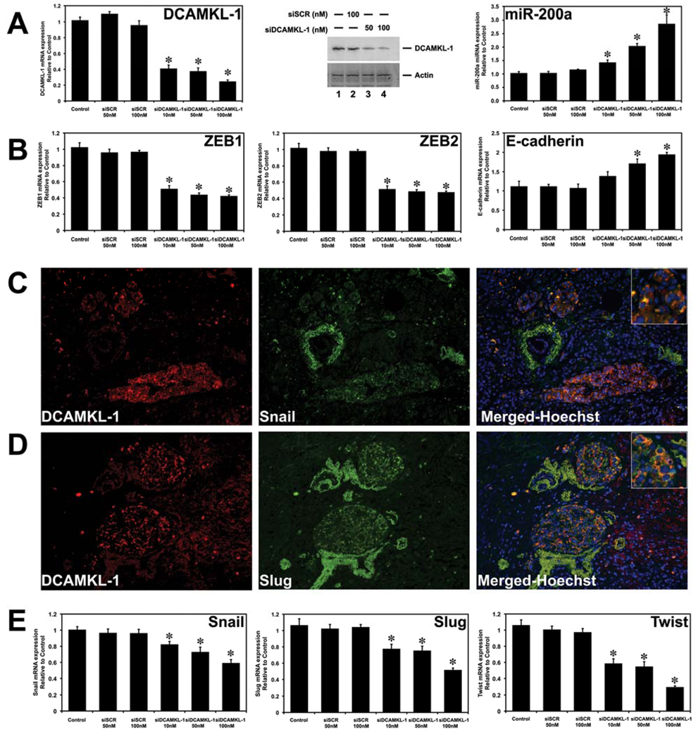
Recent report suggests that the downregulation of several miRNAs (miR-200a, miR-200b, miR-200c, miR-141 and miR-429) is an essential feature of EMT (30). Consequently, induction of these miRNAs results in inhibition of EMT (30–32). We have previously demonstrated that DCAMKL-1 negatively regulates tumor suppressor miRNA let-7a (23). To determine the potential role of DCAMKL-1 in EMT in pancreatic cancer, we performed siRNA-mediated knockdown of DCAMKL-1 and evaluated miRNA expression of several candidate miRNAs known to play a role in EMT (31, 32). One such miRNA, miR-200a inhibits EMT by repressing the transcription factors ZEB1 and ZEB2 with subsequent rescue of E-cadherin (31, 32). Here we demonstrate that siRNA-mediated knockdown of DCAMKL-1 results in upregulation of pri-miR-200a (Fig. 4A) and downregulation of ZEB1 and ZEB2 with upregulation of E-cadherin (Fig. 4B) in the AsPC-1 human pancreatic cancer cell line.
Figure 4. Knockdown of DCAMKL-1 inhibits EMT.
(A) DCAMKL-1 specific siRNA (siDCAMKL-1) decreases DCAMKL-1 mRNA expression (left panel), DCAMKL-1 protein expression (middle panel) and increases expression of pri-miR-200a (right panel) compared to scrambled siRNA (siSCR)-treated or Control untreated AsPC-1 human pancreatic cancer cells. (B) AsPC-1-siDCAMKL-1 cancer cells demonstrated decreased expression ZEB1 (left panel), ZEB2 (middle panel) and rescues/upregulates E-cadherin (right panel). (C) DCAMKL-1 (red) and Snail (green) in human pancreatic adenocarcinoma. Co-localization demonstrated in merged image and nuclei are stained blue with Hoechst dye (100X). (D) DCAMKL-1 (red) and Slug (green) in human pancreatic adenocarcinoma. Co-localization demonstrated in merged image and nuclei are stained blue with Hoechst dye (100X). (E) siRNA-mediated knockdown of DCAMKL-1 decreases Snail (left panel), Slug (middle panel) and Twist (right panel) mRNA expression in AsPC-1 cancer cells.
Insets in the images on the right in the panel C and D are magnified images. For panels A, B and E values given as mean ± SEM, and asterisks denote statistically significant differences (*p<0.01) compared with control.
The transcription factors Snail and Slug are key regulators of EMT and are expressed in pancreatic cancer but not in normal tissue, suggesting a role in the progression of human pancreatic tumors (33). In this report, we demonstrate that DCAMKL-1 co-localizes with Snail (Fig. 4C) and Slug (Fig. 4D) in human pancreatic cancer tissue. Furthermore, DCAMKL-1 knockdown results in the downregulation of Snail, Slug and Twist (Fig. 4E) in AsPC-1 cells. These data taken together suggest that knockdown of DCAMKL-1 inhibits EMT via miR-200a dependent mechanism in human pancreatic cancer.
DCAMKL-1 regulates oncogenic c-Myc and KRAS
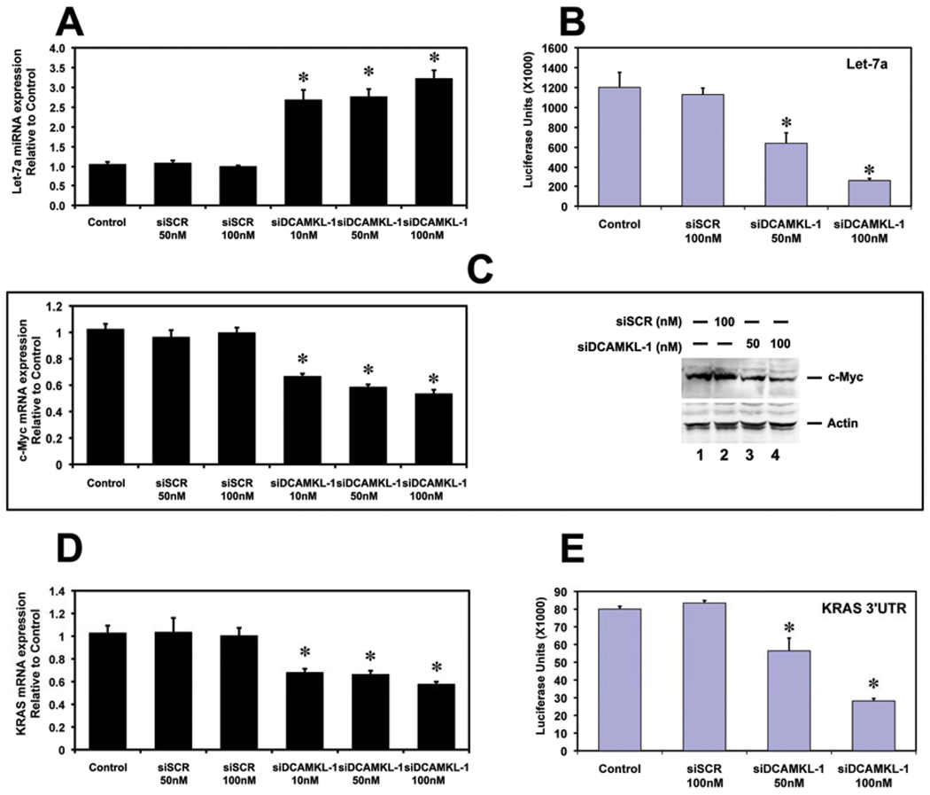
We have recently demonstrated that DCAMKL-1 is a novel putative pancreatic stem/progenitor cell marker in the normal mouse pancreas (22). Furthermore, DCAMKL-1 negatively regulates let-7a miRNA (a tumor suppressor miRNA) in normal mouse pancreas (Supplementary Figure S3A and B) and human colorectal cancer cells (23). Moreover, let-7a negatively regulates several key oncogenes including c-Myc and KRAS in various solid tumors (23, 34, 35). To determine whether DCAMKL-1 regulates let-7a miRNA in pancreatic cancer cells, control, scrambled and DCAMKL-1 siRNA-treated human pancreatic cancer cell lines (AsPC-1 and BxPC3) were analyzed for pri-miRNA expression by real-time RT-PCR. Compared to control and siSCR-treated cells, there was a 3-fold increase in pri-let-7a miRNA in DCAMKL-1 siRNA-treated cells (Fig. 5A and Supplementary Figure S4A and B). Thus, DCAMKL-1 negatively regulates pri-let-7a miRNA in human pancreatic cancer cells. To determine quantitatively the effect of siRNA-mediated knockdown of DCAMKL-1 on let-7a miRNA, we performed a luciferase reporter gene assay (23). AsPC-1 cells were transfected with a plasmid containing firefly luciferase gene with a complementary let-7a binding site in the 3’ UTR. A dose-dependent reduction in luciferase activity was observed following the knockdown of DCAMKL-1 (Fig. 5B). These data taken together suggest that DCAMKL-1 may be a posttranscriptional regulator of let-7a miRNA downstream targets in pancreatic cancer.
Figure 5. DCAMKL-1 regulates oncogenes c-Myc and KRAS via let-7a miRNA.
(A) siRNA-mediated knockdown of DCAMKL-1 results in upregulation of pri-miR-let-7a. (B) Knockdown of DCAMKL-1 decreases luciferase activity (luciferase units) following transfection with plasmid encoding luciferase containing let-7a binding site in AsPC-1 cells. (C) A decreased expression of c-Myc mRNA (left panel) and protein (right panel) was observed in AsPC-1 cells following the knockdown of DCAMKL-1. (D) AsPC-1-siDCAMKL-1 cells demonstrated a decrease in KRAS mRNA. (E) Knockdown of DCAMKL-1 decreases luciferase activity (luciferase units) following transfection with plasmid encoding luciferase containing binding sites for let-7 family members (similar to KRAS 3’ UTR) in AsPC-1 cells.
Values represented as mean ± SEM, and asterisks denote statistically significant differences (*p<0.01) compared with control.
Others and we demonstrated that c-Myc is a key downstream target of let-7a miRNA (23, 34). To demonstrate this in pancreatic cancer cell line, control, AsPC-1-siSCR and AsPC-1-siDCAMKL-1 cells were analyzed for c-Myc expression by real-time RT-PCR. Compared to control and AsPC-1-siSCR cells, there was a significant (p<0.01) 50% reduction of c-Myc mRNA expression in AsPC-1-siDCAMKL-1 cells was observed (Fig. 5C). Similarly a reduction in c-Myc protein was observed following the knockdown of DCAMKL-1 (Fig. 5C).
KRAS is a critical gene that is mutated in many cancers including pancreatic cancer and several studies have reported that up to 95% of pancreatic cancers contain KRAS mutations (36). KRAS is an another key downstream target of let-7a miRNA (35). Following knockdown of DCAMKL-1, we observed a 45% reduction in KRAS mRNA expression compared to control or AsPC-1-siSCR cells (Fig. 5D). To determine the mechanism of siRNA-mediated knockdown of DCAMKL-1 on KRAS, we transfected a KRAS-Luc reporter vector containing specific binding sites for let-7 family members within the 3’UTR of the firefly luciferase gene (similar to KRAS 3’UTR). A dose-dependent reduction in luciferase activity was observed following knockdown of DCAMKL-1 (Fig. 5E). Similar results were observed in the BxPC3 human pancreatic cancer cell line (data not shown). These data taken together demonstrate that DCAMKL-1 knockdown results in downregulation of c-Myc and KRAS, two key mediators of tumorigenesis in pancreatic cancer.
DCAMKL-1 regulates Notch-1 in pancreatic cancer via miR-144
Notch signaling is frequently dysregulated in human malignancies (36, 37). Notch plays a key role in several cellular developmental pathways including proliferation and apoptosis (38). Upregulated expression of Notch receptors and their ligands has been described in cervical, lung, colon, head and neck, renal carcinoma, acute myeloid, Hodgkins lymphoma, large-cell lymphomas, and pancreatic cancer (39–43). Notch signaling is required for initiation and progression of pancreatic ductal adenocarcinoma (36). Furthermore, inhibition of Notch signaling using a γ-secretase inhibitor (MRK-003) completely inhibited tumor development in Pdx1-Cre; LSL-KRASG12D; p53lox/+ mouse model of pancreatic neoplasia (36). Given the potential roles of Notch signaling in adult stem cell regulation and tumorigenesis (44), we investigated the effect of siRNA-mediated knockdown of DCAMKL-1 on Notch-1 in pancreatic cancer cells.
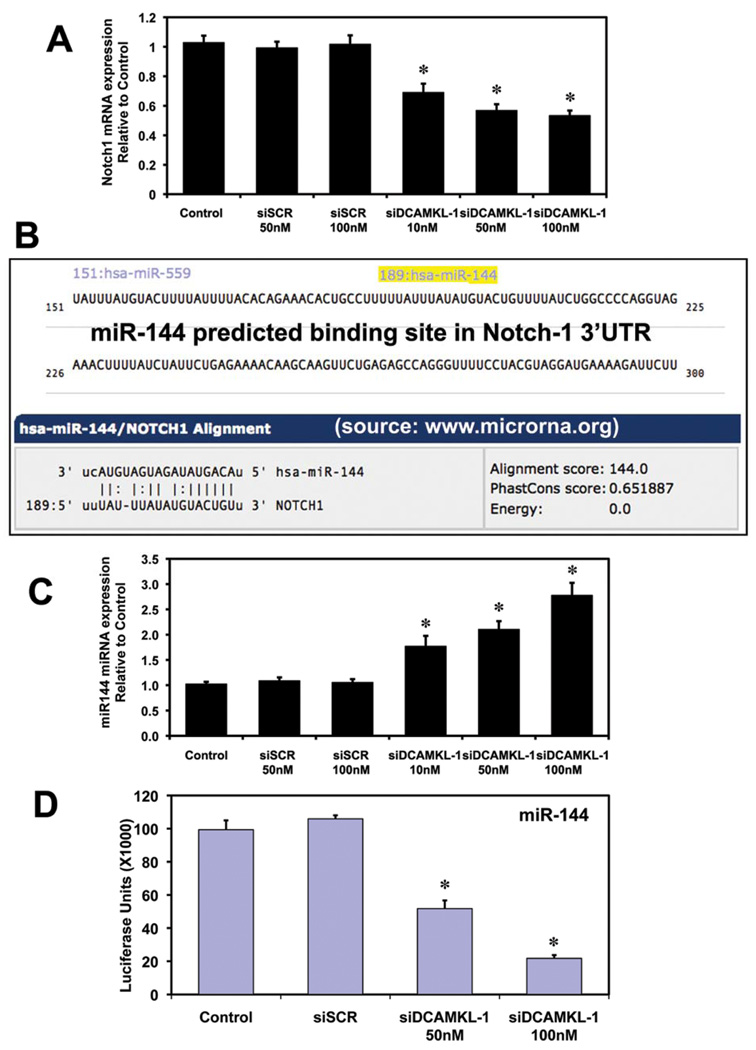
In this study, we observed a 50% reduction in Notch-1 mRNA in AsPC-1-siDCAMKL-1 cells compared to control AsPC-1 or AsPC-1-siSCR cells (Fig. 6A). Similar results were obtained in BxPC3 cells (Supplementary Figure S4C). In order to determine the mechanism by which Notch-1 is inhibited, we first performed a computational/bioinformatics (www.microrna.org: A resource for microRNA targets and expression) analysis of the Notch-1 3’UTR. We found a predicted binding site for miR-144 in the Notch-1 3’ UTR (at the 189th base pair) (Fig. 6B).
Figure 6. Knockdown of DCAMKL-1 downregulates Notch-1 via miR-144.
(A) siRNA-mediated knockdown of DCAMKL-1 decreases Notch-1 mRNA in AsPC-1 cells. (B) A putative binding site for miR-144 at 189th base pair position on Notch-1 3’ UTR (source: www.microrna.org). (C) AsPC-1-siDCAMKL-1 cells demonstrate increased expression of pri-miR-144. (D) Knockdown of DCAMKL-1 decreases luciferase activity (luciferase units) following transfection with plasmid encoding luciferase containing miR-144 binding site in AsPC-1 cells.
Values represented as mean ± SEM, and asterisks denote statistically significant differences (*p<0.01) compared with control.
To investigate the role of DCAMKL-1 in the regulation of miR-144 miRNA, control, scrambled and DCAMKL-1 siRNA-treated AsPC-1 cells were analyzed for pri-miR-144 miRNA expression by real-time RT-PCR. Compared to control and AsPC-1-siSCR cells, there was a 2.5-fold increase in pri-miR-144 miRNA expression in AsPC-1-siDCAMKL-1 cells (Fig. 6C). These data suggest that DCAMKL-1 negatively regulates pri-miR-144 miRNA in human pancreatic cancer cells. Similarly, DCAMKL-1 was also found to negatively regulate pri-miR-144 in normal mouse pancreas (Supplementary Figure S3C). To evaluate these findings quantitatively, we performed a luciferase reporter gene assay using AsPC-1 cells that were transfected with a plasmid containing the firefly luciferase gene with a complementary miR-144 binding site in the 3’ UTR. A dose-dependent reduction in luciferase activity was observed following DCAMKL-1 knockdown (Fig. 6D), indicating that DCAMKL-1 may be a posttranscriptional regulator of miR-144 miRNA downstream targets in pancreatic cancer. Taken together, these data strongly suggest that Notch-1 is a downstream target of miR-144 miRNA and that DCAMKL-1 regulates posttranscriptional control of Notch-1.
Discussion
Solid tumors are histologically heterogeneous and include tumor cells, stroma, inflammatory infiltrates, and vascular structures. The CSC hypothesis suggests that tumors are initiated and maintained by a minority subpopulation of cells within the tumor that have the capacity to self-renew and to generate the more differentiated, rapidly proliferating, cells that make up the bulk of a tumor (2, 45).
The existence of CSCs has profound implications for cancer biology and therapy due to the likelihood that eradication of CSCs is the critical determinant in achieving cure. Recent reports have demonstrated that breast and glioblastoma CSCs are radioresistant and may therefore contribute to treatment failures (46, 47). The cell surface marker CD133 is widely used for isolating CSCs from various cancers (48). Additionally, a subpopulation of CD44+CD24+ESA+ cells was identified as putative pancreatic CSCs (3, 48). However, in general, most cell surface proteins used for isolation of CSCs serve as purification markers without functional implication (2, 48). Thus it is critical to demonstrate that isolated cells from any particular cancer tissue have the functional characteristics of CSCs. Currently, this has been most convincingly demonstrated by serial transplantation in animal models (2).
We have previously demonstrated that DCAMKL-1 is upregulated in human colorectal cancers and siRNA mediated knockdown of DCAMKL-1 results in tumor growth arrest via let-7a miRNA dependent manner (23). In this report, we provide evidence that DCAMKL-1 is upregulated in pancreatic cancer and may also identify pancreatic CSCs. Interestingly, we observed co-expression of DCAMKL-1 and 14-3-3 σ, an inhibitor of Bad pro-apoptotic activity, within human pancreatic adenocarcinomas (10, 11). Co-localization of 14-4-3 σ and DCAMKL-1 is significant as it may represent a target cell within tumors where 14-3-3 σ is transcriptional activated. We also observed distinct DCAMKL-1 immunostaining in the intervening stroma between epithelial tumor elements, which co-expressed vimentin. These findings were indeed surprising in that we did not observe DCAMKL-1 in non-epithelial cells under basal conditions. We next evaluated ductal DCAMKL-1 within PanIN lesions. There we observed several elongated cells that also co-expressed vimentin and DCAMKL-1 suggesting that these cells are of mesenchymal origin. These findings suggest that DCAMKL-1 expressing cells may be undergoing EMT (27). Desmoplasia, the appearance of fibrous, mesenchymal-like tissue in the peritumor stroma, is associated with poor clinical outcome (28). Indeed, gene-profiling studies suggest that mesenchymal gene profiles in tumors are predictive of poor clinical outcome (2). Myofibroblasts have long been thought to be derived from fibroblasts, but recent data has shown that a substantial proportion of these cells are derived from EMT and are associated with tumor progression (29).
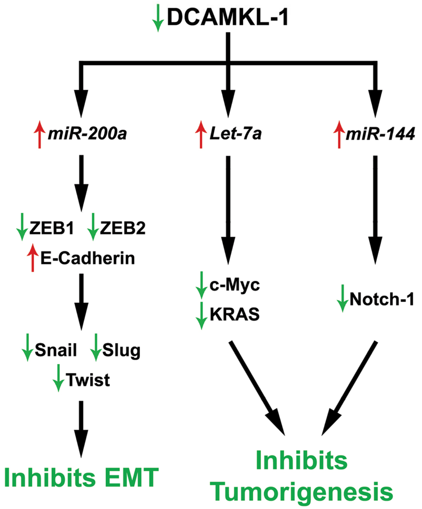
We have previously demonstrated a functional role for DCAMKL-1 in the regulation of let-7a a key tumor suppressor miRNA in many cancers including colorectal cancer. miRNAs are important regulators of mRNAs at the posttranscriptional level by targeting them for cleavage or translational repression (17). miRNAs have emerged as important developmental regulators and control critical processes such as cell fate determination and cell death (17). There is increasing evidence that several miRNAs are mutated or poorly expressed in human cancers and may act as tumor suppressors or oncogenes (18, 19). Here we report that DCAMKL-1 regulates miR-200a, let-7a and miR-144 in the AsPC-1 pancreatic cancer cell line. Each of these miRNAs has been shown to play important roles in several key aspects of tumor initiation and progression. For example, miR-200a inhibits EMT in several cancers by inhibiting transcription factors ZEB1 and ZEB2 (31, 32). Indeed in this report, knockdown of DCAMKL-1 induces pri-miR-200a resulting in down regulation of ZEB1, ZEB2, Snail, Slug and Twist in pancreatic cancer cell lines. Additionally let-7a, a tumor suppressor miRNA, has been shown to inhibit several key oncogenes. Following knockdown of DCAMKL-1, we observed a marked increase in let-7a, which resulted in downregulation of proto-oncogenes c-Myc and KRAS in pancreatic cancer cell lines using real-time RT-PCR and luciferase reporter assays. This is similar to our previous report demonstrating that DCAMKL-1 regulates c-Myc via let-7a miRNA in colorectal cancer cells (23). These data strongly support a direct regulatory role for DCAMKL-1 in cancer via miRNA dependent mechanisms. DCAMKL-1 knockdown in AsPC-1 cells resulted in a marked decrease in Notch-1 mRNA (50%), which contains a putative predicted binding site for miR-144 in the 3’UTR. miR-144 is a regulator of embryonic -hemoglobin (-E1), through targeting the 3'-UTR of Krüppel-like factor D gene and positively regulates erythroid differentiation in hematopoietic stem cells. In order to determine whether DCAMKL-1 regulates Notch-1 through a novel microRNA, we evaluated the expression of miR-144 in AsPC-1-siDCAMKL-1 cells. Here for the first time, we report that DCAMKL-1 negatively regulates Notch-1 via miR-144 dependent mechanism. These data taken together clearly demonstrate a multi-functional role for DCAMKL-1 in regulation of miRNAs that control important genes that contribute to key aspects of tumorigenesis (Fig. 7).
Figure 7. Selective blockade of DCAMKL-1 results in inhibition of EMT and tumorigenesis in CSCs of pancreatic cancer.
As recently reported, the induction of EMT in human mammary epithelial cells resulted in a “stem-cell-like” phenotype characterized by a CD44high and CD24low cell surface marker expression pattern. Furthermore, these cells formed mamospeheres, colonies in soft agar and tumors in nude mice more aggressively than non-EMT induced cells. These studies demonstrate a direct link between the induction of EMT and the gain of stem-cell-like properties (49). These recent findings lend support to our hypothesis that EMT in the stem cell population may play a critical role in tumorigenesis. Further studies are needed to clearly define the role of DCAMKL-1 and other potential stem cell proteins (BMI-1 and LGR5) in cancer progression, metastasis and EMT. Nevertheless the studies presented here provide strong evidence that DCAMKL-1 may be an important target for therapy to eradicate pancreatic cancer and perhaps other solid tumors.
The Notch signaling pathway is frequently activated in many human cancers (36, 37). Notch signaling is required for initiation and progression of pancreatic ductal adenocarcinoma (36). Inhibition of Notch signaling using a γ-secretase inhibitor (MRK-003) completely blocked tumor development in Pdx1-Cre; LSL-KRASG12D; p53lox/+ mice (36). siRNA-mediated knockdown of DCAMKL-1 in human pancreatic cancer cell lines resulted in 50% reduction in Notch-1 mRNA. These data suggests that DCAMKL-1 disruption results in inhibition of the Notch-1 pathway thereby confirming its role as a potential target in anti-cancer strategies.
Supplementary Material
Acknowledgments
Grant Support
This work was supported NIH and National Cancer Institute (NCI) grants: NIH DK-065887, DK-002822, CA-137482; Veterans Affairs Merit Award and Oklahoma Center for Advancement of Science and Technology (OCAST) grant-AR101-030 to CWH
Footnotes
Authors have no conflict of interest
References
- 1.Hoyer M, Roed H, Traberg Hansen A, et al. Phase II study on stereotactic body radiotherapy of colorectal metastases. Acta Oncol. 2006;45(7):823–830. doi: 10.1080/02841860600904854. [DOI] [PubMed] [Google Scholar]
- 2.Diehn M, Clarke MF. Cancer stem cells and radiotherapy: new insights into tumor radioresistance. J Natl Cancer Inst. 2006 Dec 20;98(24):1755–1757. doi: 10.1093/jnci/djj505. [DOI] [PubMed] [Google Scholar]
- 3.Li C, Heidt DG, Dalerba P, et al. Identification of pancreatic cancer stem cells. Cancer research. 2007 Feb 1;67(3):1030–1037. doi: 10.1158/0008-5472.CAN-06-2030. [DOI] [PubMed] [Google Scholar]
- 4.Bonnet D, Dick JE. Human acute myeloid leukemia is organized as a hierarchy that originates from a primitive hematopoietic cell. Nat Med. 1997 Jul;3(7):730–737. doi: 10.1038/nm0797-730. [DOI] [PubMed] [Google Scholar]
- 5.Al-Hajj M, Wicha MS, Benito-Hernandez A, Morrison SJ, Clarke MF. Prospective identification of tumorigenic breast cancer cells. Proc Natl Acad Sci U S A. 2003 Apr 1;100(7):3983–3988. doi: 10.1073/pnas.0530291100. [DOI] [PMC free article] [PubMed] [Google Scholar]
- 6.Singh SK, Clarke ID, Hide T, Dirks PB. Cancer stem cells in nervous system tumors. Oncogene. 2004 Sep 20;23(43):7267–7273. doi: 10.1038/sj.onc.1207946. [DOI] [PubMed] [Google Scholar]
- 7.Singh SK, Clarke ID, Terasaki M, et al. Identification of a cancer stem cell in human brain tumors. Cancer Res. 2003 Sep 15;63(18):5821–5828. [PubMed] [Google Scholar]
- 8.Singh SK, Hawkins C, Clarke ID, et al. Identification of human brain tumour initiating cells. Nature. 2004 Nov 18;432(7015):396–401. doi: 10.1038/nature03128. [DOI] [PubMed] [Google Scholar]
- 9.Prasad GL, Valverius EM, McDuffie E, Cooper HL. Complementary DNA cloning of a novel epithelial cell marker protein, HME1, that may be down-regulated in neoplastic mammary cells. Cell Growth Differ. 1992 Aug;3(8):507–513. [PubMed] [Google Scholar]
- 10.Subramanian RR, Masters SC, Zhang H, Fu H. Functional conservation of 14-3-3 isoforms in inhibiting bad-induced apoptosis. Exp Cell Res. 2001 Nov 15;271(1):142–151. doi: 10.1006/excr.2001.5376. [DOI] [PubMed] [Google Scholar]
- 11.Samuel T, Weber HO, Rauch P, et al. The G2/M regulator 14-3-3sigma prevents apoptosis through sequestration of Bax. J Biol Chem. 2001 Nov 30;276(48):45201–45206. doi: 10.1074/jbc.M106427200. [DOI] [PubMed] [Google Scholar]
- 12.Nakanishi K, Hashizume S, Kato M, Honjoh T, Setoguchi Y, Yasumoto K. Elevated expression levels of the 14-3-3 family of proteins in lung cancer tissues. Hum Antibodies. 1997;8(4):189–194. [PubMed] [Google Scholar]
- 13.Villaret DB, Wang T, Dillon D, et al. Identification of genes overexpressed in head and neck squamous cell carcinoma using a combination of complementary DNA subtraction and microarray analysis. Laryngoscope. 2000 Mar;110(3 Pt 1):374–381. doi: 10.1097/00005537-200003000-00008. [DOI] [PubMed] [Google Scholar]
- 14.Guweidhi A, Kleeff J, Giese N, et al. Enhanced expression of 14-3-3sigma in pancreatic cancer and its role in cell cycle regulation and apoptosis. Carcinogenesis. 2004 Sep;25(9):1575–1585. doi: 10.1093/carcin/bgh159. [DOI] [PubMed] [Google Scholar]
- 15.Han Z, Dimas K, Tian X, et al. 14-3-3sigma-dependent resistance to cisplatin. Anticancer Res. 2009 Jun;29(6):2009–2014. [PubMed] [Google Scholar]
- 16.Neupane D, Korc M. 14-3-3sigma Modulates pancreatic cancer cell survival and invasiveness. Clin Cancer Res. 2008 Dec 1;14(23):7614–7623. doi: 10.1158/1078-0432.CCR-08-1366. [DOI] [PMC free article] [PubMed] [Google Scholar]
- 17.Bartel DP. MicroRNAs: genomics, biogenesis, mechanism, and function. Cell. 2004 Jan 23;116(2):281–297. doi: 10.1016/s0092-8674(04)00045-5. [DOI] [PubMed] [Google Scholar]
- 18.McManus MT. MicroRNAs and cancer. Semin Cancer Biol. 2003 Aug;13(4):253–258. doi: 10.1016/s1044-579x(03)00038-5. [DOI] [PubMed] [Google Scholar]
- 19.Takamizawa J, Konishi H, Yanagisawa K, et al. Reduced expression of the let-7 microRNAs in human lung cancers in association with shortened postoperative survival. Cancer Res. 2004 Jun 1;64(11):3753–3756. doi: 10.1158/0008-5472.CAN-04-0637. [DOI] [PubMed] [Google Scholar]
- 20.Didiano D, Hobert O. Perfect seed pairing is not a generally reliable predictor for miRNA-target interactions. Nat Struct Mol Biol. 2006 Sep;13(9):849–851. doi: 10.1038/nsmb1138. [DOI] [PubMed] [Google Scholar]
- 21.May R, Riehl TE, Hunt C, Sureban SM, Anant S, Houchen CW. Identification of a novel putative gastrointestinal stem cell and adenoma stem cell marker, doublecortin and CaM kinase-like-1, following radiation injury and in adenomatous polyposis coli/multiple intestinal neoplasia mice. Stem cells (Dayton, Ohio) 2008 Mar;26(3):630–637. doi: 10.1634/stemcells.2007-0621. [DOI] [PubMed] [Google Scholar]
- 22.May RJ, Sureban SM, Lightfoot SA, et al. Identification of a Novel Putative Pancreatic Stem/Progenitor Cell Marker Dcamkl-1 in Normal Mouse Pancreas. Am J Physiol Gastrointest Liver Physiol. 2010 Jun 3; doi: 10.1152/ajpgi.00146.2010. In Press. [DOI] [PMC free article] [PubMed] [Google Scholar]
- 23.Sureban SM, May R, Ramalingam S, et al. Selective blockade of DCAMKL-1 results in tumor growth arrest by a Let-7a MicroRNA-dependent mechanism. Gastroenterology. 2009 Aug;137(2):649–659. doi: 10.1053/j.gastro.2009.05.004. 59 e1–2. [DOI] [PMC free article] [PubMed] [Google Scholar]
- 24.Jackson EL, Willis N, Mercer K, et al. Analysis of lung tumor initiation and progression using conditional expression of oncogenic K-ras. Genes Dev. 2001 Dec 15;15(24):3243–3248. doi: 10.1101/gad.943001. [DOI] [PMC free article] [PubMed] [Google Scholar]
- 25.Mohammed A, Janakiram NB, Li Q, et al. The Epidermal Growth Factor Receptor Inhibitor Gefitinib Prevents the Progression of Pancreatic Lesions to Carcinoma in a Conditional LSL-KrasG12D/+ Transgenic Mouse Model. Cancer Prev Res (Phila) 2010 Nov;3(11):1417–1426. doi: 10.1158/1940-6207.CAPR-10-0038. [DOI] [PMC free article] [PubMed] [Google Scholar]
- 26.Logsdon CD, Simeone DM, Binkley C, et al. Molecular profiling of pancreatic adenocarcinoma and chronic pancreatitis identifies multiple genes differentially regulated in pancreatic cancer. Cancer Res. 2003 May 15;63(10):2649–2657. [PubMed] [Google Scholar]
- 27.Turley EA, Veiseh M, Radisky DC, Bissell MJ. Mechanisms of disease: epithelial-mesenchymal transition--does cellular plasticity fuel neoplastic progression? Nature clinical practice. 2008 May;5(5):280–290. doi: 10.1038/ncponc1089. [DOI] [PMC free article] [PubMed] [Google Scholar]
- 28.Poste G, Greig R. On the genesis and regulation of cellular heterogeneity in malignant tumors. Invasion & metastasis. 1982;2(3):137–176. [PubMed] [Google Scholar]
- 29.Polakis P. Wnt signaling and cancer. Genes Dev. 2000 Aug 1;14(15):1837–1851. [PubMed] [Google Scholar]
- 30.Zhang H, Li Y, Lai M. The microRNA network and tumor metastasis. Oncogene. 2010 Feb 18;29(7):937–948. doi: 10.1038/onc.2009.406. [DOI] [PubMed] [Google Scholar]
- 31.Xia H, Ng SS, Jiang S, et al. miR-200a-mediated downregulation of ZEB2 and CTNNB1 differentially inhibits nasopharyngeal carcinoma cell growth, migration and invasion. Biochem Biophys Res Commun. 2010 Jan 1;391(1):535–541. doi: 10.1016/j.bbrc.2009.11.093. [DOI] [PubMed] [Google Scholar]
- 32.Korpal M, Lee ES, Hu G, Kang Y. The miR-200 family inhibits epithelial-mesenchymal transition and cancer cell migration by direct targeting of E-cadherin transcriptional repressors ZEB1 and ZEB2. J Biol Chem. 2008 May 30;283(22):14910–14914. doi: 10.1074/jbc.C800074200. [DOI] [PMC free article] [PubMed] [Google Scholar]
- 33.Hotz B, Arndt M, Dullat S, Bhargava S, Buhr HJ, Hotz HG. Epithelial to mesenchymal transition: expression of the regulators snail, slug, and twist in pancreatic cancer. Clin Cancer Res. 2007 Aug 15;13(16):4769–4776. doi: 10.1158/1078-0432.CCR-06-2926. [DOI] [PubMed] [Google Scholar]
- 34.Sampson VB, Rong NH, Han J, et al. MicroRNA let-7a down-regulates MYC and reverts MYC-induced growth in Burkitt lymphoma cells. Cancer Res. 2007 Oct 15;67(20):9762–9770. doi: 10.1158/0008-5472.CAN-07-2462. [DOI] [PubMed] [Google Scholar]
- 35.Johnson SM. RAS is regulated by the let-7 microRNA family. Cell. 2005;120:635–647. doi: 10.1016/j.cell.2005.01.014. [DOI] [PubMed] [Google Scholar]
- 36.Plentz R, Park JS, Rhim AD, et al. Inhibition of gamma-secretase activity inhibits tumor progression in a mouse model of pancreatic ductal adenocarcinoma. Gastroenterology. 2009 May;136(5):1741–1749. doi: 10.1053/j.gastro.2009.01.008. e6. [DOI] [PMC free article] [PubMed] [Google Scholar]
- 37.Koch U, Radtke F. Notch and cancer: a double-edged sword. Cell Mol Life Sci. 2007 Nov;64(21):2746–2762. doi: 10.1007/s00018-007-7164-1. [DOI] [PMC free article] [PubMed] [Google Scholar]
- 38.Ohishi K, Katayama N, Shiku H, Varnum-Finney B, Bernstein ID. Notch signalling in hematopoiesis. Semin Cell Dev Biol. 2003 Apr;14(2):143–150. doi: 10.1016/s1084-9521(02)00183-0. [DOI] [PubMed] [Google Scholar]
- 39.Leethanakul C, Patel V, Gillespie J, et al. Distinct pattern of expression of differentiation and growth-related genes in squamous cell carcinomas of the head and neck revealed by the use of laser capture microdissection and cDNA arrays. Oncogene. 2000 Jun 29;19(28):3220–3224. doi: 10.1038/sj.onc.1203703. [DOI] [PubMed] [Google Scholar]
- 40.Rae FK, Stephenson SA, Nicol DL, Clements JA. Novel association of a diverse range of genes with renal cell carcinoma as identified by differential display. Int J Cancer. 2000 Dec 1;88(5):726–732. doi: 10.1002/1097-0215(20001201)88:5<726::aid-ijc7>3.0.co;2-h. [DOI] [PubMed] [Google Scholar]
- 41.Tohda S, Nara N. Expression of Notch1 and Jagged1 proteins in acute myeloid leukemia cells. Leuk Lymphoma. 2001 Jul;42(3):467–472. doi: 10.3109/10428190109064603. [DOI] [PubMed] [Google Scholar]
- 42.Wang Z, Zhang Y, Li Y, Banerjee S, Liao J, Sarkar FH. Down-regulation of Notch-1 contributes to cell growth inhibition and apoptosis in pancreatic cancer cells. Mol Cancer Ther. 2006 Mar;5(3):483–493. doi: 10.1158/1535-7163.MCT-05-0299. [DOI] [PubMed] [Google Scholar]
- 43.Miyamoto Y, Maitra A, Ghosh B, et al. Notch mediates TGF alpha-induced changes in epithelial differentiation during pancreatic tumorigenesis. Cancer Cell. 2003 Jun;3(6):565–576. doi: 10.1016/s1535-6108(03)00140-5. [DOI] [PubMed] [Google Scholar]
- 44.Fan X, Matsui W, Khaki L, et al. Notch pathway inhibition depletes stem-like cells and blocks engraftment in embryonal brain tumors. Cancer Res. 2006 Aug 1;66(15):7445–7452. doi: 10.1158/0008-5472.CAN-06-0858. [DOI] [PubMed] [Google Scholar]
- 45.Tang C, Ang BT, Pervaiz S. Cancer stem cell: target for anti-cancer therapy. Faseb J. 2007 Jul 11; doi: 10.1096/fj.07-8560rev. [DOI] [PubMed] [Google Scholar]
- 46.Bao S, Wu Q, McLendon RE, et al. Glioma stem cells promote radioresistance by preferential activation of the DNA damage response. Nature. 2006 Dec 7;444(7120):756–760. doi: 10.1038/nature05236. [DOI] [PubMed] [Google Scholar]
- 47.Phillips TM, McBride WH, Pajonk F. The response of CD24(-/low)/CD44+ breast cancer-initiating cells to radiation. J Natl Cancer Inst. 2006 Dec 20;98(24):1777–1785. doi: 10.1093/jnci/djj495. [DOI] [PubMed] [Google Scholar]
- 48.Ischenko I, Seeliger H, Kleespies A, et al. Pancreatic cancer stem cells: new understanding of tumorigenesis, clinical implications. Langenbecks Arch Surg. 2010 Jan;395(1):1–10. doi: 10.1007/s00423-009-0502-z. [DOI] [PubMed] [Google Scholar]
- 49.Mani SA, Guo W, Liao MJ, et al. The epithelial-mesenchymal transition generates cells with properties of stem cells. Cell. 2008 May 16;133(4):704–715. doi: 10.1016/j.cell.2008.03.027. [DOI] [PMC free article] [PubMed] [Google Scholar]
Associated Data
This section collects any data citations, data availability statements, or supplementary materials included in this article.


