

Abstract
After much effort in surface chemistry development and optimization by several groups, fluorescent semiconductor nanocrystals probes, also known as quantum dots or qdots, are now entering the realm of biological applications with much to offer to biologists. The road to success has been paved with hurdles but from these efforts has stemmed a multitude of original surface chemistries that scientists in the biological fields can draw from for their specific biological applications. The ability to easily modulate the chemical nature of qdot surfaces by employing one or more of the recently developed qdot coatings, together with their exceptional photophysics have been key elements for qdots to acquire a status of revolutionary fluorescent bio-probes. Indeed, the unique properties of qdots not only give biologists the opportunity to explore advanced imaging techniques such as single molecule or lifetime imaging but also to revisit traditional fluorescence imaging methodologies and extract yet unobserved or inaccessible information in vitro or in vivo.
Keywords: Biomimetic material, Confocal microscopy, Fluorescence, Molecular imaging, Nanoparticle, Surface modification
1. Qdots and their properties
Qdots are nanometers sized crystalline clusters (1–10 nm) that are synthesized from a variety of semiconductor materials (for detailed reviews on the synthesis and properties of qdots see Refs. [1,2]). At such small scales, qdots retain some of the bulk properties of the material from which they are derived, but also adopt new properties that directly depend on their size. In term of photophysics, this translates into a composition-, shape- and size-dependant luminescence with absorption and emission bands that scale with the bulk band gap energy of the material and the final diameter of the qdot clusters. Qdots are characterized by large absorption spectra, but narrow and symmetric emission bands (full width at half maximum of 25–35 nm) that can span the light spectrum from the ultraviolet (UV) to the infrared (IR) (400–1350 nm). In addition, they possess an excellent photostability (many orders of magnitude longer than conventional organic fluorophores) and quantum yield (the ratio of emitted to absorbed photons) as high as 90%. They also have large absorption cross sections and long fluorescence lifetimes (>10 ns). With all these features, qdots have rapidly emerged as potential new fluorescent probes for the imaging of biological samples. Indeed they offer many advantages over conventional fluorophores for imaging techniques such as two-photon or time-gated microscopy, while allowing multicolor, long term and high sensitivity fluorescence imaging. However biological applications using qdots have been slowed down by the difficulties encounter to efficiently process these inorganic semiconductor into biocompatible probes. Over the years, different solubilization strategies have been devised to allow the successful use of qdots as fluorescence bio-probes.
2. A wide choice of surface coatings
The first demonstrations of qdots utilization for biomedical applications by Bruchez et al. [3] and Chan et al. [4] in 1998 have been followed by 2 years with very few publications using qdots as bio-probes, eventually expanding exponentially after 2000 (Fig. 1). Beyond being a testimony to the pioneering nature of both articles, this lag time also stresses the difficulties that had to be overcome to efficiently use these new fluorescent probes for biological questions, particularly in live cells and in vivo imaging. Indeed the complexity of biological environments imposes stringent conditions on the stability and efficacy of reporter probes. During biochemical processes, molecular interactions rely on the conformational flexibility of bio-molecules to attain electrostatic, hydrophobic and steric matching of a substrate or ligand with its biological target. Qdots, which are relatively large and rigid spheres of inorganic material, might appear at first rather ill-suited when probing such shape-sensitive processes. The solution resides in interfacing one or more “soft” organic layers with the inorganic qdots. Over the past few years scientists have rivaled in ingenuity in developing robust and versatile surface chemistries and providing biocompatible organic interfaces that (i) solubilize and stabilize qdots in biological buffers, (ii) maintain their original colloidal and photophysical properties, (iii) keep their size relatively small and (iv) provide reactive groups for subsequent conjugation to bio-molecules. The aqueous solubilization of qdots synthesized from hydrophobic solvents usually involves either shielding or exchange of surface hydrophobic ligands with amphiphilic ones. Both approaches have advantages and inconveniences. Surface shielding chemistries, such as encapsulation in phospholipid micelles [5] or coating with amphiphilic polymers [6-8], retain the original hydrophobic qdot surface ligands and efficiently preserve the qdot photophysical properties. In particular, the fluorescence quantum yield is minimally affected. These approaches are readily applicable to other nanomaterials presenting similar hydrophobic surfactants on their surface, but often result in qdots with final sizes three to four times larger than the original particles [7,9], bringing them in the same size range as the smallest fluorescent polystyrene microspheres (20 nm). While large size is a lesser issue for in vitro applications, it might be detrimental for entry and specific interactions in crowded biological environments, as for instance in live cells and in vivo applications. This problem was recently illustrated by Howarth et al. who showed that large size qdots have difficulties to access neuronal synapses in hypocampal neurons [10].
Fig. 1.
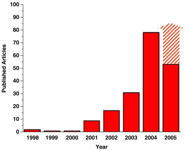
Evolution, of the number of publications describing the use of quantum dot probes in biological applications (until July 2005).
In contrast, surface chemistries replacing the original surface ligands usually produce particles with smaller final diameter (8–15 nm for CdSe/ZnS particles originally 4–9 nm in diameter). For CdSe/ZnS qdots in particular, surface modifications often involve coordination of thiolated hydrophilic ligands on the qdot ZnS layer. Early approaches using mono-thiol ligands are now known to result in qdots with poor stability in biological buffers owing to the detachment over time of these ligands [11]. More robust surface chemistries involve the use of di-thiol ligands and further coating with engineered proteins [12], cross-linking of ligands after surface exchange in the case of silica [13] or dendrimer coating [14] and the use of polymers with multiple anchoring point to the qdot surface such as oligomeric phosphine ligands [15] or polycysteinyl peptides developed in our laboratory [16] that provide enhanced stability. With the notable exception of the phosphine ligand chemistry, exchange surface chemistries require tailoring of the amphiphilic ligands for each new nanomaterial, making such approaches much less general than shielding chemistries. When trying to adapt a specific surface exchange chemistry to other materials, surface coating with peptides is particularly interesting since they are amenable to molecular evolution. Peptide display libraries in bio-engineered phage [17] or bacteria [18] have indeed been shown to be very powerful tools to rapidly screen and select unique peptide sequences capable of binding to semiconductor or metallic materials. With the appropriate combinatorial techniques (a peptide library can consist of more than a billion random sequences), the unique versatility of the 21 natural amino-acid peptide code can, in principle, be harvested via accelerated evolution to select peptide sequences that specifically bind to any type of nanomaterials. Beyond screening peptides for binding and stabilization of various semiconductor qdots (InP, CdTe, PbSe, etc.), one can also envision the same approach to select peptides enhancing the quantum yield or reducing the fluorescence intermittency of qdots to improve their photophysical properties. This very practical goal is supported by our recent discovery that peptide coatings can lead to significant increase in quantum yield for CdSe qdots with graded CdS/ZnS shells [19].
Whether based on ligand shielding or exchange, all qdot surface chemistries are designed to provide reactive groups such as amine (−NH2), carboxyl (−COOH) or mercapto (−SH) groups for direct conjugation to biomolecules. A growing set of functions (streptavidin, protein A, biotin, etc.) is available for easy and customized conjugation to nearly all biomolecules of interest. “Ready-to-use” qdots equipped with those functions or with antibodies can now be purchased from different commercial sources. With the peptide-coating approach, it is easy to construct a sequence that will present any of the previously cited reactive group at the surface. For instance, we have recently introduced thiol derivatized qdots. These qdots are coated with peptides presenting a terminal cysteine, and are efficiently modified with biocytin maleimide (Fig. 2) or used to conjugate full-length antibodies by reaction with hetero-bifunctional cross-linking reagents such as succinimidyl-4-(N-maleimidomethyl)cyclohexane-1-carboxylate (SMCC). An alternative to cross-linking qdots to full length proteins is to derivatize them with polypeptides sequences capable of folding into active binding domains such as Src-homology 2 (SH2) domains (domain composed of a beta-sheet surrounded by two alpha helices that binds specifically to peptides containing phosphotyrosines) or PDZ domains (a beta-sandwich with two alpha-helices involved in high affinity binding at the c-terminal residues of transmembrane receptors and ion channels) which are involved in intracellular signaling. Polycysteinyl peptides with more than 50 amino acids can be attached on the surface of qdots, and application to small proteins domains, which are about 100 amino acids long, should provide qdots with targeting and binding functions while keeping their size relatively small.
Fig. 2.

Activation of peptide-coated quantum dots with biocytin. Quantum dots (CdSe/CdS/ZnS emitting at 630 nm) were coated with phytochelatin-related peptides modified with a N-terminal cysteine and reacted with maleimide-biocytin after reduction with dithiothreitol (1 mm for 1 h). Conjugation of biocytin to the quantum dots was verified by a gel mobility shift assay following incubation with 1 mg/ml of streptavidin as previously described [16]. Cha: 3-cyclohexylalanine.
3. Making the most of qdots size and shape
Owing to their structure and size, qdots have a large surface to volume ratio and therefore present a large number of surface attachment points that can be exploited to engineer multifunctional qdot probes. A common example of useful function demonstrated with nearly all published qdot solubilization chemistries is the incorporation of polyethylene glycol (PEG) molecules in the amphiphilic organic surface layer. PEG not only enhances the aqueous solubility of the qdots but also reduces non-specific adhesion to biological cells. The non-toxic and excellent solvating properties of PEG polymers have previously been employed in drug development to improve bio-distribution and circulation time in vivo and to limit immunogenicity. Pegylation has proven to be fully compatible with qdot surface chemistries [20] and is bound to play a prevalent role when optimizing in vivo pharmacokinetics of qdot bio-probes [21].
Combining multiple functions on qdot surfaces could also be advantageous in different situations. An example is in vivo targeting of cancer cells, where this could help create probes with enhanced targeting selectivity. Tumor cells often over-express different cell surface markers that are usually absent or found only in low copy numbers in normal cells. The large surface area of qdots may be used as a scaffold to graft multiple ligands specific for these over-expressed markers. Assuming that these markers colocalize on the cell membrane (or are present at a large enough density), the qdot probes should exhibit a large avidity for the colocalized target markers and therefore have an enhanced relative affinity for tumor cells versus normal cells. Non-specific binding to healthy tissue could be further reduced if low affinity ligands are chosen. Multi-potent qdot probes might also prove useful to develop multi-contrast imaging as was recently demonstrated with in vivo positron emission tomography (PET) and fluorescence imaging using qdots labeled with radioactive peptides [22].
On the other hand, the presence of multiple reactive groups on the surface of qdots can be problematic when trying to achieve a one-to-one qdot-to-ligand ratio. Multiple ligand per qdot can indeed result in unwanted side effects such as target aggregation. This can interfere with many biological processes where molecules are activated upon mutual cross-linking by ligands. In such cases, controlling the number of ligand per qdots becomes absolutely necessary to avoid activation upon binding and to study bio-molecules in their “resting” state. Although highly desirable, single-ligand derivatization of qdots has to our knowledge not yet been achieved.
4. A new tool for fluorescence imaging of biological samples
With a host of surface chemistries and the availability of commercial qdots, biomedical applications using qdots have flourished. The multitude of successful uses in immunofluorescence assays, biotechnology detection, live cells imaging, single-molecule biophysics or in vivo animal imaging is a testimony of the great excitement generated by these new fluorescent probes and their tremendous potential to revolutionize fluorescence imaging techniques. Excellent reviews on the subject have recently been published on the current applications of qdot as bio-probes [22-24].
The combination of a large absorption cross section (extinction coefficient), good quantum yield and large saturation intensity makes qdots brighter than fluorescence dyes or fluorescent proteins. The sensitivity of detection in fluorescence-based assays is therefore significantly enhanced [6]. However, one should keep in mind that qdots are bulky probes and, when bound to a target, a qdot might prevent the access of another qdot to a neighboring target molecule due to steric hindrance. For labeling of molecules expressed in high copy numbers and relatively localized, this may result in a decrease of the total number of labeled molecules, compared to a staining performed with smaller fluorophores, and lead to a decrease of the overall signal intensity and a worsening of the detection sensitivity. Qdots’ brightness is therefore best used when high sensitivity detection of low copy numbers of molecules is desired, or when the molecules to be detected are sparsely distributed. In particular, qdots are excellent probes for single-molecule fluorescence microscopy, which is one of the most exciting new application offered to biologists. This was nicely demonstrated by Dahan et al. [25], who followed the lateral dynamics of qdot-labeled single glycine receptors in the membrane of neurons. Taking advantages of the brightness and high photostability of the probes they were able to observe the diffusion and entry of glycine receptors in neuronal synapses with a spatial resolution of 5–10 nm. With qdot probes, biologists equipped with a standard epifluorescence microscope and a good CCD camera can easily perform single-molecule imaging and use this powerful technique to detect biological events usually hidden in ensemble measurements or inaccessible because of the fast photobleaching of conventional organic fluorophores [2].
Qdots are orders of magnitudes more photostable than dyes. High fluorescence photostability allows immunostained samples to be repeatedly imaged and crisp, high-resolution three-dimensional images to be acquired. Resistance to photobleaching becomes especially advantageous in live cell experiments, where whole cells or even molecules can be observed and tracked for hours or days. Dubertret et al. convincingly demonstrated cell lineage tracing by injecting qdots in a single Xenopus frog cell at an early embryonic stage and by following daughter cells over days [5].
In marked contrast with fluorophores, qdots have broadband photon absorption spectrum and narrow, symmetrical and tunable emission bands which facilitate the simultaneous detection of multiple signals. To date, qdots emitting in the visible part of the spectrum (e.g. CdSe/ZnS) have been the most commonly used. Highly optimized chemical synthesis for these semiconductor materials have made the production of these qdots fairly easy and safe [1,26]. CdSe qdots’ range of emission wavelength matches perfectly the detection range of typical imaging and fluorescence devices (such as CCD cameras and photomultiplier tubes). With the extension of qdot probes to new biological applications, the demand for new types of semiconductor materials has grown. Significant advances have been made in the production and solubilization of qdots emitting in the near infra-red (NIR) and IR spectral range (CdTe/CdSe, InAs, etc.). Currently, the available palette of semiconductor materials allows the synthesis of qdot probes with emission wavelength ranging from 400 nm to several micrometers. Their broad excitation spectrum mentioned previously allows a single excitation wavelength to simultaneously excite all qdots emitting from the UV to the IR. Multicolor imaging of biological samples is therefore significantly simplified [27], and improved multicolor colocalization studies are made possible with the elimination of many chromatic aberration and alignment issues encountered with standard fluorophore microscopy [28]. A recent example of dynamic multicolor imaging, made possible by the long-term stability of qdots is the interesting study of time-dependent colocalization of cell surface receptors involved in signal transduction by Lidke et al. [29].
The narrow and tunable emission spectra of qdots have also recently been exploited to customize donor emission in fluorescence resonance energy transfer (FRET) assays between qdot donors and fluorescent dye acceptors [30]. FRET measurements with qdots are able to convey good qualitative information about molecular associations in ensemble measurements and appear to have great potential as nanoscale biosensors. However there are still some problems to solve to be able to extract accurate distance measurements by FRET, as would be needed to study dynamic changes in the conformation of biomolecules. One problem stems from the fact that, unlike chromophores, qdots from a single synthesis batch are structurally and spectrally slightly different from one another. This is due to the large numbers of atoms qdots are made of, and to the various steps involved in their synthesis which can result in small variations and defects in either the core, shell or the biocompatible organic layers. These defects have significant influences on the qdot photophysical properties. An example is the spectral heterogeneity within a qdot batch illustrated in Fig. 3. This heterogeneity would translate into a corresponding heterogeneity of the Förster radius R0, which would affect the precision of single-molecule FRET measurements using qdots, unless the actual spectrum of each individual qdot can be measured. Another potentially annoying characteristic of qdot is their environment-dependent fluorescence intermittency, also known as blinking [2,31]. Qdot blinking is associated to charge trapping and un-trapping at surface defects during excitation and results in an alternation (at all timescales) of bright and dark states during which no photons are emitted. Qdot blinking therefore results in the random loss of distance information at all time scales, preventing a recovery of the complete conformational dynamics of the observed single molecule. In addition, blinking is strongly correlated with spectral jumping (change in the emission peak position) [32] that can affect the FRET efficiency. An intimate understanding and control of the photophysic is therefore required to harvest the full potential of qdots as FRET probes. Improvements such as the near-elimination of blinking in the presence of β-mercaptoethanol or dithiothreitol have been reported [33]. Although live cells imaging at the high concentration of β-mercaptoethanol used (1 mm) might not be possible, this is already a tremendous amelioration for FRET applications in vitro [34].
Fig. 3.

Spectral heterogeneity within a single qdot batch. Left: Confocal image of spin cast green CdSe/ZnS qdots. Right: Distribution of spectra peak positions of individual qdots (blue) and Gaussian fit of the distribution (black: full width half maximum distribution: FWHM 14.6 nm) compared to the average spectrum of all qdots (green) and its Gaussian fit (red). The individual spectra have a smaller FWHM (FWHM: 15.2±2.6 nm) (inset) than that of the average spectrum (FWHM: 20.3 nm). Image scale bar: 1 μm, exposition: 100 ms/pixel, excitation: 200 nW at 488 nm. Data acquisition was performed as described in Ref. [28].
Time-gated and lifetime imaging of biological samples labeled with qdot probes are two other promising techniques taking advantage of the long fluorescence lifetime of qdots. Qdots have a fluorescence lifetime (time of fluorescence emission decay after excitation) of 20–30 ns that is significantly longer than the ubiquitous short-lived autofluorescence of cells (2 ns) or organic fluorophores lifetimes (1–4 ns). Autofluorescence is the most common source of background and reduces detection sensitivity. The fluorescence decay of qdots is long enough that by the time the autofluorescence signal of a specimen has vanished, qdots still emit photons. In time-gated imaging, early incoming photons are discarded to filter-out the autofluorescence signal and only retain the qdot signal. This results in a dramatic contrast enhancement for cellular imaging [35]. In a similar approach, fluorescence lifetime imaging microscopy can exploit differences in fluorescence decay rates to discriminate qdot signal from that of other fluorophores within the same specimen [36]. The combination of multi-lifetime and multicolor imaging of fluorescence dyes with qdot probes offers yet unprecedented multiplexing capabilities for fluorescence imaging of biological samples.
5. Live cell imaging: passing the plasma membrane
Most live cells studies with qdots have, understandably, focused on membrane markers since they are easy to access and do not require passage of the probes through the highly regulated and organized cell membrane. The obvious (but difficult) next step is to extend targeting to cytoplasmic molecules. Although some interesting attempts have been performed using membrane translocation peptides [37], electroporation or transfection reagents [38], qdots often tend to accumulate in vesicles or appear non-homogeneously distributed in the cytoplasm. As of today, there seems to be no real success to overcome this complication and the best technique for cytoplasmic translocation of qdots is the direct injection in living cells (Fig. 4). This approach has allowed the targeting of qdots to sub-cellular compartments such as mitochondria or the nucleus using targeting peptides [38,39]. Cell injection, albeit useful for single-cell observation, is very time consuming when large numbers of cells are to be labeled. New methods to homogenously distribute qdots in the cytoplasm of cells would therefore be welcome.
Fig. 4.

Various approaches to translocate quantum dots inside cells. While direct microinjection of peptide coated CdSe/CdS/ZnS quantum dots (620 nm, 1 pl of a 100 nm solution) in the cytoplasm (a) or the nucleus (b) gives homogenous labeling, other approaches with polyethylenimine polymers (c) often employed for DNA transfection or translocation peptides (d) such as Chariot peptides result in accumulation in endocytic vesicles. Pictures are merged fluorescence (red) and bright field images of Hela cells. N: nucleus. Scale bars: 5 μm.
6. In vivo imaging with qdots
Applications of qdot probes to animal imaging have been surprisingly rapid, sometime overlooking fundamental yet unsolved issues such as cytotoxicity. Nonetheless the results are spectacular. Qdots have allowed high-sensitivity and high-contrast imaging in deep tissues in vivo not only in mice but also in larger species. Intravenous injection in mice was performed to image blood vessel [40], to target tissue-specific vascular markers [41], or to image lymph nodes [42]. The first targeting of grafted tumors in vivo was also demonstrated with PEG and antibody-coated qdots [8]. All these experiments advocate the use of PEG surface ligands to enhance the circulation time, reduce the dosage and improve the targeting specificity of qdots in vivo as mentioned earlier. In one case, qdots could be found in the bone marrow and the lymph nodes of mice several months after injection [21]. While Gao et al. [8] had to resort to spectral unmixing algorithms to identify the proper location of targeted qdots emitting in the visible spectrum, Kim et al. [42] injected NIR CdTe/CdSe qdots emitting at 850 nm to perform nearly background-free imaging of lymph nodes 1 cm deep in tissues. The significant advances made in the production and solubilization of NIR and IR emitting qdots are particularly exciting for deep tissue imaging in vivo. Combining these approaches with time-gated microscopy to separate short autofluorescence lifetime signals of the animal from that of the much longer lifetime of qdots could result in detection sensitivities approaching that of radio-labeled probes.
7. On the cytotoxicity of qdot probes
As the range of biomedical applications with qdots expands to measurements in vivo, legitimate questions concerning their short and long term cytotoxicity have been raised. Indeed the composition of cadmium chalcogenide qdot probes poses potentially serious health risks that should not be overlooked. Metal cadmium, like the very potent ions of mercury and lead, is strongly poisonous. Cadmium has a half-life of about 20 years in humans and is a suspected carcinogen that can accumulate in the liver and kidney and biodistributes in all tissues since there are no known active mechanisms to excrete cadmium from the human body [43]. Even if cadmium is present in qdots in an inorganic crystalline form, possible toxicity by leakage of these ions following chemical degradation (e.g. in cell endosomes) or photodegradation (e.g. during excitation) is of serious concern. Unfortunately, there have been a very limited number of studies specially designed to assess thoroughly the toxicity of qdots, and most of the reports describing the potential toxic effects of qdots have used non-standardized methods and have often been performed by scientists that are not from the fields of toxicology or health science. In addition, assessing the potential toxicity of qdot probes appears to be a relatively complicated matter since no qdots are similar and different composition, coating and solubilization chemistries might display different toxicities. It seems fair to assume that qdot toxicity will depend on multiple physical, chemical and environmental factors, such as qdot size, charge, surface coating, concentration, chemical composition, colloidal and chemical stability, production of free radicals or singlet oxygen upon excitation and exposition route, all of which might be determining factors of qdot short and long term toxic effects in vivo [44]. Early reports using qdots have not found significantly detrimental effects on the normal function or the morphology of cells, at least within the short time frame of the performed experiments. However, recent reports [45-47] have indicated that bare CdSe qdots are indeed toxic to cells through the release of free cadmium ions. While overcoating of CdSe cores with a protective ZnS shell decreases cytotoxicity, the nature of the amphiphilic surface organic coating itself appears to critically influence the toxic properties of the particles. In this respect, natural biocompatible surface coatings for qdots such as peptides [16] or sugars [48] might be less nocuous to cells than other approaches. Nevertheless, lack of morphological changes for cells is not a sufficient parameter to assess toxicity as demonstrated by Hsieh et al. who recently reported that in human bone marrow mesenchymal stem cells the expression of specific genes is suppressed after transfection of qdots, despite no observable changes in cell proliferation [49]. Although the jury is still out to determine if qdots are safe probes, a range of concentrations exists at which potential interference with normal physiological processes can be reduced. Improving the detection efficiency and the brightness of qdots will allow the use of lower concentrations and reduce potential cytotoxic risks. As discussed above, the use of NIR emitting qdots is very advantageous since their high visibility in vivo requires lesser material. As reported by Kim et al. [42] only 400 pmol of CdTe/CdSe qdots were injected for the in vivo imaging of lymph nodes in a pig. This amount is far below the dose known to induce cadmium poisoning in animals. Non-heavy metal semi-conductor materials such as InGaP qdots also offer an interesting alternative with respect to cytotoxicity issues.
The potential cytotoxicity of qdots could, on the other hand, be of interest when employing them as therapeutic agents, for example in the destruction of cancer cells. Several groups have suggested that cadmium ion release, but also free radicals and singlet-oxygen production by qdots upon UV excitation might be harvested for photodynamic therapy [50,51]. Although much work still needs to be done in this domain, band-gap engineering of qdots and development of thin surface coatings to optimize direct energy transfer to oxygen or to surface conjugated photosensitizing agents is a very exciting avenue to explore.
8. The next step: beyond imaging
Although they are mainly employed for fluorescence imaging applications, qdots have a yet untapped potential in bioelectronics. Indeed, beyond their exceptional photophysical properties, qdots have also unique electronic properties that arise from quantum confinement in such small semiconductor clusters. Many efforts have been launched to interface qdots with neurons [52,53], in an attempt to trigger neuronal functions by qdot-mediated stimulation. Although no clear proof of the feasibility of such projects has yet emerged, a thin and carefully designed surface coating together with qdots optimized for large separation of charges after excitation (e.g. type II CdTe/CdSe qdots) might indeed open the doors to a new generation of light-activable qdot probes, where photo-generated carriers (electrons or hole) may undergo electron transfer, produce heat or generate dipole moments sufficiently strong to interact with membrane proteins and trigger changes in local membrane potentials.
As mentioned earlier in this article, the various surface chemistries developed over the years to interface “soft” biological materials with inorganic qdots have not only been essential for the successful use of qdots in biological imaging, but have also provided new tools in materials science for the controlled assembly of nanomaterials. Indeed, these interfaces, when used as molecular glue between inorganic nanoscaled building blocks, offer means to control 2D and 3D assemblies of nanoscaled objects by taking advantage of the unmatched ability of biological molecules to spatially and dynamically self-assemble and self-organize into complex molecular superstructures. Programmable nanomaterials scaffolding, driven by self-assembling peptides, protein domains interaction, DNA hybridization or carbohydrate/carbohydrate recognition, hold great promises for the creation and emergence of new biomaterials and the expansion of biomimetic nanotechnology as recently discussed by N. Seeman and A. Belcher [54].
9. Conclusion
Within a few years, qdots have gained recognition as very versatile bio-probes for fluorescence imaging in vitro and in vivo. They are available in a large assortment of emission wavelengths and in a variety of surface chemistries and conjugation strategies that permit derivatization with virtually any biologically active molecules. While they are not the perfect fluorophores, and more needs to be done to better understand and improve their photophysics and to further assess their cytotoxicity, qdots have found their place in the existing repertory of bioimaging tools. They will, without doubt, be a probe of choice for long-term, high-sensitivity and multicontrast imaging of molecular dynamics in biological samples.
Acknowledgements
This work was supported by the NIH (Grant 5-R01-EB000312), the Keck Foundation (Grant 04074070) and DARPA/AFOSR (Grant FA955004-10048). Fluorescent microscopy images of Fig. 4 were obtained at the CNSI Advanced Light Microscopy/Spectroscopy Shared Facility at UCLA.
References
- [1].Alivisatos AP. Semiconductor clusters, nanocrystals, and quantum dots. Science. 1996;271(5251):933–7. [Google Scholar]
- [2].Michalet X, Pinaud F, Lacoste TD, Dahan M, Bruchez MP, Alivisatos AP, et al. Properties of fluorescent semiconductor nanocrystals and their application to biological labeling. Single Molec. 2001;2(4):261–76. [Google Scholar]
- [3].Bruchez M, Moronne M, Gin P, Weiss S, Alivisatos AP. Semiconductor nanocrystals as fluorescent biological labels. Science. 1998;281(5385):2013–6. doi: 10.1126/science.281.5385.2013. [DOI] [PubMed] [Google Scholar]
- [4].Chan WCW, Nie SM. Quantum dot bioconjugates for ultrasensitive nonisotopic detection. Science. 1998;281(5385):2016–8. doi: 10.1126/science.281.5385.2016. [DOI] [PubMed] [Google Scholar]
- [5].Dubertret B, Skourides P, Norris DJ, Noireaux V, Brivanlou AH, Libchaber A. In vivo imaging of quantum dots encapsulated in phospholipid micelles. Science. 2002;298(5599):1759–62. doi: 10.1126/science.1077194. [DOI] [PubMed] [Google Scholar]
- [6].Wu XY, Liu HJ, Liu JQ, Haley KN, Treadway JA, Larson JP, et al. Immunofluorescent labeling of cancer marker Her2 and other cellular targets with semiconductor quantum dots. Nat Biotechnol. 2003;21(1):41–6. doi: 10.1038/nbt764. [DOI] [PubMed] [Google Scholar]
- [7].Pellegrino T, Manna L, Kudera S, Liedl T, Koktysh D, Rogach AL, et al. Hydrophobic nanocrystals coated with an amphiphilic polymer shell: a general route to water soluble nanocrystals. Nano Lett. 2004;4(4):703–7. [Google Scholar]
- [8].Gao X, Cui Y, Levenson RM, Chung LWK, Nie S. In vivo cancer targeting and imaging with semiconductor quantum dots. Natl Biotechnol. 2004;22(8):969–76. doi: 10.1038/nbt994. [DOI] [PubMed] [Google Scholar]
- [9].Doose S, Tsay JM, Pinaud F, Weiss S. Comparison of photophysical and colloidal properties of biocompatible semiconductor nanocrystals using fluorescence correlation spectroscopy. Anal Chem. 2005;77(7):2235–42. doi: 10.1021/ac050035n. [DOI] [PubMed] [Google Scholar]
- [10].Howarth M, Takao K, Hayashi Y, Ting AY. Targeting quantum dots to surface proteins in living cells with biotin ligase. Proc Natl Acad Sci USA. 2005;102(21):7583–8. doi: 10.1073/pnas.0503125102. [DOI] [PMC free article] [PubMed] [Google Scholar]
- [11].Chan WCW, Maxwell DJ, Gao XH, Bailey RE, Han MY, Nie SM. Luminescent quantum dots for multiplexed biological detection and imaging. Curr Opin Biotech. 2002;13(1):40–6. doi: 10.1016/s0958-1669(02)00282-3. [DOI] [PubMed] [Google Scholar]
- [12].Mattoussi H, Mauro JM, Goldman ER, Anderson GP, Sundar VC, Mikulec FV, et al. Self-assembly of CdSe-ZnS quantum dot bioconjugates using an engineered recombinant protein. J Am Chem Soc. 2000;122(49):12142–50. [Google Scholar]
- [13].Gerion D, Pinaud F, Williams SC, Parak WJ, Zanchet D, Weiss S, et al. Synthesis and properties of biocompatible water-soluble silica-coated CdSe/ZnS semiconductor quantum dots. J Phys Chem B. 2001;105(37):8861–71. [Google Scholar]
- [14].Wang YA, Li JJ, Chen HY, Peng XG. Stabilization of inorganic nanocrystals by organic dendrons. J Am Chem Soc. 2002;124(10):2293–8. doi: 10.1021/ja016711u. [DOI] [PubMed] [Google Scholar]
- [15].Kim S, Bawendi MG. Oligomeric ligands for luminescent and stable nanocrystal quantum dots. J Am Chem Soc. 2003;125(48):14652–3. doi: 10.1021/ja0368094. [DOI] [PubMed] [Google Scholar]
- [16].Pinaud F, King D, Moore HP, Weiss S. Bioactivation and cell targeting of semiconductor CdSe/ZnS nanocrystals with phytochelatin-related peptides. J Am Chem Soc. 2004;126(19):6115–23. doi: 10.1021/ja031691c. [DOI] [PMC free article] [PubMed] [Google Scholar]
- [17].Whaley SR, English DS, Hu EL, Barbara PF, Belcher AM. Selection of peptides with semiconductor binding specificity for directed nanocrystal assembly. Nature. 2000;405:665–8. doi: 10.1038/35015043. [DOI] [PubMed] [Google Scholar]
- [18].Brown S. Engineered iron oxide-adhesion mutants of the Escherichia coli phage ë receptor. Proc Natl Acad Sci USA. 1992;89:8651–5. doi: 10.1073/pnas.89.18.8651. [DOI] [PMC free article] [PubMed] [Google Scholar]
- [19].Tsay JM, Doose S, Pinaud F, Weiss S. Enhancing the photoluminescence of peptide-coated nanocrystals with shell composition and UV irradiation. J Phys Chem B. 2005;109(5):1669–74. doi: 10.1021/jp046828f. [DOI] [PMC free article] [PubMed] [Google Scholar]
- [20].Bentzen EL, House F, Tomlinson ID, Rosenthal SJ, Crowe JE, Wright DD. Application of semiconductor fluorescent nanocrystals as optical probes for rapid early viral detection. Proc SPIE. 2005;5704:45–56. [Google Scholar]
- [21].Ballou B, Lagerholm BC, Ernst LA, Bruchez MP, Waggoner AS. Noninvasive imaging of quantum dots in mice. Bioconj Chem. 2004;15(1):79–86. doi: 10.1021/bc034153y. [DOI] [PubMed] [Google Scholar]
- [22].Michalet X, Pinaud FF, Bentolila LA, Tsay JM, Doose S, Li JJ, et al. Quantum dots for live cells, in vivo imaging, and diagnostics. Science. 2005;307(5709):538–44. doi: 10.1126/science.1104274. [DOI] [PMC free article] [PubMed] [Google Scholar]
- [23].Medintz IL, Uyeda HT, Goldman ER, Mattoussi H. Quantum dot bioconjugates for imaging, labelling and sensing. Nat Mater. 2005;4(6):435–46. doi: 10.1038/nmat1390. [DOI] [PubMed] [Google Scholar]
- [24].Gao X, Yang L, Petros JA, Marshall FF, Simons JW, Nie S. In vivo molecular and cellular imaging with quantum dots. Curr Opin Biotechnol. 2005;16(1):63–72. doi: 10.1016/j.copbio.2004.11.003. [DOI] [PubMed] [Google Scholar]
- [25].Dahan M, Levi S, Luccardini C, Rostaing P, Riveau B, Triller A. Diffusion dynamics of glycine receptors revealed by single-quantum dot tracking. Science. 2003;302(5644):442–5. doi: 10.1126/science.1088525. [DOI] [PubMed] [Google Scholar]
- [26].Peng ZA, Peng X. Formation of high-quality CdTe, CdSe, and CdS nanocrystals using CdO as precursor. J Am Chem Soc. 2001;123(1):183–4. doi: 10.1021/ja003633m. [DOI] [PubMed] [Google Scholar]
- [27].Jaiswal JK, Mattoussi H, Mauro JM, Simon SM. Long-term multiple color imaging of live cells using quantum dot bioconjugates. Nat Biotechnol. 2003;21(1):47–51. doi: 10.1038/nbt767. [DOI] [PubMed] [Google Scholar]
- [28].Lacoste TD, Michalet X, Pinaud F, Chemla DS, Alivisatos AP, Weiss S. Ultrahigh-resolution multicolor colocalization of single fluorescent probes. Proc Natl Acad Sci USA. 2000;97(17):9461–6. doi: 10.1073/pnas.170286097. [DOI] [PMC free article] [PubMed] [Google Scholar]
- [29].Lidke DS, Nagy P, Heintzmann R, Arndt-Jovin DJ, Post JN, Grecco HE, et al. Quantum dot ligands provide new insights into erbB/HER receptor-mediated signal transduction. Nat Biotechnol. 2004;22(2):198–203. doi: 10.1038/nbt929. [DOI] [PubMed] [Google Scholar]
- [30].Medintz IL, Clapp AR, Mattoussi H, Goldman ER, Fisher B, Mauro JM. Self-assembled nanoscale biosensors based on quantum dot FRET donors. Nat Mater. 2003;2(9):630–8. doi: 10.1038/nmat961. [DOI] [PubMed] [Google Scholar]
- [31].Kuno M, Fromm DP, Hamann HF, Gallagher A, Nesbitt DJ. Nonexponential “blinking” kinetics of single CdSe quantum dots: a universal power law behavior. J Chem Phys. 2000;112(7):3117–20. [Google Scholar]
- [32].Neuhauser RG, Shimizu KT, Woo WK, Empedocles SA, Bawendi MG. Correlation between fluorescence intermittency and spectral diffusion in single semiconductor quantum dots. Phys Rev Lett. 2000;85(15):3301–4. doi: 10.1103/PhysRevLett.85.3301. [DOI] [PubMed] [Google Scholar]
- [33].Hohng S, Ha T. Near-complete suppression of quantum dot blinking in ambient conditions. J Am Chem Soc. 2004;126(5):1324–5. doi: 10.1021/ja039686w. [DOI] [PubMed] [Google Scholar]
- [34].Hohng S, Ha T. Single-molecule quantum-dot fluorescence resonance energy transfer. Chemphyschem. 2005;6(5):956–60. doi: 10.1002/cphc.200400557. [DOI] [PubMed] [Google Scholar]
- [35].Dahan M, Laurence T, Pinaud F, Chemla DS, Alivisatos AP, Sauer M, et al. Time-gated biological imaging by use of colloidal quantum dots. Opt Lett. 2001;26(11):825–7. doi: 10.1364/ol.26.000825. [DOI] [PubMed] [Google Scholar]
- [36].Grecco HE, Lidke KA, Heintzmann R, Lidke DS, Spagnuolo C, Martinez OE, et al. Ensemble and single particle photophysical properties (two-photon excitation, anisotropy, FRET, lifetime, spectral conversion) of commercial quantum dots in solution and in live cells. Microsc Res Tech. 2004;65(4–5):169–79. doi: 10.1002/jemt.20129. [DOI] [PubMed] [Google Scholar]
- [37].Hoshino A, Fujioka K, Oku T, Nakamura S, Suga M, Yamaguchi Y, et al. Quantum dots targeted to the assigned organelle in living cells. Microbiol Immunol. 2004;48(12):985–94. doi: 10.1111/j.1348-0421.2004.tb03621.x. [DOI] [PubMed] [Google Scholar]
- [38].Derfus AM, Chan WCW, Bhatia SN. Intracellular delivery of quantum dots for live cell labeling and organelle tracking. Adv Mater. 2004;16(12):961. [Google Scholar]
- [39].Chen FQ, Gerion D. Fluorescent CdSe/ZnS nanocrystal-peptide conjugates for long-term, nontoxic imaging and nuclear targeting in living cells. Nano Lett. 2004;4(10):1827–32. [Google Scholar]
- [40].Larson DR, Zipfel WR, Williams RM, Clark SW, Bruchez MP, Wise FW, et al. Water-soluble quantum dots for multiphoton fluorescence imaging in vivo. Science. 2003;300(5624):1434–6. doi: 10.1126/science.1083780. [DOI] [PubMed] [Google Scholar]
- [41].Akerman ME, Chan WCW, Laakkonen P, Bhatia SN, Ruoslahti E. Nanocrystal targeting in vivo. Proc Natl Acad Sci USA. 2002;99(20):12617–21. doi: 10.1073/pnas.152463399. [DOI] [PMC free article] [PubMed] [Google Scholar]
- [42].Kim S, Lim YT, Soltesz EG, De Grand AM, Lee J, Nakayama A, et al. Near-infrared fluorescent type II quantum dots for sentinel lymph node mapping. Nat Biotechnol. 2004;22(1):93–7. doi: 10.1038/nbt920. [DOI] [PMC free article] [PubMed] [Google Scholar]
- [43].Nath R, Prasad R, Palinal VK, Chopra RK. Molecular basis of cadmium toxicity. Prog Food Nutr Sci. 1984;8(1-2):109–63. [PubMed] [Google Scholar]
- [44].Ron H. A toxicological review of quantum dots: Toxicity depends on physico-chemical and environmental factors. doi:10.1289/ehp.8284 (available at http://dx.doi.org)
- [45].Derfus AM, Chan WCW, Bhatia SN. Probing the cytotoxicity of semiconductor quantum dots. Nano Lett. 2004;4(1):11–8. doi: 10.1021/nl0347334. [DOI] [PMC free article] [PubMed] [Google Scholar]
- [46].Hoshino A, Fujioka K, Oku T, Suga M, Sasaki YF, Ohta T, et al. Physicochemical properties and cellular toxicity of nanocrystal quantum dots depend on their surface modification. Nano Lett. 2004;4(11):2163–9. [Google Scholar]
- [47].Kirchner C, Liedl T, Kudera S, Pellegrino T, Javier AM, Gaub HE, et al. Cytotoxicity of colloidal CdSe and CdSe/ZnS nanoparticles. Nano Lett. 2005;5(2):331–8. doi: 10.1021/nl047996m. [DOI] [PubMed] [Google Scholar]
- [48].Osaki F, Kanamori T, Sando S, Sera T, Aoyama Y. A quantum dot conjugated sugar ball and its cellular uptake on the size effects of endocytosis in the subviral region. J Am Chem Soc. 2004;126(21):6520–1. doi: 10.1021/ja048792a. [DOI] [PubMed] [Google Scholar]
- [49].Hsieh S-C, Wang F-F, Lin C-S, Chen Y-J, Hung S-C, Wang Y-J. The inhibition of osteogenesis with human bone marrow mesenchymal stem cells by CdSe/ZnS quantum dot labels. Biomaterials. 2005 doi: 10.1016/j.biomaterials.2005.09.004. [2005/09/23], doi:10.1016/j.biomaterials.2005.09.004. [DOI] [PubMed] [Google Scholar]
- [50].Bakalova R, Ohba H, Zhelev Z, Ishikawa M, Baba Y. Quantum dots as photosensitizers? Nat Biotechnol. 2004;22(11):1360–1. doi: 10.1038/nbt1104-1360. [DOI] [PubMed] [Google Scholar]
- [51].Samia ACS, Chen XB, Burda C. Semiconductor quantum dots for photodynamic therapy. J Am Chem Soc. 2003;125(51):15736–7. doi: 10.1021/ja0386905. [DOI] [PubMed] [Google Scholar]
- [52].Winter JO, Liu TY, Korgel BA, Schmidt CE. Recognition molecule directed interfacing between semiconductor quantum dots and nerve cells. Adv Mater. 2001;13(22):1673–7. [Google Scholar]
- [53].Winter JO, Gomez N, Korgel BA, Schmidt CE. Quantum dots for electrical stimulation of neural cells. Proc SPIE. 2005;5705:235–46. [Google Scholar]
- [54].Seeman NC, Belcher AM. Emulating biology: building nanostructures from the bottom up. Proc Natl Acad Sci USA. 2002;99(Suppl 2):6451–5. doi: 10.1073/pnas.221458298. [DOI] [PMC free article] [PubMed] [Google Scholar]