Abstract
In a genetic screen we isolated mutations in CG10260, which encodes a phosphatidylinositol 4-kinase (PI4KIIIalpha), and found that PI4KIIIalpha is required for Hippo signaling in Drosophila ovarian follicle cells. PI4KIIIalpha mutations in the posterior follicle cells lead to oocyte polarization defects similar to those caused by mutations in the Hippo signaling pathway. PI4KIIIalpha mutations also cause misexpression of well-established Hippo signaling targets. The Merlin-Expanded-Kibra complex is required at the apical membrane for Hippo activity. In PI4KIIIalpha mutant follicle cells, Merlin fails to localize to the apical domain. Our analysis of PI4KIIIalpha mutants provides a new link in Hippo signal transduction from the cell membrane to its core kinase cascade.
Keywords: Drosophila, Hippo signaling, Merlin, PI4kinase, Oocyte polarity
INTRODUCTION
The Hippo signaling pathway has been identified as a tumor suppressor pathway that is conserved from Drosophila to mammals (Edgar, 2006; Pan, 2007). At its core is a series of phosphorylation events that lead to the inhibition of the transcriptional regulator Yorkie (Yki). The proteins involved in these phosphorylation events include the Sterile 20-like kinase Hippo (Hpo), the scaffold protein Salvador (Sav), the DBF family kinase Warts (Wts) and its associated protein Mats. Phosphorylation of Yki prevents it from being transported into the nucleus and activating the transcription of genes that promote cell proliferation and inhibit apoptosis. Loss of hpo, wts, sav or mats leads to Yki hyperactivation and causes tissue overgrowth (Justice et al., 1995; Xu et al., 1995; Tapon et al., 2002; Harvey et al., 2003; Pantalacci et al., 2003; Udan et al., 2003; Wu et al., 2003; Huang et al., 2005; Lai et al., 2005; Dong et al., 2007; Wei et al., 2007).
Several upstream inputs of the Hippo pathway have been identified (Grusche et al., 2010). Merlin and Expanded, two FERM (4.1, Ezrin, Radixin and Moesin) domain-containing proteins, are required for Hippo pathway activity (McCartney et al., 2000; Hamaratoglu et al., 2006). FERM domain-containing proteins are important signaling mediators at the membrane-cytoskeleton interface (McClatchey and Fehon, 2009). The scaffold protein Kibra interacts with Merlin and Expanded both genetically and physically and the Merlin-Expanded-Kibra apical complex promotes Hippo activity (Baumgartner et al., 2010; Genevet et al., 2010; Yu et al., 2010). Fat, an atypical cadherin localized on the apical cell membrane, modulates the Hippo signaling pathway (Bennett and Harvey, 2006; Cho et al., 2006; Silva et al., 2006; Willecke et al., 2006; Rogulja et al., 2008). In addition, components of cortical cell polarity complexes, such as Crumbs, send input to the Hippo pathway through Expanded (Grzeschik et al., 2010; Robinson et al., 2010). It remains to be determined whether other components of the apical cell membrane and cytoskeleton participate in the regulation of the Hippo pathway. Specifically, it is unclear how the Merlin-Expanded-Kibra complex is apically localized and regulated.
The Hippo pathway is involved in other developmental processes in addition to proliferation control (Mikeladze-Dvali et al., 2005; Emoto et al., 2006). During Drosophila oogenesis, Hippo signaling activity is required for oocyte polarization (Meignin et al., 2007; Polesello and Tapon, 2007; Yu et al., 2008). The Drosophila oocyte is a highly polarized cell with distinct anterior-posterior (AP) and dorsal-ventral (DV) axes. The polarity is manifested in the structure of the cytoskeleton and the asymmetric distribution of cortical proteins and maternal RNAs. Residing in an egg chamber, the oocyte is surrounded by a layer of epithelial cells called follicle cells (FCs). Interactions between the oocyte and the FCs are crucial for the establishment and maintenance of oocyte polarity (reviewed by van Eeden and St Johnston, 1999; Roth and Lynch, 2009). During mid-oogenesis, multiple signaling pathways, including Notch, EGFR, JAK/STAT and Hippo, are required in the posterior follicle cells (PFCs) for sending an unidentified signal to initiate an oocyte repolarization process. In response, the oocyte nucleus migrates from the posterior to the dorsal-anterior corner of the oocyte, establishing the DV asymmetry of the egg and embryo (Gonzalez-Reyes et al., 1995; Roth et al., 1995; Deng et al., 2001; Lopez-Schier and St Johnston, 2001; Xi et al., 2003; Meignin et al., 2007; Polesello and Tapon, 2007; Yu et al., 2008). Mutations in Hippo components in the PFCs lead to defects in this oocyte repolarization event, at least in part by interfering with Notch signaling (Meignin et al., 2007; Polesello and Tapon, 2007; Yu et al., 2008).
In a genetic screen to identify Drosophila genes required in FCs for oocyte polarization, we isolated alleles of CG10260, which encodes a phosphatidylinositol 4-kinase (PI4KIIIalpha) that catalyzes the production of phophatidylinositol-4-phosphate (PIP4), an important cell membrane phospholipid and a precursor for other phosphoinositide species such as PI(4,5)P2 (PIP2). Loss of PI4KIIIalpha in the PFCs leads to oocyte polarization defects similar to those caused by mutations in the Hippo pathway. Moreover, PI4KIIIalpha mutations affect the expression of the Hippo signaling targets expanded (ex) and diap1 (thread – FlyBase) in the FCs. Importantly, the apical localization of Merlin is lost in PI4KIIIalpha mutant FCs, indicating a potential direct link between membrane composition and Hippo signaling.
MATERIALS AND METHODS
Fly stocks and genetics
Six PI4KIIIalpha mutant alleles were isolated from a previously described genetic screen (Denef et al., 2008). Duplication, deficiency and P-element lines were from the Bloomington Stock Center. sav[3] FRT82B flies (Tapon et al., 2002) were a kind gift from Dr Ken Irvine (Rutgers University, NJ, USA). Reporter lines used to assay various signaling pathways and other transgenic fly lines included kekkon-lacZ (Pai et al., 2000), 10×STAT-GFP (Bach et al., 2007), ex-lacZ (Boedigheimer and Laughon, 1993), diap1-lacZ (Hay et al., 1995), Kin-lacZ (Clark et al., 1994) and Ubi-PH-PLCδ-GFP (Gervais et al., 2008). FC clones were generated using the FRT/UAS-FLP/GAL4 system (Duffy et al., 1998). Eye disc clones were generated using FRT/eyFlp. Genotypes of dissected females were:
PI4KIIIalphaGS27 FRT19A/Ubi-GFP FRT19A; e22c-Gal4, UAS-Flp/+;
PI4KIIIalphaFQ88 FRT19A/Ubi-GFP FRT19A; e22c-Gal4, UAS-Flp/+;
PI4KIIIalphaGS27 FRT19A/Ubi-GFP FRT19A; e22c-Gal4, UAS-Flp/+; Kin-lacZ/+;
PI4KIIIalphaGS27 FRT19A/FRT19A; e22c-Gal4, UAS-Flp/+; 10×STAT-GFP/+;
PI4KIIIalphaGS27 FRT19A/Ubi-GFP FRT19A; e22c-Gal4, UAS-Flp/kekkon-lacZ;
PI4KIIIalphaGS27 FRT19A/Ubi-GFP FRT19A; e22c-Gal4, UAS-Flp/ex-lacZ;
PI4KIIIalphaGS27 FRT19A/Ubi-GFP FRT19A; e22c-Gal4, UAS-Flp/+; diap1-lacZ/+;
PI4KIIIalphaGS27 FRT19A/FRT19A; e22c-Gal4, UAS-Flp/Ubi-PH-PLCδ-GFP;
hsFLP; sav3 FRT82B/Ubi-GFP FRT82B; and
PI4KIIIalphaGS27 FRT19A/Ubi-GFP FRT19A; ey-Flp/+.
Immunofluorescence staining and microscopy
Ovaries were dissected, fixed and stained following standard procedures. Primary antibodies used were: mouse anti-Gurken (1D12, 1:10, DSHB), mouse anti-Cut (2B10, 1:20, DSHB), mouse anti-Hindsight (1G9, 1:20, DSHB), rabbit anti-phospho-Histone H3 (Ser28) (1:500, Millipore), rabbit anti-β-galactosidase (β-gal) (1:1000, Millipore), rabbit anti-Staufen [1:2000 (St Johnston et al., 1991)], chicken anti-GFP (1:1000, Aves Labs), guinea pig anti-Expanded [1:200 (Maitra et al., 2006)], guinea pig anti-Merlin [1:500 (McCartney and Fehon, 1996)], rabbit anti-Kibra [1:100 (Genevet et al., 2010)], rabbit anti-phospho-ERM (Cell Signaling, 1:100) and guinea pig anti-Cad99C [1:2000 (D'Alterio et al., 2005)]. Alexa Fluor 568- and 647-conjugated secondary antibodies were from Molecular Probes and used at 1:1000. Alexa Fluor 546-phalloidin (1:1000) and Hoechst (1 μg/ml; Molecular Probes) were used to stain actin and DNA, respectively. Images were taken on Zeiss LSM510 and LSM700 confocal microscopes.
Mutation mapping and sequencing
The lethality of the PI4KIIIalpha complementation group was rescued by Dp(1;Y)w+303. We used 15 deficiency lines to further map the lethality to the 3A4-3A8 chromosomal region. Deficiency lines used were: Df(1)dm75e19, Df(1)N-8, Df(1)N-264-105, Df(1)N-81k1, Df(1)w-N71a, Df(1)ED6630, Df(1)w258-42, Df(1)w258-11, Df(1)JC19, Df(1)X12, Df(1)64c18, Df(1)TEM7, Df(1)ED6584, Df(1)ED411 and Df(1)ED11354 (for details, see http://flybase.org). According to public databases, the 3A4-3A8 region contains 16 genes. After performing complementation tests with known genes in the region, we sequenced PCR products from the coding regions of the remaining candidate genes. DNA for sequencing was derived from two independent genomic preparations from homozygous mutant first/second instar larvae (identified by the lack of fluorescence from mutant stocks balanced over an FM7,Kr>GFP chromosome). Sequences were compared with the control sequences of the starting chromosome (yw FRT19A) to identify molecular lesions.
RESULTS AND DISCUSSION
PI4KIIIalpha mutations affect oocyte polarization during mid-oogenesis
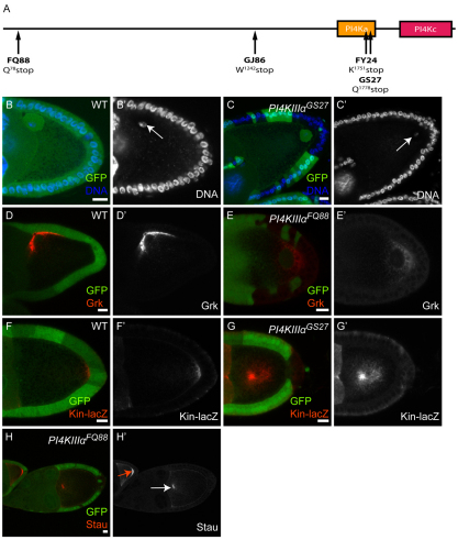
DV asymmetry of the Drosophila oocyte is established during mid-oogenesis through a repolarization process initiated in the PFCs. In response to an unknown signal from the PFCs the oocyte nucleus migrates from the posterior end to the dorsal-anterior corner of the oocyte. As a consequence, the Gurken (Grk) protein no longer accumulates at the posterior cortex of the oocyte, but is now found in the dorsal-anterior membrane overlying the oocyte nucleus where it activates EGFR to initiate DV patterning (Gonzalez-Reyes et al., 1995; Roth et al., 1995). In a genetic screen directed at FC components affecting this repolarization process (Denef et al., 2008), we isolated a complementation group with six lethal mutant alleles, initially named after a representative allele, GS27. When the PFCs were mutant for the GS27 gene product, the oocyte nucleus frequently remained at the posterior end of the oocyte (Fig. 1B,C; 47.7%, n=111). This phenotype was confirmed by the abnormal posterior localization of Grk in late egg chambers (Fig. 1D,E).
Fig. 1.
Mutations in PI4KIIIalpha lead to oocyte polarity defects. (A) CG10260, the Drosophila PI4KIIIalpha homolog. Alleles PI4KIIIalphaFQ88, PI4KIIIalphaGJ86, PI4KIIIalphaFY24 and PI4KIIIalphaGS27 contain early stop codons at Gln78, Trp1242, Lys1751 and Gln1778, respectively. Protein domains are annotated with CDD search (Marchler-Bauer et al., 2009). PI4Ka, phosphoinositide 4-kinase (PI4K) accessory domain; PI4Kc, PI4K, type III, alpha isoform, catalytic domain. (B-E′) Wild-type egg chambers (B,D) and egg chambers containing PI4KIIIalphaGS27 (C) or PI4KIIIalphaFQ88 (E) mutant posterior follicle cells (PFCs), marked by the absence of GFP (green) and stained for DNA (blue, B,C) or Grk (red, D,E). The DNA (B′,C′) and Grk (D′,E′) channels are also shown separately. The oocyte nucleus is located in the dorsal-anterior corner in a wild-type egg chamber (B′, arrow). In mutant PFCs, the oocyte nucleus remains at the posterior of the oocyte (C′, arrow). Similarly, Grk localization is disrupted in the mutant egg chamber (E). (F-G′) A wild-type egg chamber (F) and an egg chamber containing PI4KIIIalphaGS27 mutant PFCs (G) expressing Kin-lacZ and stained for β-gal (red). The β-gal channel is shown separately in F′,G′. In the wild-type egg chamber, Kin-β-gal localizes to the posterior of the oocyte (F). In the egg chamber containing a large PI4KIIIalphaGS27 PFC clone, Kin-β-gal is mislocalized to the center of the oocyte (G). (H,H′) Egg chambers stained for Staufen (red), which forms a tight crescent at the posterior of a wild-type egg chamber (H′, red arrow), but is mislocalized to the center of the oocyte in an egg chamber containing a PI4KIIIalphaFQ88 PFC clone (H′, white arrow). Scale bars: 10 μm.
We mapped the lethality of the GS27 complementation group through duplication and deficiency mapping to the X-chromosomal region 3A4-3A8, which contains 16 genes. Sequencing of candidate genes showed that four alleles of the GS27 complementation group contained mutations that lead to premature stop codons in the coding region of CG10260 (Fig. 1A), a predicted phosphatidylinositol 4-kinase (http://flybase.org). Phosphatidylinositol 4-kinases (PI4Ks) catalyze the generation of PIP4. Phosphoinositides, including PIP4, are important phospholipids in the cell membrane that participate in numerous signaling events (Skwarek and Boulianne, 2009). Four classes of PI4Ks have been identified in mammalian cells that localize to different cellular compartments and are likely to perform non-redundant functions (Balla and Balla, 2006). Three PI4K genes have been annotated in the fly genome: four wheel drive (fwd; PI4KIIIbeta) (Polevoy et al., 2009), CG2929 (PI4KIIalpha) (Raghu et al., 2009) and CG10260 (PI4KIIIalpha).
To investigate the oocyte polarization defects caused by PI4KIIIalpha mutations, we checked the localization of well-established oocyte polarity markers. The microtubule cytoskeleton is polarized in the oocyte. We examined the microtubule plus-end marker Kinesin (Kin, or Khc) fused to β-gal (Kin-β-gal), which normally forms a crescent at the posterior of the oocyte after stage 8 (Clark et al., 1994) (Fig. 1F). When the PFCs were mutant for PI4KIIIalpha, Kin-β-gal either localized to the center of the oocyte or was diffuse in the oocyte (Fig. 1G; 66.7%, n=24). Staufen localizes to the posterior pole of wild-type oocytes after stage 8 and is required for the localization of maternal RNAs (St Johnston et al., 1991). In PFC clones mutant for PI4KIIIalpha, Staufen also frequently mislocalized to the center of the oocyte or became dispersed in the oocyte (Fig. 1H; 73.4%, n=74). Therefore, in combination with the mislocalization of the oocyte nucleus, our results demonstrate that PI4KIIIalpha is required in the PFCs for all aspects of the establishment of correct oocyte polarity.
PI4KIIIalpha mutations and mutations in Hippo pathway components produce similar phenotypes during oogenesis
Oocyte polarization relies on the integrity of four signaling pathways in the PFCs: Notch, JAK/STAT, EGFR and Hippo (Gonzalez-Reyes et al., 1995; Roth et al., 1995; Lopez-Schier and St Johnston, 2001; Xi et al., 2003; Meignin et al., 2007; Polesello and Tapon, 2007; Yu et al., 2008). To examine whether the polarization defect we observed in PI4KIIIalpha mutants was caused by disruption of one of these signaling pathways, we examined well-established downstream targets of each pathway in PI4KIIIalpha mutants.
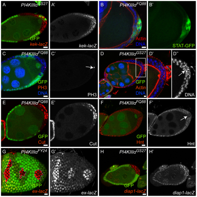
The EGFR signaling reporter kekkon-lacZ (kek-lacZ) is highly expressed in the PFCs at stage 7 and 8 as a result of EFGR activation by Grk (Pai et al., 2000). In PFCs mutant for PI4KIIIalpha, the kek-lacZ expression level was comparable to that of wild-type PFCs, indicating that EFGR signaling was unaffected (Fig. 2A; n>50). The JAK/STAT signaling reporter 10×STAT92E-GFP (Bach et al., 2007) is normally turned on in the PFCs during stage 7 and 8 in response to JAK/STAT activation. We detected apparently normal levels of GFP in the nuclei of PI4KIIIalpha mutant PFCs, suggesting that JAK/STAT signaling was also intact (Fig. 2B; n>30).
Fig. 2.
Effects of PI4KIIIalpha mutations on different signaling pathways. Mutant cells are marked by the absence of GFP (green), except in B where we generated unmarked clones in order to visualize the 10×STAT-GFP reporter and PI4KIIIalpha mutant cells were identified by the loss of their monolayered epithelial structure outlined by actin staining. (A,A′) A Drosophila egg chamber containing PI4KIIIalpha mutant PFCs, expressing the EGFR signaling reporter kekkon-lacZ and stained for β-gal (red in A, gray in A′). (B,B′) An egg chamber containing PI4KIIIalpha mutant PFCs, expressing the JAK/STAT signaling reporter 10×STAT-GFP (green). Both EGFR and JAK/STAT signaling pathways were correctly activated and transduced in the PI4KIIIalpha mutant PFCs, as indicated by the normal levels of β-gal staining (A,A′) and GFP signal (B,B′). (C-F′) Egg chambers containing PI4KIIIalpha mutant PFCs stained for phosphorylated Histone H3 (PH3, red, C), Actin (red, D), DNA (blue, C,D), Cut (red, E) and Hnt (red, F); the PH3, Cut and Hnt channels are also shown separately (C′,E′,F′); the boxed region in D is shown at higher magnification in D′ and D″ (DNA channel only). PI4KIIIalpha mutant PFCs remained in the mitotic cycle after stage 6, as indicated by the presence of PH3-positive cells (C′, arrow) and multilayered cells with smaller nuclei (D″, arrow). PI4KIIIalpha mutant PFCs failed to downregulate Cut (E) and to upregulate Hnt (F) after stage 6. These results indicate that Notch signaling is compromised in PI4KIIIalpha mutant PFCs. (D,E) Note that PI4KIIIalpha mutant cells at the lateral side of the egg chambers show normal epithelial structure (D) and correctly downregulated Cut expression (E), as in the wild-type cells. (G-H′) Egg chambers containing PI4KIIIalpha mutant FCs, expressing the Hippo signaling reporters ex-lacZ (G) and diap1-lacZ (H), stained for β-gal (red in G,H; gray in G′,H′). Upregulation of both reporters indicates that Hippo signaling is disrupted in PI4KIIIalpha mutant FCs. Scale bars: 10 μm.
Notch signaling is required for FCs to exit the mitotic cell cycle at stage 6 and switch to an endocycle (Deng et al., 2001; Lopez-Schier and St Johnston, 2001). PI4KIIIalpha mutant PFCs maintained a mitotic cell cycle after stage 6, as indicated by the sustained staining of the mitotic marker phosphorylated Histone H3 (PH3), which is only seen up to stage 6 in wild-type FCs (Fig. 2C; n>30). Consistent with a failure to exit the mitotic cycle, the PI4KIIIalpha mutant PFCs often lost their monolayered epithelial structure and had smaller nuclei than neighboring cells (Fig. 2D). We also examined the expression of two Notch signaling targets, Cut and Hindsight (Hnt; Pebbled – FlyBase). In wild-type FCs, Cut expression is downregulated whereas Hnt expression is upregulated upon Notch activation at stage 6 (Sun and Deng, 2005; Sun and Deng, 2007). PI4KIIIalpha mutant PFCs frequently failed to downregulate Cut (Fig. 2E; 81.6%, n=76) and upregulate Hnt (Fig. 2F; 66.7%, n=57) expression. Interestingly, PI4KIIIalpha mutant cells on the lateral side of the egg chambers showed no defect in Notch signaling (Fig. 2D-E). These results suggest that PI4KIIIalpha mutations compromise Notch signaling in the PFCs only.
The phenotypes described above are similar to those caused by mutations in Hippo pathway components (Meignin et al., 2007; Polesello and Tapon, 2007; Yu et al., 2008). In particular, the observation that only PFCs appear affected is characteristic of mutations in the Hippo pathway, which are reported to affect Notch signaling only in this group of FCs (Meignin et al., 2007; Polesello and Tapon, 2007; Yu et al., 2008). When we checked the expression of a Hippo pathway target, ex, using the enhancer trap line ex-lacZ (Boedigheimer and Laughon, 1993), we detected a much higher level of β-gal in PI4KIIIalpha mutant FCs than in wild-type cells (Fig. 2G; 81.4%, n=86). This upregulation was observed in all FCs, regardless of their position. Another Hippo pathway target, Diap1, monitored with the enhancer trap line diap1-lacZ (Hay et al., 1995), was mildly upregulated in the PI4KIIIalpha mutant FCs (Fig. 2H; 43.4%, n=53). These results indicate that the polarization defect in the PI4KIIIalpha mutants is likely to be caused by defective Hippo signaling.
Merlin localization is affected in PI4KIIIalpha mutant follicle cells and eye disc cells
Multiple lines of evidence suggest that the apical localization of the Expanded-Merlin-Kibra complex is crucial for Hippo signaling activity (Baumgartner et al., 2010; Genevet et al., 2010; Grzeschik et al., 2010; Robinson et al., 2010; Yu et al., 2010) as it is proposed to function as a platform to bring the core Hippo components into close proximity and facilitate the phosphorylation reactions (Baumgartner et al., 2010; Genevet et al., 2010; Grzeschik et al., 2010; Robinson et al., 2010; Yu et al., 2010). In addition, it has been reported that Expanded directly interacts with Yki and functions to sequester Yki in the cytoplasm (Badouel et al., 2009).
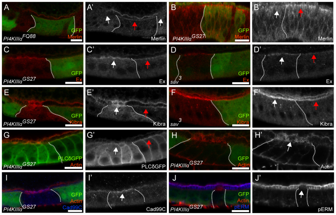
To investigate how mutations in PI4KIIIalpha lead to defective Hippo signaling, we examined the apical localization of the Merlin-Expanded-Kibra complex. The complex is confined to the apical domain in wild-type FCs. In the PI4KIIIalpha mutant cells, we observed a loss of apical Merlin staining (Fig. 3A; n>30), whereas Expanded and Kibra were upregulated at the apical membrane (Fig. 3C,E; n>30). In addition to being Hippo pathway regulators, Expanded and Kibra are also targets of the Hippo signaling pathway. Mutations in Hippo pathway components lead to upregulation of Expanded and Kibra (Fig. 3D,F; n>30) (Hamaratoglu et al., 2006; Baumgartner et al., 2010; Genevet et al., 2010; Yu et al., 2010). In addition, it has been reported that the apical sorting of Merlin, Expanded and Kibra occur independently of each other (McCartney et al., 2000; Baumgartner et al., 2010; Genevet et al., 2010; Yu et al., 2010). Therefore, the absence of Merlin from the apical membrane in PI4KIIIalpha mutant cells is the likely cause of the signaling defect, and the upregulation of Expanded and Kibra would be an expected secondary consequence of the disrupted Hippo signaling.
Fig. 3.
Merlin mislocalization from the apical membrane in PI4KIIIalpha mutant follicle cells and eye disc cells. Clone boundaries are marked by dotted white lines. Mutant cells are marked by the absence of GFP (green), except in G where we generated unmarked clones in order to visualize the Ubi-PH-PLCδ-GFP reporter. PI4KIIIalpha mutant cells were identified by the abnormal actin structure on the apical domain. FCs and eye disc cells are orientated with the apical side up. (A-B′) The follicular epithelium (A) and the imaginal eye disc epithelium (B) containing PI4KIIIalpha mutant cells stained for Merlin (red in A,B; gray in A′,B′). (C-F′) Egg chambers containing PI4KIIIalpha (C,E) or sav (D,F) mutant FCs stained for Expanded (red in C,D; gray in C′,D′) and Kibra (red in E,F; gray in E′,F′). Merlin, Expanded and Kibra localization in wild-type cells is indicated with red arrows. Merlin disappears from the apical and junctional region of the mutant cells (A′,B′, white arrow), whereas Expanded (C′, white arrow) and Kibra (E′, white arrow) are upregulated but remain localized. Upregulation of Expanded (D′, white arrow) and Kibra (F′, white arrow) is similarly observed in sav mutant FCs. (G-J′) Egg chambers containing PI4KIIIalpha mutant cells, expressing the PI(4,5)P2 reporter Ubi-PH-PLCδ-GFP (G), stained for GFP (green in G; gray in G′), actin (red in G-J; gray in H′), Cad99C (blue in I; gray in I′) and phospho-ERM proteins (blue in J; gray in J′). The PI(4,5)P2 reporter is absent from the apical membrane of the mutant cells (G′, white arrow), in contrast to the wild-type cells (G′, red arrow). PI4KIIIalpha mutant cells exhibit abnormal actin spikes on the apical side (H′, white arrow) as marked by the microvillus marker Cad99C (I′, white arrow). The apical microvilli region of PI4KIIIalpha mutant cells shows depletion of phospho-ERM proteins (J′, white arrow). Scale bars: 10 μm.
When we examined PI4KIIIalpha mutant clones in the imaginal eye discs of early second instar larvae, we also observed an absence of Merlin from the apical and junctional region (Fig. 3B; n>10). However, we did not observe an overgrowth phenotype typical of Hippo pathway mutations (data not shown). In fact, adults with mutant eye clones had smaller eyes than wild-type adults. Eye discs from late L2 larvae exhibited pyknotic nuclei staining in PI4KIIIalpha mutant clones, indicating death of the mutant cells (data not shown).
Multiple classes of PI4Ks exist in eukaryotic cells that participate in producing various phosphoinositide species in distinct cellular compartments (Balla and Balla, 2006). Three PI4K genes have been annotated in the fly genome. When we examined the intracellular distribution and level of PIP2 using a Ubi-PH-PLCδ-GFP reporter (Gervais et al., 2008), we observed a complete absence of PIP2 from PI4KIIIalpha mutant FCs in rare cases (2 out of 40 clones). In most cases, the PIP2 reporter was specifically lost from the apical plasma membrane in the mutant cells (Fig. 3G; 82.5%, n=40). The yeast homolog of PI4KIIIalpha, Stt4p, localizes to patches on the plasma membrane where it is required for normal actin cytoskeleton organization (Audhya et al., 2000; Audhya and Emr, 2002). When we examined the actin cytoskeleton of PI4KIIIalpha mutant FCs by phalloidin staining, they exhibited abnormal actin-enriched spike structures on their apical domain (Fig. 3H; n>100) that were positively marked by the microvillus marker Cad99C (D'Alterio et al., 2005) (Fig. 3I; n>30), suggesting that the spikes were malformed microvilli. As mutations in the Hippo pathway have been reported to lead to apical domain expansion (Justice et al., 1995; Wu et al., 2003; Genevet et al., 2009), one possibility is that the malformed microvilli are caused by defective Hippo signaling. However, the morphology of the actin-enriched spikes in PI4KIIIalpha mutant cells is distinct from that caused by mutations in the Hippo pathway (Fig. 3H), suggesting that the loss of PI4KIIIalpha might also have a Hippo-independent effect on apical membrane structure.
How could PI4KIIIalpha mutations cause Merlin mislocalization? Expanded and Merlin are ERM (Ezrin, Radixin and Moesin)-related proteins, which are key linkers of the plasma membrane and cytoskeleton. Classical ERM proteins bind to PIP2 in the membrane to switch from a closed to an open conformation for their activation (Nakamura et al., 1999; Fievet et al., 2004; Fehon et al., 2010). Significantly, in PI4KIIIalpha mutant cells, phosphorylated ERM proteins were absent from the apical microvilli region as indicated by a phospho-ERM-specific antibody (Fig. 3J; n>20). The malformed microvillus structure might therefore indicate a general failure of ERM protein activation in the PI4KIIIalpha mutant cells (Takeuchi et al., 1994). For Merlin, the closed conformation is the active form, opposite to other ERM proteins (Okada et al., 2007; McClatchey and Fehon, 2009). Nevertheless, Merlin undergoes a similar conformational switch to the other ERM proteins (Gonzalez-Agosti et al., 1999) and contains an ERM PIP2-binding site (Barret et al., 2000). Given our observations, it is possible that PIP2 binding activates and/or stabilizes Merlin in the apical membrane, and a depletion of this lipid species due to the absence of PI4KIIIalpha might directly lead to the loss of Merlin.
In summary, we have shown that PI4KIIIalpha is required in the FCs for Merlin localization and Hippo signaling. PI4KIIIalpha mutations in the PFCs lead to a Notch signaling defect and the subsequent failure of oocyte repolarization, which are precisely the phenotypes reported for Hippo mutations in the FCs. This effect is likely to be caused by a change in lipid composition in the membrane. How the abnormal actin structures are generated in the mutant cells, and whether they have a direct role in Merlin localization, remain to be investigated.
Acknowledgements
We thank I. Clark, D. St Johnston, E. A. Bach, K. D. Irvine, R. G. Fehon, D. Godt, N. Tapon, the Developmental Studies Hybridoma Bank and the Bloomington Stock Center for fly stocks and antibodies; G. Barcelo for technical assistance; J. Goodhouse for help with confocal microscopy; members of the laboratories of T.S. and E. Wieschaus for advice and feedback; and Y. C. Wang and A. C. Martin for helpful comments on the manuscript. This work was supported by the Howard Hughes Medical Institute and US Public Health Service Grant RO1 GM 077620. Deposited in PMC for release after 6 months.
Footnotes
Competing interests statement
The authors declare no competing financial interests.
References
- Audhya A., Emr S. D. (2002). Stt4 PI 4-kinase localizes to the plasma membrane and functions in the Pkc1-mediated MAP kinase cascade. Dev. Cell 2, 593-605 [DOI] [PubMed] [Google Scholar]
- Audhya A. M., Foti M., Emr S. D. (2000). Distinct roles for the yeast phosphatidylinositol 4-kinases, Stt4p and Pik1p, in secretion, cell growth, and organelle membrane dynamics. Mol. Biol. Cell 11, 2673-2689 [DOI] [PMC free article] [PubMed] [Google Scholar]
- Bach E. A., Ekas L. A., Ayala-Camargo A., Flaherty M. S., Lee H., Perrimon N., Baeg G. H. (2007). GFP reporters detect the activation of the Drosophila JAK/STAT pathway in vivo. Gene Expr. Patterns 7, 323-331 [DOI] [PubMed] [Google Scholar]
- Badouel C., Gardano L., Amin N., Garg A., Rosenfeld R., Le Bihan T., McNeill H. (2009). The FERM-domain protein expanded regulates Hippo pathway activity via direct interactions with the transcriptional activator Yorkie. Dev. Cell 16, 411-420 [DOI] [PubMed] [Google Scholar]
- Balla A., Balla T. (2006). Phosphatidylinositol 4-kinases: old enzymes with emerging functions. Trends Cell Biol. 16, 351-361 [DOI] [PubMed] [Google Scholar]
- Barret C., Roy C., Montcourrier P., Niggli V. (2000). Mutagenesis of the phosphatidylinositol 4,5-bisphosphate (PIP(2)) binding site in the NH(2)-terminal domain of ezrin correlates with its altered cellular distribution. J. Cell Biol. 151, 1067-1080 [DOI] [PMC free article] [PubMed] [Google Scholar]
- Baumgartner R., Poernbacher I., Buser N., Hafen E., Stocker H. (2010). The WW domain protein Kibra acts upstream of Hippo in Drosophila. Dev. Cell 18, 309-316 [DOI] [PubMed] [Google Scholar]
- Bennett F. C., Harvey K. F. (2006). Fat cadherin modulates organ size in Drosophila via the Salvador/Warts/Hippo signaling pathway. Curr. Biol. 16, 2101-2110 [DOI] [PubMed] [Google Scholar]
- Boedigheimer M., Laughon A. (1993). Expanded: a gene involved in the control of cell proliferation in imaginal discs. Development 118, 1291-1301 [DOI] [PubMed] [Google Scholar]
- Cho E., Feng Y., Rauskolb C., Maitra S., Fehon R., Irvine K. D. (2006). Delineation of a fat tumor suppressor pathway. Nat. Genet. 38, 1142-1150 [DOI] [PubMed] [Google Scholar]
- Clark I., Giniger E., Ruohola-Baker H., Jan L. Y., Jan Y. N. (1994). Transient posterior localization of a kinesin fusion protein reflects anteroposterior polarity of the Drosophila oocyte. Curr. Biol. 4, 289-300 [DOI] [PubMed] [Google Scholar]
- D'Alterio C., Tran D. D., Yeung M. W., Hwang M. S., Li M. A., Arana C. J., Mulligan V. K., Kubesh M., Sharma P., Chase M., et al. (2005). Drosophila melanogaster Cad99C, the orthologue of human Usher cadherin PCDH15, regulates the length of microvilli. J. Cell Biol. 171, 549-558 [DOI] [PMC free article] [PubMed] [Google Scholar]
- Denef N., Chen Y., Weeks S. D., Barcelo G., Schüpbach T. (2008). Crag regulates epithelial architecture and polarized deposition of basement membrane proteins in Drosophila. Dev. Cell 14, 354-364 [DOI] [PMC free article] [PubMed] [Google Scholar]
- Deng W. M., Althauser C., Ruohola-Baker H. (2001). Notch-Delta signaling induces a transition from mitotic cell cycle to endocycle in Drosophila follicle cells. Development 128, 4737-4746 [DOI] [PubMed] [Google Scholar]
- Dong J., Feldmann G., Huang J., Wu S., Zhang N., Comerford S. A., Gayyed M. F., Anders R. A., Maitra A., Pan D. (2007). Elucidation of a universal size-control mechanism in Drosophila and mammals. Cell 130, 1120-1133 [DOI] [PMC free article] [PubMed] [Google Scholar]
- Duffy J. B., Harrison D. A., Perrimon N. (1998). Identifying loci required for follicular patterning using directed mosaics. Development 125, 2263-2271 [DOI] [PubMed] [Google Scholar]
- Edgar B. A. (2006). From cell structure to transcription: Hippo forges a new path. Cell 124, 267-273 [DOI] [PubMed] [Google Scholar]
- Emoto K., Parrish J. Z., Jan L. Y, Jan Y. N. (2006). The tumour suppressor Hippo acts with the NDR kinases in dendritic tiling and maintenance. Nature 443, 210-213 [DOI] [PubMed] [Google Scholar]
- Fehon R. G., McClatchey A. I., Bretscher A. (2010). Organizing the cell cortex: the role of ERM proteins. Nat. Rev. Mol. Cell Biol. 11, 276-287 [DOI] [PMC free article] [PubMed] [Google Scholar]
- Fievet B. T., Gautreau A., Roy C., Del Maestro L., Mangeat P., Louvard D., Arpin M. (2004). Phosphoinositide binding and phosphorylation act sequentially in the activation mechanism of ezrin. J. Cell Biol. 164, 653-659 [DOI] [PMC free article] [PubMed] [Google Scholar]
- Genevet A., Polesello C., Blight K., Robertson F., Collinson L. M., Pichaud F., Tapon N. (2009). The Hippo pathway regulates apical-domain size independently of its growth-control function. J. Cell Sci. 122, 2360-2370 [DOI] [PMC free article] [PubMed] [Google Scholar]
- Genevet A., Wehr M. C., Brain R., Thompson B. J., Tapon N. (2010). Kibra is a regulator of the Salvador/Warts/Hippo signaling network. Dev. Cell 18, 300-308 [DOI] [PMC free article] [PubMed] [Google Scholar]
- Gervais L., Claret S., Januschke J., Roth S., Guichet A. (2008). PIP5K-dependent production of PIP2 sustains microtubule organization to establish polarized transport in the Drosophila oocyte. Development 135, 3829-3838 [DOI] [PubMed] [Google Scholar]
- Gonzalez-Agosti C., Wiederhold T., Herndon M. E., Gusella J., Ramesh V. (1999). Interdomain interaction of merlin isoforms and its influence on intermolecular binding to NHE-RF. J. Biol. Chem. 274, 34438-34442 [DOI] [PubMed] [Google Scholar]
- Gonzalez-Reyes A., Elliott H., St Johnston D. (1995). Polarization of both major body axes in Drosophila by gurken-torpedo signalling. Nature 375, 654-658 [DOI] [PubMed] [Google Scholar]
- Grusche F. A., Richardson H. E., Harvey K. F. (2010). Upstream regulation of the Hippo size control pathway. Curr. Biol. 20, R574-R582 [DOI] [PubMed] [Google Scholar]
- Grzeschik N. A., Parsons L. M., Allott M. L., Harvey K. F., Richardson H. E. (2010). Lgl, aPKC, and Crumbs regulate the Salvador/Warts/Hippo pathway through two distinct mechanisms. Curr. Biol. 20, 573-581 [DOI] [PubMed] [Google Scholar]
- Hamaratoglu F., Willecke M., Kango-Singh M., Nolo R., Hyun E., Tao C., Jafar-Nejad H., Halder G. (2006). The tumour-suppressor genes NF2/Merlin and Expanded act through Hippo signalling to regulate cell proliferation and apoptosis. Nat. Cell Biol. 8, 27-36 [DOI] [PubMed] [Google Scholar]
- Harvey K. F., Pfleger C. M., Hariharan I. K. (2003). The Drosophila Mst ortholog, hippo, restricts growth and cell proliferation and promotes apoptosis. Cell 114, 457-467 [DOI] [PubMed] [Google Scholar]
- Hay B. A., Wassarman D. A., Rubin G. M. (1995). Drosophila homologs of baculovirus inhibitor of apoptosis proteins function to block cell death. Cell 83, 1253-1262 [DOI] [PubMed] [Google Scholar]
- Huang J., Wu S., Barrera J., Matthews K., Pan D. (2005). The Hippo signaling pathway coordinately regulates cell proliferation and apoptosis by inactivating Yorkie, the Drosophila homolog of YAP. Cell 122, 421-434 [DOI] [PubMed] [Google Scholar]
- Justice R. W., Zilian O., Woods D. F., Noll M., Bryant P. J. (1995). The Drosophila tumor suppressor gene warts encodes a homolog of human myotonic dystrophy kinase and is required for the control of cell shape and proliferation. Genes Dev. 9, 534-546 [DOI] [PubMed] [Google Scholar]
- Lai Z. C., Wei X., Shimizu T., Ramos E., Rohrbaugh M., Nikolaidis N., Ho L. L., Li Y. (2005). Control of cell proliferation and apoptosis by mob as tumor suppressor, mats. Cell 120, 675-685 [DOI] [PubMed] [Google Scholar]
- Lopez-Schier H., St Johnston D. (2001). Delta signaling from the germ line controls the proliferation and differentiation of the somatic follicle cells during Drosophila oogenesis. Genes Dev. 15, 1393-1405 [DOI] [PMC free article] [PubMed] [Google Scholar]
- Maitra S., Kulikauskas R. M., Gavilan H., Fehon R. G. (2006). The tumor suppressors Merlin and Expanded function cooperatively to modulate receptor endocytosis and signaling. Curr. Biol. 16, 702-709 [DOI] [PubMed] [Google Scholar]
- Marchler-Bauer A., Anderson J. B., Chitsaz F., Derbyshire M. K., DeWeese-Scott C., Fong J. H., Geer R. C., Gonzales N. R., Gwadz M., He S. (2009). CDD: specific functional annotation with the conserved domain database. Nucleic Acids Res. 37, D205-D210 [DOI] [PMC free article] [PubMed] [Google Scholar]
- McCartney B. M., Fehon R. G. (1996). Distinct cellular and subcellular patterns of expression imply distinct functions for the Drosophila homologues of moesin and the neurofibromatosis 2 tumor suppressor, merlin. J. Cell Biol. 133, 843-852 [DOI] [PMC free article] [PubMed] [Google Scholar]
- McCartney B. M., Kulikauskas R. M., LaJeunesse D. R., Fehon R. G. (2000). The neurofibromatosis-2 homologue, Merlin, and the tumor suppressor expanded function together in Drosophila to regulate cell proliferation and differentiation. Development 127, 1315-1324 [DOI] [PubMed] [Google Scholar]
- McClatchey A. I., Fehon R. G. (2009). Merlin and the ERM proteins-regulators of receptor distribution and signaling at the cell cortex. Trends Cell Biol. 19, 198-206 [DOI] [PMC free article] [PubMed] [Google Scholar]
- Meignin C., Alvarez-Garcia I., Davis I., Palacios I. M. (2007). The salvador-warts-hippo pathway is required for epithelial proliferation and axis specification in Drosophila. Curr. Biol. 17, 1871-1878 [DOI] [PMC free article] [PubMed] [Google Scholar]
- Mikeladze-Dvali T., Wernet M. F., Pistillo D., Mazzoni E. O., Teleman A. A., Chen Y. W., Cohen S., Desplan C., et al. (2005). The growth regulators warts/lats and melted interact in a bistable loop to specify opposite fates in Drosophila R8 photoreceptors. Cell 122, 775-787 [DOI] [PubMed] [Google Scholar]
- Nakamura F., Huang L., Pestonjamasp K., Luna E. J., Furthmayr H. (1999). Regulation of F-actin binding to platelet moesin in vitro by both phosphorylation of threonine 558 and polyphosphatidylinositides. Mol. Biol. Cell 10, 2669-2685 [DOI] [PMC free article] [PubMed] [Google Scholar]
- Okada T., You L., Giancotti F. G. (2007). Shedding light on Merlin's wizardry. Trends Cell Biol. 17, 222-229 [DOI] [PubMed] [Google Scholar]
- Pai L. M., Barcelo G., Schüpbach T. (2000). D-cbl, a negative regulator of the Egfr pathway, is required for dorsoventral patterning in Drosophila oogenesis. Cell 103, 51-61 [DOI] [PubMed] [Google Scholar]
- Pan D. (2007). Hippo signaling in organ size control. Genes Dev. 21, 886-897 [DOI] [PubMed] [Google Scholar]
- Pantalacci S., Tapon N., Léopold P. (2003). The Salvador partner Hippo promotes apoptosis and cell-cycle exit in Drosophila. Nat. Cell Biol. 5, 921-927 [DOI] [PubMed] [Google Scholar]
- Polesello C., Tapon N. (2007). Salvador-warts-hippo signaling promotes Drosophila posterior follicle cell maturation downstream of notch. Curr. Biol. 17, 1864-1870 [DOI] [PubMed] [Google Scholar]
- Polevoy G., Wei H. C., Wong R., Szentpetery Z., Kim Y. J., Goldbach P., Steinbach S. K., Balla T., Brill J. A. (2009). Dual roles for the Drosophila PI 4-kinase four wheel drive in localizing Rab11 during cytokinesis. J. Cell Biol. 187, 847-858 [DOI] [PMC free article] [PubMed] [Google Scholar]
- Raghu P., Coessens E., Manifava M., Georgiev P., Pettitt T., Wood E., Garcia-Murillas I., Okkenhaug H., Trivedi D., Zhang Q., et al. (2009). Rhabdomere biogenesis in Drosophila photoreceptors is acutely sensitive to phosphatidic acid levels. J. Cell Biol. 185, 129-145 [DOI] [PMC free article] [PubMed] [Google Scholar]
- Robinson B. S., Huang J., Hong Y., Moberg K. H. (2010). Crumbs regulates Salvador/Warts/Hippo signaling in Drosophila via the FERM-domain protein expanded. Curr. Biol. 20, 582-590 [DOI] [PMC free article] [PubMed] [Google Scholar]
- Rogulja D., Rauskolb C., Irvine K. D. (2008). Morphogen control of wing growth through the Fat signaling pathway. Dev. Cell 15, 309-321 [DOI] [PMC free article] [PubMed] [Google Scholar]
- Roth S., Lynch J. A. (2009). Symmetry breaking during Drosophila oogenesis. Cold Spring Harb. Perspect. Biol. 1, a001891 [DOI] [PMC free article] [PubMed] [Google Scholar]
- Roth S., Neuman-Silberberg F. S., Barcelo G., Schüpbach T. (1995). Cornichon and the EGF receptor signaling process are necessary for both anterior-posterior and dorsal-ventral pattern formation in Drosophila. Cell 81, 967-978 [DOI] [PubMed] [Google Scholar]
- Silva E., Tsatskis Y., Gardano L., Tapon N., McNeill H. (2006). The tumor-suppressor gene fat controls tissue growth upstream of expanded in the hippo signaling pathway. Curr. Biol. 16, 2081-2089 [DOI] [PubMed] [Google Scholar]
- Skwarek L. C., Boulianne G. L. (2009). Great expectations for PIP: phosphoinositides as regulators of signaling during development and disease. Dev. Cell 16, 12-20 [DOI] [PubMed] [Google Scholar]
- St Johnston D., Beuchle D., Nüsslein-Volhard C. (1991). Staufen, a gene required to localize maternal RNAs in the Drosophila egg. Cell 66, 51-63 [DOI] [PubMed] [Google Scholar]
- Sun J., Deng W. M. (2005). Notch-dependent downregulation of the homeodomain gene cut is required for the mitotic cycle/endocycle switch and cell differentiation in Drosophila follicle cells. Development 132, 4299-4308 [DOI] [PMC free article] [PubMed] [Google Scholar]
- Sun J., Deng W. M. (2007). Hindsight mediates the role of notch in suppressing hedgehog signaling and cell proliferation. Dev. Cell 12, 431-442 [DOI] [PMC free article] [PubMed] [Google Scholar]
- Takeuchi K., Sato N., Kasahara H., Funayama N., Nagafuchi A., Yonemura S., Tsukita S., Tsukita S. (1994). Perturbation of cell adhesion and microvilli formation by antisense oligonucleotides to ERM family members. J. Cell Biol. 125, 1371-1384 [DOI] [PMC free article] [PubMed] [Google Scholar]
- Tapon N., Harvey K. F., Bell D. W., Wahrer D. C., Schiripo T. A., Haber D. A., Hariharan I. K. (2002). Salvador Promotes both cell cycle exit and apoptosis in Drosophila and is mutated in human cancer cell lines. Cell 110, 467-478 [DOI] [PubMed] [Google Scholar]
- Udan R. S., Kango-Singh M., Nolo R., Tao C., Halder G. (2003). Hippo promotes proliferation arrest and apoptosis in the Salvador/Warts pathway. Nat. Cell Biol. 5, 914-920 [DOI] [PubMed] [Google Scholar]
- van Eeden F., St Johnston D. (1999). The polarisation of the anterior-posterior and dorsal-ventral axes during Drosophila oogenesis. Curr. Opin. Genet. Dev. 9, 396-404 [DOI] [PubMed] [Google Scholar]
- Wei X., Shimizu T., Lai Z. C. (2007). Mob as tumor suppressor is activated by Hippo kinase for growth inhibition in Drosophila. EMBO J. 26, 1772-1781 [DOI] [PMC free article] [PubMed] [Google Scholar]
- Willecke M., Hamaratoglu F., Kango-Singh M., Udan R., Chen C. L., Tao C., Zhang X., Halder G. (2006). The fat cadherin acts through the hippo tumor-suppressor pathway to regulate tissue size. Curr. Biol. 16, 2090-2100 [DOI] [PubMed] [Google Scholar]
- Wu S., Huang J., Dong J., Pan D. (2003). Hippo encodes a Ste-20 family protein kinase that restricts cell proliferation and promotes apoptosis in conjunction with salvador and warts. Cell 114, 445-456 [DOI] [PubMed] [Google Scholar]
- Xi R., McGregor J. R., Harrison D. A. (2003). A gradient of JAK pathway activity patterns the anterior-posterior axis of the follicular epithelium. Dev. Cell 4, 167-177 [DOI] [PubMed] [Google Scholar]
- Xu T., Wang W., Zhang S., Stewart R. A., Yu W. (1995). Identifying tumor suppressors in genetic mosaics: the Drosophila lats gene encodes a putative protein kinase. Development 121, 1053-1063 [DOI] [PubMed] [Google Scholar]
- Yu J., Poulton J., Huang Y. C., Deng W. M. (2008). The hippo pathway promotes Notch signaling in regulation of cell differentiation, proliferation, and oocyte polarity. PLoS One 3, e1761 [DOI] [PMC free article] [PubMed] [Google Scholar]
- Yu J., Zheng Y., Dong J., Klusza S., Dang W. M., Pan D. (2010). Kibra functions as a tumor suppressor protein that regulates Hippo signaling in conjunction with Merlin and Expanded. Dev. Cell 18, 288-299 [DOI] [PMC free article] [PubMed] [Google Scholar]


