Abstract
Cantharidin is an intoxicant found in beetles in the Meloidae (Coleoptera) family. Ingestion may result in haematemesis, impaired level of consciousness, electrolyte disturbance, haematurea and renal impairment. Here, we report two paediatric cases of meloid beetle ingestion resulting in cantharidin poisoning and the clinical presentation of the ensuing intoxication.
Keywords: Cantharidin, Intoxication, Meloidae, Paediatric, Blister beetle, Case report, Saudi Arabia
Cantharidin, from the Greek word Kantharos (‘beetle’),1 is an intoxicant produced by beetles in the family Meloidae (Coleoptera). The Spanish fly (Lytta Cantharis vesicatoria Linnaeus, 1758) is the most well known species. Spanish flies and other meloids have historically been used medicinally as aphrodisiacs, skin irritants, vesicants and abortifacients.2
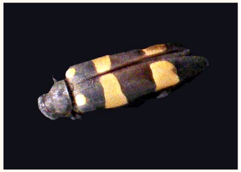
Many species of Meloidae produce cantharidin,2 including Hycleus maculiventris (Klug, 1845). The species recovered in both cases reported here [Figure 1] was identified by Dr. Marco Bolonga, University of Roma Tre, Italy who has great experience in studying the Meloidae of the Arabian Peninsula.
Figure 1.
Hycleus maculiventris (1.5 cm in length) insect ingested by the child
Cantharidin, a bicyclic terpenoid, has an inhibitory effect on protein phosphatase 1 (PP1) and protein phosphatase 2A (PP2A). It is stored in the haemolymph, genitalia and other tissues.3,4 The systemic manifestations of a paediatric cantharidin poisoning have been reported previously.5,7 Our purpose in reporting the clinical presentations of these two cases is to increase awareness of the signs and symptoms of meloid beetle ingestion.
Case Reports
CASE ONE
An otherwise healthy eight month-old Saudi male child ingested a beetle, later identified in the infant vomitus as Hycleus maculiventris [Figure 1]. Thirty minutes after ingestion, he developed haematemesis and four hours later he presented to the Emergency Room (ER) at Aseer Central Hospital, Abha, Saudi Arabia, with impaired consciousness. There was no history of fever, ecchymosis, diarrhea, seizure, photophobia or weakness. There was also no history of cardiovascular or respiratory systems involvement.
In the ER, he was stuporous with a Glasgow coma score (GCS) of 12/15. His heart rate was 140/min, respiratory rate 30/min, temperature 38°C, and oxygen saturation 92% in room air.
On physical examination, his respiratory, cardiovascular, and gastrointestinal systems assessments were normal. There were no skin lesions or musculoskeletal abnormalities. A central nervous system evaluation revealed hypertonia and hyperreflexia of both upper and lower limbs. The child was admitted to the paediatric intensive care unit and shortly after admission he developed gross haematuria.
A blood work-up revealed a white cell count of 17,700/cc; red blood cell count of 4,100,000/cc; platelet count of 131,000/cc, and haemoglobin of 10.1 gm/dl. Blood urea nitrogen was initially 47 mg/dl and serum creatinine was 0.4 mg/dl. On day two, his kidney function deteriorated, with urea increasing to 68 mg/dl and serum creatinine increasing to 1.2 mg/dl. Serum electrolytes and blood sugar were within normal ranges. Initial venous blood gas was pH 7.32; PC02 29.5; PO2 61.2; HCO3 14.9; ABE -10 and oxygen pulse oxymeter was 95%. Serum aspartate aminotransferase (AST) was 64 u/l; serum alanine aminotransferase (ALT) 20 u/l; alkaline phosphatase 253 u/l (normal range [NR] = 145–420); creatine phosphokinase (CPK) 226 u/l (NR = 0–170), and lactate dehydrogenase 409 u/l (NR = 135–225). His total bilirubin was 0.4 mg/dl; direct bilirubin 0.0; total serum protein 5.4 gm/dl and albumin 3.4 gm/dl. The serum calcium level was 6 mg/dl, and urinalysis showed numerous red blood cells, granular casts, proteins and glucose.
Over the next 4 days, the child’s condition improved steadily with a return to normal consciousness. The haematuria cleared up gradually with renormalisation of complete blood count (CBC) and renal function.
CASE TWO
An otherwise healthy eleven months-old Saudi female ingested a beetle which the father found in the child’s vomitus and brought to the hospital for recognition, it was later identified as Hycleus maculiventris [Figure 1].
Two hours after ingestion of the insect, she developed haematemesis associated with decreased consciousness and was brought to the local hospital. Her temperature was 40°C. She developed an attack of generalised tonic-clonic convulsion which was aborted by a diazepam injection of 0.3 mg/kg/dose followed by a loading dose of phenobarbitone 15 mg/kg intravenously.
The initial blood work-up showed a white blood cell count of 45,000/cc; platelets of 51,000/cc; red blood cells (RBCs) of 4,600,000/cc, and haemoglobin of 11 gm/dl. Her blood urea nitrogen was 79 mg/dl and serum creatinine 2.1 mg/dl.
She was managed at the local hospital with concerns about possible encephalitis. She was started on ceftriaxone 100 mg/kg/day in two divided doses; phenobarbitone 5 mg/kg/day in two divided doses, and calcium gluconate intravenously at a dose of 70 mg elemental ca/kg. She was kept nil per os (nothing per mouth) and given intravenous fluids, and 05% dextrose with ½ normal saline. Mannitol of 0.25 g/kg was given for possible increased intracranial pressure, since there was no cranial scanning (computed tomography [CT] scan) facility at the local hospital.
Two days later, she was referred to our hospital for brain imaging and proper intensive care management. Her initial assessment revealed that she was moderately dehydrated. Her temperature was 38°C rectally, respiratory rate was 40/min, pulse rate 156/min, and blood pressure 90/55 mmHg. She was not pale or jaundiced and not in distress.
A central nervous system (CNS) examination revealed a conscious, non-irritable child. Her cranial nerves, motor and sensory systems evaluation were normal. Examination of chest (cardio vascular system), abdomen, skin and musculoskeletal systems were within normal limits as well.
Results of repeated blood tests revealed a white blood cell count of 84,800/cc, and a platelet count of 43,000/cc. Her initial blood gas was pH 7.28; PC02 27.2; HCO3-13; ABE -10. This abnormality was corrected over 24 hours. The patient started to have haematuria and her blood urea nitrogen peaked to 143 mg/dl, while serum creatinine increased to 2.4 mg/dl. The serum sodium level was 123 mmol/l; potassium 5.6 mmol/l; calcium 5.3 mg/dl; phosphorus 6.1 mg/dl and chloride 92 mmol/dl. A CT scan of the brain was normal. The patient remained on ceftriaxone and acyclovir was added to her management. The results of a septic work up, including a cerebral spinal fluid (CSF) examination (including protein, sugar, culture and cell count), were normal. The antibiotics were discontinued and the patient managed conservatively with intravenous fluids and phenobarbitone. Over the following three days all symptoms resolved with normalisation of her laboratory values. By the end of the 6 day she was back to normal status and was discharged in good condition.
Discussion
These cases describe the clinical presentations of cantharidin intoxication following ingestion of blister beetles. Once the insect is disturbed it exudes cantharidin in a milky oral fluid, however the adult beetle reflexively secretes the toxin from the leg joints.8 The amount of cantharidin found in the blister beetle varies from 0.2 mg to 0.7 mg among different species. Some species like E. immaculate have larger amounts of 4.8 mg per beetle. Females have a significantly lower concentration.9
Symptoms begin within 2–4 hours of ingestion with sudden onset of haematemesis of fresh blood, which is likely due to local irritation by the toxin. Haematemesis is either associated with, or followed by, agitation impaired consciousness or convulsions. These symptoms are followed by haematuria either on the first or second day.5,7
Fever was a presenting feature in both cases, similar to observations reported in other cases.7 Presentation is also associated with leukocytosis and thrombocytopenia with a mild drop in haemoglobin. Initially, there was only mild impairment of renal function, but over 24 hours the impairment became more pronounced, with creatinine increasing to 2.1 mg/dl. In spite of renal impairment, the blood pressure remained in within the normal range.
The exact amount of cantharidin which causes intoxication in children is unknown, but a minimal amount of cantharidin causes severe intoxication. The fatal dose is estimated to range from 10–65 mg with a minimal lethal dose being approximately 1 mg/kg. Fatality usually result from renal failure.4 Serum electrolytes may also be affected, manifesting as hyponatraemia, hyperkalaemia and hypocalcaemia.8 It is not clear why electrolytes are affected, but it might be due to renal impairment secondary to the local toxin effect. There was a mild elevation of AST to 64 u/L, but there was no jaundice associated with this presentation.
Gross and microscopic haematurea with a granular cast is a prominent feature of cantharidin toxicity, and was also present in the cases discussed here.5,7
There is no specific antidote for cantharidin intoxication. Management, therefore, has to be supportive in nature, including IV fluid at maintenance rate and correction of electrolyte and blood gas abnormalities. The use of intravenous proton pump inhibitors or H2 blockers may enhance the healing of the ulceration that results from the local effect of the toxin on the gastric mucosa.
These clinical presentations are similar to those described for adults10,13 and animals.5,11,12 There is currently no clear explanation for these clinical presentations, but they are likely secondary to the local toxic effect of cantharidin on different organ systems.
For topical exposure, the affected area should be cleaned with acetone, ether, fatty soap or alcohol, which helps to dissolve and dilute the cantharidin. Topical steroids may be applied to intact skin if it is symptomatic.14
Conclusion
There is no pathognomonic feature of cantharidin intoxication in children, but a careful history and examination, combined with the sequence of events, should alert the medical team to this rare event.
Acknowledgement
We would like to express our thanks to Dr Marco Bologna, University of Roma Tre, Italy, the taxonomist who identified the beetle.
References
- 1.Cantharidin and meloids: a review of classical history, biosynthesis, and function. [Accessed Feb 2010]. From www.colostate.edu.
- 2.Dolson CJ, Tattersall RN. Cantharides wasp, bee, and scorpion stings. Clin Toxicol. 1969;30:475–84. [Google Scholar]
- 3.Beasley V, editor. International Veterinary Information Service. Veterinary Toxicology. [Accessed Feb 2010]. From www.ivis.org.
- 4.Rauh R, Kahl S, Boechzelt H, Bouer R, Kaina B, Efferth T. Molecular biology of cantharidin in cancer cells. Chin Med. 2007;2:8. doi: 10.1186/1749-8546-2-8. [DOI] [PMC free article] [PubMed] [Google Scholar]
- 5.Wertelecki W, Vietti TJ, Kulapongs P. Cantharidin Poisoning from ingestion of a “Blister Beetle”. Pediatrics. 1967;39:287–9. [PubMed] [Google Scholar]
- 6.Mallari RQ, Saif M, Elbualy MS, Sapru A. Ingestion of a Blister Beetle (Mecoidae Family) Pediatrics. 1996;98:458–9. [PubMed] [Google Scholar]
- 7.Tagwireyi D, Ball DE, Loga PJ, Moyo S. Cantharidin poisoning due to “Blister beetle” ingestion. Toxicon. 2000;38:1865–9. doi: 10.1016/s0041-0101(00)00093-3. [DOI] [PubMed] [Google Scholar]
- 8.Carrel JE, McCairel MH, Slagel AJ, Doom JP, Bril J, McCormick JP. Cantharidin production in a blister beetle. Experientia. 1993;49:171–4. doi: 10.1007/BF01989424. [DOI] [PubMed] [Google Scholar]
- 9.Capinera JL, Gardner DR, Stermitz FR. J Echonom Entemol. 1985;78:1052–5. [Google Scholar]
- 10.Al-Rumikan A, Al-Hamdan NA. Indirect cantharidin food poisoning caused by eating wild birds. Saudi Epidemiol Bull. 1999;6:26. [Google Scholar]
- 11.Karras DJ, Farrel SE, Harrigan RA, Henretig FM, Gealt L. Poisoning from “Spanish fly” (cantharidin) Am J Emerg Med. 1996;14:478–83. doi: 10.1016/S0735-6757(96)90158-8. [DOI] [PubMed] [Google Scholar]
- 12.Fisch HP, Reutter FW, Gloor F. Lesions of the kidney and the efferent urinary tract due to cantharidin (German) Schweiz Med Wochenschr. 1978;108:1664–7. [PubMed] [Google Scholar]
- 13.Ewart WB, Rabkin SW, Mintenko PA. Poisoning by cantharides. CMA Journal. 1978;118:1199. [PMC free article] [PubMed] [Google Scholar]
- 14.Lisa Moed BA, Tor A, Shwayder MD, Mary Wu Chang. A blistering defence of an ancient medicine. Arch Dermatol. 2001;137:1357–60. doi: 10.1001/archderm.137.10.1357. [DOI] [PubMed] [Google Scholar]


