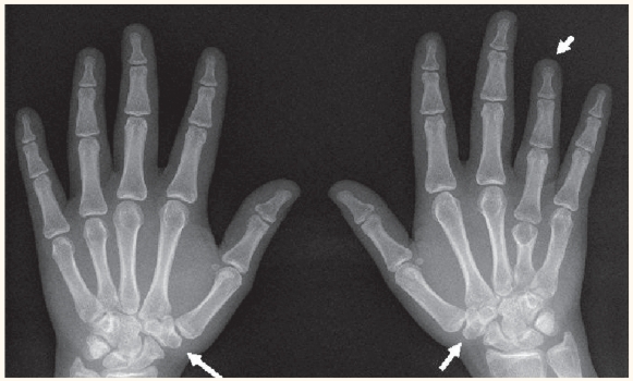
This is an images of the hands of a 23 year-old Omani girl who presented to the Family Medicine Clinic of Sultan Qaboos University Hospital, Oman, with complaints of bilateral wrist pain for the previous four weeks [Figure 1]. The pain was aggravated by activities especially those requiring weight lifting. The history revealed no other joint involvement, no previous trauma, fever, morning stiffness, or weight loss. On examination, she was of average height and weight. Her wrist examination showed mild tenderness and swelling over the lateral aspect of her wrists proximal to the second metacarpal base, especially in her left wrist. In addition, her left fourth metacarpal was relatively shorter than rest of her metacarpals. However, the range of movements at the wrist and the metacarpophalangeal joints were full, but were associated with pain at the extremes of movement. The diagnosis after X-ray was congenital fusion of carpal bones.
Figure 1.
X-ray of both hands, posterior-anterior view. The upper arrow shows the short left fourth metacarpal while the lower arrows show bilateral fusion of the trapezium and trapezoid.
Fusion or synostosis of various carpal bones is possible in different combinations. It can either be a part of a syndrome or can occur as an isolated anomaly.2 The term ‘fusion’ can be considered as a misnomer as it is not truly a fusion of carpal bones, rather it is an absence of joint cavitation, and chondrification of the joint interzone. This leads to the phenomenon of carpal synostosis which becomes apparent only after the bones ossify. Carpal fusions occur as normal variations in about 0.1% of the population. Lunate-triquetral fusion is the most common fusion anomaly of the carpus.2 This bone anomaly is mostly bilateral, but more common on the left side when unilaterally present.3 Among the various population groups studied, the highest incidence is seen in people of African descent with a with a higher female to male ratio (2:1) and a strong familial tendency.
Carpal coalition is usually asymptomatic and often discovered incidentally. There have been reports of symptomatic pisohamate coalition, scapholunate triquetral coalition, and scaphoid trapezium synchondrosis.1,2,5 The loss of movement between the fused bones leads to a compensatory increase in motion at surrounding joints. This predisposes the person to recurrent sprains causing pain, especially under stressful conditions. Occasionally, this may also result in an overgrowth of bone called carpal bossing.1,2 Increased demand on the joint, especially in high activity level people like sportsmen, may lead to progressive stress loading and early degenerative arthritis or pseudoarthrosis. Generally, isolated carpal fusions involve two bones of the same row, while the syndrome-associated fusions, such as Ellis van Creveld syndrome, Osteochondritis dissecans, foetal alcohol syndrome, symphalangia, diastrophic dwarfism, gonadal dysgenesis, and Poland syndrome, are quite often multiple.1,2 Acquired fusion can occur secondary to arthritis, trauma or as a result of surgery for joint stabilisation.2
Radiological findings may vary in appearance and fusion may present as partial or complete union of the carpal bone or there may be narrowing of the joint space without adjacent sclerosis or osteophytosis.2 For asymptomatic cases, treatment is rarely required. Symptomatic treatment might be needed for the minority of patients who present with symptoms.
References
- 1.Terrence JJJ. Congenital fusion of the trapezium and trapezoid. Rom J Morphol Embryol. 2008;49:417–9. [PubMed] [Google Scholar]
- 2.Singh P, Tuli A, Choudhry R, Mangal A. Intercarpal fusion – a review. J Anat Soc India. 2003;52:183–8. [Google Scholar]
- 3.Cockshott WP. Carpal fusion. Am J Roentgenol Radium Ther Nucl Med. 1963;89:1260–71. [PubMed] [Google Scholar]
- 4.Resnick D, Niwayama G. Diagnosis of Bone and Joint Disorders. 2nd Ed. Philadelphia: WB Saunders Co.; 1988. pp. 3560–1. [Google Scholar]
- 5.Knezevich S, Gottesman M. Symptomatic scapholunatotriquetral carpal coalition with fusion of the capitatometacarpal joint: Report of a case. Clin Orthop Relat Res. 1990;251:153–6. [PubMed] [Google Scholar]
- 6.Simmons BP, McKenzie WD. Symptomatic carpal coalition. J Hand Surg. 1985;10:190–3. doi: 10.1016/s0363-5023(85)80103-9. [DOI] [PubMed] [Google Scholar]
- 7.Chouhdry R, Tuli A, Chimmalgi M, Anand M. Os capitate trapezoid: a case report. Surg Radiol Anat. 1998;20:373–5. doi: 10.1007/BF01630624. [DOI] [PubMed] [Google Scholar]
- 8.Friedman T, Reed M, Elliott AM. The carpal fusion in Poland syndrome. Skeletal Radiol. 2009;38:585–91. doi: 10.1007/s00256-008-0638-x. [DOI] [PubMed] [Google Scholar]


