Abstract
Objective:
To ascertain the relationship between serum leptin levels and related variables (weight, Body Mass Index (BMI) and fat percentage) in a group of Omani obese and non-obese healthy subjects.
Methods:
Leptin levels were assessed in serum samples from 35 obese Omanis and 20 non-obese healthy subjects.
Results:
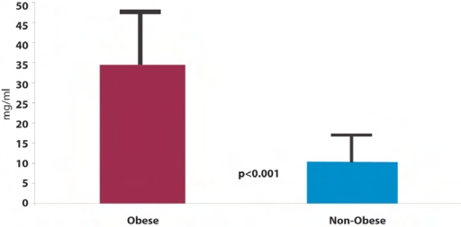
There was a significant difference (p< 0.001) in serum leptin between the obese group (34.78 + 13.96 ng/ml) and the control non-obese subjects (10.6 ± 4.2 ng/ml). Leptin levels were higher in females compared to males. There was a significantly positive correlation between leptin levels in obese subjects with weight (p=0.002), body fat percentage (p=0.0001) and BMI (p=0.001).
Conclusions:
We concluded that serum leptin levels are higher in the Omani obese group and correlate positively with body fatness and obesity.
Keywords: Leptin, Obesity, Body weight, Oman, Body Mass Index
The name Leptin is derived from the leptose, meaning thin.1,3,5 It is secreted by the adipose tissue in proportion to adipose mass and therefore its circulating levels increase with weight gain and decrease with weight loss.1,2 A large amount of research has been conducted on leptin since its discovery in 1994 and it is now possible to evaluate the physiological significance of leptin.1,2,3,4 Leptin is considered to play a wide range of functions in humans such as decreasing appetite and thereby food intake, stimulating and maintaining energy expenditure and acting as a metabolic hormone in a wide range of processes by binding to receptors in the brain.5 Leptin functions primarily as an anti-obesity hormone. Its serum concentrations in healthy individuals positively correlate with body fat content,3 but it correlates negatively when energy intake is reduced and energy stores in fat are declining.6
In most obese subjects, leptin levels are high and correlate with the Body Mass Index (BMI) and the percentage of body fat,3 Leptin levels are found to be correlated with a number of endocrine substances such as insulin, glucocorticoids, thyroid hormones and testosterone.7 Similar studies also indicate that it may be involved in mediating some endocrine-related processes, like the onset of puberty.8 Impaired or modified receptor functions may be associated with certain medical conditions other than obesity, such as cardiovascular disease9 and certain types of cancer.10 Research findings also indicate that leptin can act as a growth factor in the foetus and in the neonate.7
It has been reported that obesity is a major risk factor for the development of diabetes mellitus11, therefore, leptin might play a role in this disease. Many studies have documented a high prevalence of diabetes and metabolic syndrome among the Omani population.12,13,14 No data are available in the literature for levels of serum leptin among the Omani population. Studying leptin levels in Omanis may be relevant in the light of reported associations of leptin serum status with obesity and diabetes. Certain social economic and environmental factors may be unique in the Omani population.
The objective of this study was to report leptin profiles in Omanis and to ascertain the relationship between serum leptin levels and related variables (weight, BMI and fat percentage) between two groups of Omanis, obese and non-obese healthy subjects.
METHODS
This study was approved by the Sultan Qaboos University Medical Research and Ethics Committee (Project No. MREC 155).
SUBJECTS
A group of 35 Omani subjects, who were attending our obesity clinic (25 female and 10 males, age range 22–49 years) and were considered to be obese on the basis of BMI. BMI was defined as weight in kilogrames divided by height in meters (kg/m2). A BMI value of > or equal to 30 was used as an indicator of obesity. Fat percentage was measured by a Tanita Fat Analyser (USA) using the Bioelectrical Impedance method. Twenty healthy non-obese subjects of similar age range (20–50 years) served as a control group. None of the subjects studied had any clinical or laboratory (e.g: fasting blood glucose, urea & electrolytes, liver and thyroid function tests) signs of disease associated with obesity and were taking no drugs.
SERUM LEPTIN ASSAY
An in vitro sandwich Enzyme-Linked Immunosorbent Assay (ELISA) (DRG Instrument GmbH, Germany) was employed for the quantitative measurement of human leptin in serum. The microtiter wells were coated with a monoclonal anti-leptin antibody. Test and control samples were incubated in the coated wells with a specific rabbit anti-leptin antibody. After incubation the unbound material was washed off and anti-rabbit peroxidase conjugate was added for detection of the bound leptin. The intensity of colour developed was proportional to the concentration of leptin in the sample.
STATISTICAL ANALYSIS
We used a Statistical Product and Service Solutions (SPSS) software program for statistical analysis of data. A p value of < 0.05 was considered statistically significant.
RESULTS
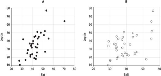
As seen in Figure 1, there was a significant difference (p=0.001) in serum leptin levels (mean + S.E.) between the obese group (34.78 + 13.96 ng/ml) and levels in the control (non-obese) subjects (10.6 + 4.2 ng/ml). A strongly significant positive correlation was observed between leptin levels with weight (r=0.5, p =0.002), BMI (r= 0.51, p =0.001) and body fat percentage (r=0.7, p =0.0001) [Figure 2].
Figure 1:
Leptin levels (mean + SE) in obese (BMI>30kg/m2) and non-obese Omani subjects
Figure 2:
Correlation between leptin levels (ng/ml) with (A) Fatness (kg) and (B) BMI (kg/m2) in obese Omani patients
Leptin concentrations and body fat percentages were higher in women than in men within the obese group Table 1. A strong positive correlation (r=0.528, p <0.007) was present when leptin concentration and BMI of the female obese group was compared.
Table 1:
Comparison between charateristics of obese subjects (25 female: 10 male; mean ± SE)
| Character | Female(n=25) | Male(n=10) |
|---|---|---|
| Age (years) | 29.2 ± 1.6 | 30.0 ± 3.1 |
| Leptin(ng/ml) | 38.2 ± 2.5 | 27.0 ± 4.9 |
| Weight (Kg) | 97.9 ± 3.9 | 108.4 ± 7.6 |
| BMI (kg/m2) | 39.6 ± 1.5 | 39.0 ± 2.9 |
| %Fat | 44.1 ± 1.0 | 38.3 ± 3.9 |
DISCUSSION
In this study, we have described significantly high serum leptin levels among obese Omani subjects compared to the non-obese group, though the number studied here is small. To our knowledge, this has not been reported previously in the Omani population. Normal ranges of leptin (in Caucasian and Arab residents of Kuwait),15,26 which have been reported in previous studies, are comparable to the range observed in the non-obese group in the current study. In a study carried out by Concidine et al.,3 it was found that obese humans had (on average) leptin levels four times higher than non-obese individuals.
The present study confirms results of previous reports16,17 regarding a gender difference in leptin levels between males and females. Females have higher serum leptin concentrations than males, which confirms the previously reported range in female Kuwaitis.26 The reason for this is unclear; the larger amounts of body fat mass, predominantly subcutaneous fat, in women and the effect of sex steroid hormones which increase leptin production have been suggested.18 Since leptin reflects mainly the amount of body fat, this seemes to be a logical explanation for the observed gender differences. Gender differences and their relationship to body composition were examined in several studies and leptin levels were still found to be significantly higher in women, even after correction was made for BMI or fat mass.19
The current study shows that serum leptin concentrations were increased in relation to increased body fat content. The positive correlation between body fat and serum leptin is probably explained primarily by the increased release of leptin from large fat cells.19 In fact, some studies have proposed that leptin can serve as an indicator of fat content and that its levels increase exponentially with increasing body fat percentage.3 Leptin levels decrease by reduction of body fat even though BMI values remain unchanged.20
In our study, we found a significant relationship between circulating leptin and BMI, an observation that is associated with the relationship of leptin to total body weight as BMI and fatness.2 In all previous investigations, leptin levels have been shown to be high and correlated to BMI.2,3
In agreement with our findings, regarding the existence of a positive correlation between the circulating leptin levels and the body weight, other studies have reported the relation of spontaneous or enforced weight changes and serum leptin levels.3 Whatever the initial concentration, weight loss due to food restriction was found to be associated with a decrease in serum leptin in both obese and normal weight humans.2
Based on the above findings, leptin concentrations can be considered as a marker of the extent of obesity in human(s). The one relatively common clinical condition, in which a significantly elevated serum leptin concentration occurs without an increase in body mass, is chronic renal failure, probably as a result of decreased renal clearance.21 A number of researchers however, have claimed subtle leptin abnormalities in conditions such as diabetes22 and thyroid disease.23 In addition, leptin seems to be involved in the pathogenesis of some obesity-associated conditions such as anovulatory infertility24 and Type 2 diabetes mellitus.25
Although the number of individuals in this study was relatively small, it was found that circulating leptin levels were high and positively correlated to body weight, the percentage fat and BMI among obese Omani subjects. The pattern of this increase may reflect the differences in lifestyle, diet, physical activity and other cultural and economic factors that are specific for people in this part of the world. The significance of this study in relation to high and increased prevalence of diabetes in Oman requires further evaluation.
REFERENCES
- 1.Zhang Y, Proenca R, Maffei M, Barone M, Leopald L, Friedman JM. Positional cloning of mouse ob gene and its human homologue. Nature. 1994;737:425–432. doi: 10.1038/372425a0. [DOI] [PubMed] [Google Scholar]
- 2.Maffei M, Halaas J, Ravussin E, Pratly RE, Lee GH, Zhang Y, Fei H, Kim S, Lallone R, Ranganathan S. Leptin levels in human and rodent; measurement of plasma leptin and ob RNA in obese and weight-reduced subjects. Nat Med. 1995;1:1155–1161. doi: 10.1038/nm1195-1155. [DOI] [PubMed] [Google Scholar]
- 3.Considine RV, Sinha MK, Heiman M, Kriauciunas A, Stephens T, Nyce M, Ohannesian JP, Marco CC, Mc-Kee J, Bauer L. Serum immunoreactive-leptin concentrations in normal-weight and obese humans. N Engl J Med. 1996;334:292–295. doi: 10.1056/NEJM199602013340503. [DOI] [PubMed] [Google Scholar]
- 4.Chow VT, Phoon MC. Measurement of serum leptin concentrations in university undergraduates by competitive ELISA reveals correlations with body mass index and sex. Adv Physiol Educ. 2003;27:70–77. doi: 10.1152/advan.00001.2003. [DOI] [PubMed] [Google Scholar]
- 5.Trayhurn P, Hoggard N, Mercer JD, Rayner DV. Leptin: fundamental aspects. Int. J. Obes. Relat Metab Disord. 1999;23:22–28. doi: 10.1038/sj.ijo.0800791. [DOI] [PubMed] [Google Scholar]
- 6.Boden G, Chen X, Mozzoli M, Ryan I. Effect of fasting on serum leptin in normal human subjects. J Clin Endocrinol Metab. 1996;81:3419–3443. doi: 10.1210/jcem.81.9.8784108. [DOI] [PubMed] [Google Scholar]
- 7.Janeckova R(a) The role of leptin in human physiology and pathophysiology. Physiol Res. 2001;50:443–459. [PubMed] [Google Scholar]
- 8.Janeckova R(b) New findings on the role of leptons in regulation of the reproductive system in humans. Cesk Fysiol. 2002;50:25–30. [PubMed] [Google Scholar]
- 9.Chu N, Spiegelman D, Hotamisligil G, Rifai N, Stampfer M, Rimm E. Plasma insulin, leptin and soluble TNF receptors levels in relation to obesity-related atherogenic and thrombogenic cardiovascular disease risk factors among men. Atherosclerosis. 2001;157:495–503. doi: 10.1016/s0021-9150(00)00755-3. [DOI] [PubMed] [Google Scholar]
- 10.Larmore K, O’Connor D, Sherman T, Funanage V, Hassink S, Oerter Klein K. Leptin and estradiol as relate to changes in pubertal status and body weight. Med Sci Monit. 2002;8:206–210. [PubMed] [Google Scholar]
- 11.Lahlou N, Issa T, Leboue Y, Carel J, Camoin L, Roger M. Leptin might play a role in diabetes. Diabetes. 2002;51:1980–1985. doi: 10.2337/diabetes.51.6.1980. [DOI] [PubMed] [Google Scholar]
- 12.Asfour M, Lambourne A, Al-Bahlani S, Al-Asfoor D, Bold A, Mahtab H, King H. High prevalence of diabetes mellitus and impaired glucose tolerance in the Sultanate of Oman. Results of the 1991 national survey. Diabet Med. 1995;12:1122–1125. doi: 10.1111/j.1464-5491.1995.tb00431.x. [DOI] [PubMed] [Google Scholar]
- 13.Al-Lawati JA, Al Riyami AM, Mohammed AJ, Jousilahti P. Increasing prevalence of diabetes mellitus in Oman. Diabet Med. 2002;19:954–957. doi: 10.1046/j.1464-5491.2002.00818.x. [DOI] [PubMed] [Google Scholar]
- 14.Al-Asfoor DH, Al-Lawati JA, Mohammed AJ. Body fat distribution and the risk of non-insulin-dependant diabetes mellitus in the Omani population. East Mediterr Health J. 1999;5:14–20. [PubMed] [Google Scholar]
- 15.Haluzik M, Kabrt J, Nedvidkora J, Svobodova J, Kotrlikora E, Papezova H. Relationship of serum leptin levels and selected nutritional parameters in patients with protein-caloric malnutrition. Nutrition. 1999;15:829–833. doi: 10.1016/s0899-9007(99)00177-x. [DOI] [PubMed] [Google Scholar]
- 16.Rosenbaum M, Nicolson M, Hirsch J, Heymsfield SB, Gallagher D, Chew F, Leibel R. Effects of gender, body composition, and menopause on plasma concentrations of leptin. J Clin Endocrinol Metab. 1996;81:3424–3427. doi: 10.1210/jcem.81.9.8784109. [DOI] [PubMed] [Google Scholar]
- 17.Licinio J, Negrao AB, Mantzoros C, Kaklamani V, et al. Sex differences in circulating human leptin pulse amplitude: clinical implications. J Clin C Endocrinol Metab. 1998;83:4140–4147. doi: 10.1210/jcem.83.11.5291. [DOI] [PubMed] [Google Scholar]
- 18.Cassabiell X, Pineiro V, Peino R, et al. Gender differences in both spontaneous and stimulated leptin secretion by human omental adipose tissue in vitro: dexamethasone and estradiol stimulate leptin release in women, but not in men. J Clin Endocrinol Metab. 1998;83:2149–2155. doi: 10.1210/jcem.83.6.4849. [DOI] [PubMed] [Google Scholar]
- 19.Lonnqvist F, Nordfors L, Jasson M, Thorne A, Shalling M, Arner P. Leptin secretion from adipose tissue in women. Relationship to plasma levels and gene expression. J Clin Investig. 1997;99:2398–2404. doi: 10.1172/JCI119422. [DOI] [PMC free article] [PubMed] [Google Scholar]
- 20.Stejskal D, Jedelsky L, Ruzicka V, Bartek J. Leptin, insulin a proinsulin - vzajemne vztahy. Vnitrini Lekarstvi. 1998;44:361–365. [PubMed] [Google Scholar]
- 21.Nordfors L, Lonnqvist F, Heimburger O, Danielsson A, Schalling M, Stenvinkel P. Low leptin gene expression and hyperleptinemia in chronic renal failure. Kidney Int. 1998;54:1267–1275. doi: 10.1046/j.1523-1755.1998.00088.x. [DOI] [PubMed] [Google Scholar]
- 22.Rudberg S, Persson B. Serum leptin levels in young females with insulin-dependent diabetes and the relationship to hyperandrogenicity and microalbuminuria. Horm Res. 1998;50:297–302. doi: 10.1159/000023294. [DOI] [PubMed] [Google Scholar]
- 23.Valcavi R, Zini M, Peino R, Casaneuva FF, Dieguez C. Influence of thyroid status on serum immunoreactive leptin levels. J Clin Endocrinol Metab. 1997;82:1632–1634. doi: 10.1210/jcem.82.5.3954. [DOI] [PubMed] [Google Scholar]
- 24.Karlsson C, Lindell K, Svensson E, Bergh C, Lind P, Billig H, Carlsson LM, Carlsson B. Expression of functional leptin receptors in the human ovary. J Clin Endocrinol Metab. 1997;82:4144–4148. doi: 10.1210/jcem.82.12.4446. [DOI] [PubMed] [Google Scholar]
- 25.Morton N, Emilsson V, de Groot P, Pallett AL, Cawthorne M. Leptin signalling in pancreatic and clonal insulin-secreting cells. J Mol Endocrinol. 1999;22:173–184. doi: 10.1677/jme.0.0220173. [DOI] [PubMed] [Google Scholar]
- 26.Al-Shoumer Kamal AS, Vasanthy Bagavathy AK, Makhlouf Hanan A, Al-Zaid Mona M. Leptin levels in Arabs with primary hyperthyroidism. Ann of Saudi Med. 2000;20:113–118. doi: 10.5144/0256-4947.2000.113. [DOI] [PubMed] [Google Scholar]


