Abstract
Situs inversus totalis (SIT) is an uncommon anomaly characterized by transposition of organs to the opposite side of the body in a mirror image of normal. We report on an adult woman, born and resident in Brazilian Amazonia, presenting acute pain located at the left hypochondrium and epigastrium. During clinical and radiological evaluation, the patient was found to have SIT and multiple stones cholelithiasis. Laparoscopic cholecystectomy was safely performed with the three-port technique in a reverse fashion. In conclusion, this case confirms that three-port laparoscopic cholecystectomy is a safe and feasible surgical approach to treat cholelithiasis even in rare and challenging conditions like SIT.
Key Words: Situs inversus totalis, Laparoscopic cholecystectomy, Three-port technique
Introduction
Situs inversus totalis (SIT) is a rare congenital malformation characterized by transposition of organs to the opposite side of the body in the mirror image of normal [1]. It occurs in 1:5,000 to 1:10,000 of hospital admissions [2], and although most patients are asymptomatic, it can be associated with a number of other conditions such as cardiac anomalies and Kartagener's syndrome [1, 3]. However, there is no clear evidence whether SIT predisposes to cholelithiasis [3].
The aim of this report is to describe the case of symptomatic cholelithiasis managed by three-port laparoscopic cholecystectomy in a woman from Brazilian Amazonia with SIT.
Case Report
A 43-year-old woman, born and resident in Brazilian Amazonia, presented with a 15-day history of dolorous syndrome localized at the left hypochondrium and epigastrium with no other concomitant digestive problems. On physical examination, she was afebrile, without jaundice, and abdominal examination was unremarkable. During clinical assessment, the patient was found to have apex beat in the right hemithorax.
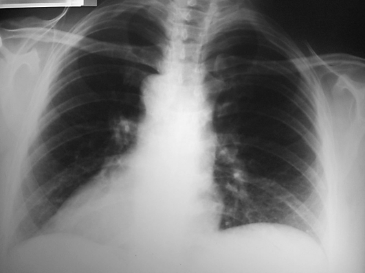
Laboratory study was normal. Chest radiography revealed dextrocardia (fig. 1). Abdominal ultrasonography showed situs inversus and hyperechogenic images in the gallbladder lumen, compatible with multiple free stones. No intra- or extrahepatic bile duct dilatations were observed. As no other structural abnormality was identified, diagnosis of SIT and cholelithiasis was made and the dolorous syndrome was attributed to it. Based on this diagnosis, the patient underwent three-port laparoscopic cholecystectomy by a left-hand surgeon.
Fig. 1.
Chest radiography showing dextrocardia.
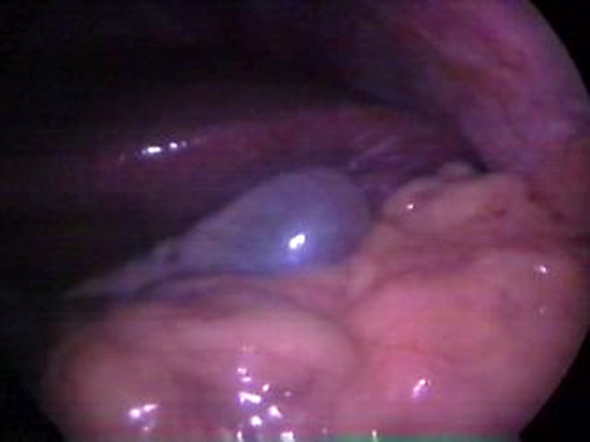
Preoperative preparation was made as routinely. However, the surgical team and the laparoscopic devices were located as a mirror image configuration from the one used in orthotopic patients. The operating surgeon and the video monitor assistant were positioned on the patient's right side and the TV monitor was placed on the upper left side of the patient. Three ports were used: a 10-mm trocar was inserted infraumbilically through which the zero viewing endoscope was introduced. Another 10-mm trocar was inserted 3 cm below the xiphisternum at midline; and finally, a 5-mm trocar at the left hypochondrium hemiclavicular line 5 cm below the costal margin. The infraumbilical port was used to create the pneumoperitoneum (12 mm Hg) with carbon dioxide (CO2). On laparoscopic examination, the gallbladder was distended and inflamed (fig. 2). The operating surgeon held the dissecting instruments with his left hand through the 10-mm trocar while holding the gallbladder at the infundibulum with a grasper through the 5-mm trocar, moving the infundibulum right and left, back and forth to display the Calot triangle. The cystic duct was then isolated, clipped and divided followed by the cystic artery. The gallbladder was dissected from the liver bed and subsequently extracted through the infraumbilical port. The patient had an uneventful recovery and was discharged home on the first postoperative day.
Fig. 2.
Laparoscopic aspect during cholecystectomy. Distended gallbladder situated at the left hypochondrium.
Discussion
Since Campos and Sipes [4] described the first case of laparoscopic cholecystectomy in a patient with situs inversus, this uncommon malformation has been challenging and amazing many surgeons. Due to the contralateral disposition of the viscera, the diagnosis and surgical approach of these patients may be more difficult than that of orthotopic patients.
According to previous reports, SIT is not a contraindication for laparoscopic cholecystectomy [1, 5]. However, the procedure often requires more time to rearrange the equipment in the operating room and extra care to recognize the mirror image anatomy [6]. The anatomical variations and, mainly, the contralateral disposition of the biliary tree demand an accurate dissection and exposition of the biliary structures to avoid iatrogenic lesions.
There is widespread acceptance that four-port technique is the standard procedure of traditional laparoscopic cholecystectomy. However, technical improvements have permitted the execution of less invasive procedures, using only three or even two ports to perform laparoscopic cholecystectomy. These modifications actually reduced postoperative pain, analgesia requirement, length of hospital stay and costs [7, 8]. No other reports in the international literature were found describing the three-port technique in patients with SIT, though. In our case, only three ports were necessary to perform a safe dissection and to obtain a good exposure of the gallbladder and the biliary tree.
Technical aspects of laparoscopic cholecystectomy in patients with SIT privilege left-handed surgeons. The dissection of the biliary tree can be carried out with either the right or the left hand [9], however for right-handed surgeons using the unskilled and nondominant left hand, the manipulation may be cumbersome and not precise [2]. In the present case, the left-handed surgeon performed the dissection of the Calot triangle by holding the dissecting instruments with the left hand through the 10-mm trocar and a grasper with the right hand to maneuver the gallbladder. The patient had a normal follow-up and, 5 months after surgery, remains asymptomatic.
In conclusion, this case confirms that three-port laparoscopaic cholecystectomy is a safe and feasible surgical approach even in patients with SIT presenting symptomatic cholelithiasis, despite the reverse anatomical relationships. However, to overcome technical difficulties and avoid iatrogenic lesions, the procedure must be carried out by an experienced laparoscopic surgeon, especially if the surgeon is left-handed, used to the three-port technique in orthotopic patients.
References
- 1.Schiffino L, Mouro J, Levardo H, Dubois F. Colecistectomia per la via laparoscopica in situs inversus totalis. Minerva Chir. 1993;48:1019–1023. [PubMed] [Google Scholar]
- 2.Machado NO, Chopra P. Laparoscopic cholecystectomy in a patient with situs inversus totalis: feasibility and technical difficulties. JSLS. 2006;10:386–391. [PMC free article] [PubMed] [Google Scholar]
- 3.Demetriades H, Botsios D, Dervenis C, Evagelou J, Agelopoulos S, Dadoukis J. Laparoscopic cholecystectomy in two patients with symptomatic cholelithiasis and situs inversus totalis. Dig Surg. 1999;16:519–521. doi: 10.1159/000018780. [DOI] [PubMed] [Google Scholar]
- 4.Campos L, Sipes E. Laparoscopic cholecystectomy in a 39-year-old female with situs inversus. J Laparoendosc Surg. 1991;1:123–126. doi: 10.1089/lps.1991.1.123. [DOI] [PubMed] [Google Scholar]
- 5.Bedioui H, Chebbi F, Ayadi S, Makni A, Fteriche F, Ksantini R, et al. Cholécystectomie laparoscopique chez un patient porteur d'un situs inversus. Ann Chir. 2006;131:398–400. doi: 10.1016/j.anchir.2005.12.014. [DOI] [PubMed] [Google Scholar]
- 6.McKay D, Blake G. Laparoscopic cholecystectomy in situs inversus totalis: a case report. BMC Surg. 2005;5:1–2. doi: 10.1186/1471-2482-5-5. [DOI] [PMC free article] [PubMed] [Google Scholar]
- 7.Trichak S. Three-port vs. standard four-port laparoscopic cholecystectomy. Surg Endosc. 2003;17:1434–1436. doi: 10.1007/s00464-002-8713-1. [DOI] [PubMed] [Google Scholar]
- 8.Al-Azawi D, Houssein R, Rayis AB, McMahon D, Hehir DJ. Three-port vs. four-port laparoscopic cholecystectomy in acute and chronic cholecystitis. BCM Surg. 2007;7:8. doi: 10.1186/1471-2482-7-8. [DOI] [PMC free article] [PubMed] [Google Scholar]
- 9.Oms LM, Badia JM. Laparoscopic cholecystectomy in situs inversus totalis. The importance of being left-handed. Surg Endosc. 2003;17:1859–1861. doi: 10.1007/s00464-003-9051-7. [DOI] [PubMed] [Google Scholar]


