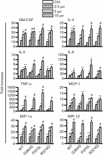
Figure 2.

Enhancement of the mRNA expressions of various cytokines and chemokines. LAD2 cells (1 × 10⁶ cells) were incubated with 2·5–10 μm wild-type catestatin (WT), Gly364Ser (G364S), Pro370Leu (P370L) or Arg374Gln (R374Q), or diluent 0·01% acetic acid (Ctrl, control) for 1 hr. Following incubation, total RNA was extracted and converted into cDNA, and quantitative real-time PCR was performed to analyse changes in the gene expression of granulocyte–macrophage colony-stimulating factor (GM-CSF), interleukin-4 (IL-4), IL-5, IL-8, tumour necrosis factor-α (TNF-α), monocyte chemoattractant protein-1 (MCP-1/CCL2), macrophage inflammatory protein-1α (MIP-1α/CCL3) and MIP-1β/CCL4. Each bar shows the mean ± SD of three different experiments. Values represent fold increases in gene expression above untreated cells (Ctrl, control). *P < 0·05, **P < 0·01, #P < 0·001.
