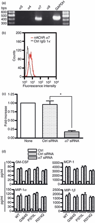
Figure 7.

Expression of the α7 nicotinic acetylcholine receptor (nAChR) on mast cells and its involvement in catestatin-induced activation of mast cells. (a) Total RNA (2 μg) isolated from LAD2 cells was analysed for expression of the α3, α4, α7 and α9 subunits of nAChR by RT-PCR. Aliquots of the PCR products were run on 2% agarose gels and visualized by ethidium bromide staining. One representative of three separate experiments is shown. (b) Expression of the α7 nAChR protein in mast cells. Cells (1 × 10⁶ cells) were incubated with an α7 nAChR antibody (nAChR α7) or isotype control rat IgG1κ antibody (Ctrl IgG1κ) for 30 min, and following staining with FITC-conjugated goat anti-rat IgG, the expression of the α7 nAChR was evaluated by FACS. (c) Cells (3 × 10⁶ cells) were transfected with 400 nmα7 nAChR siRNA or control siRNA using the Amaxa Cell Line Nucleofector kit V, program T-030. After 24 hr of gene silencing, α7 nAChR mRNA expression was evaluated by quantitative real-time PCR. Values are the mean ± SD of three separate experiments and were compared between α7 nAChR siRNA-transfected (α7 siRNA) and control siRNA-transfected cells (Ctrl siRNA). None: non-transfected mast cells. *P < 0·001. (d) In addition, cells were transfected with 400 nmα7 nAChR siRNA or control siRNA for 48 hr. Transfected cells (1 × 10⁶ cells) were stimulated with 10 μm wild-type catestatin (WT), Gly364Ser (G364S), Pro370Leu (P370L), Arg374Gln (R374Q), or diluent 0·01% acetic acid (Ctrl, control) for 6 hr, and the amounts of granulocyte–macrophage colony-stimulating factor (GM-CSF), monocyte chemoattractant protein-1 (MCP-1/CCL2), macrophage inflammatory protein-1α (MIP-1α/CCL3) and MIP-1β/CCL4 released into the culture supernatants were determined by an ELISA. Each bar represents the mean ± SD of three separate experiments.
