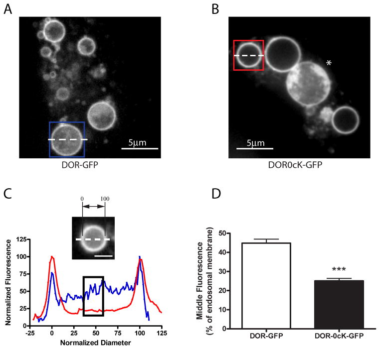
Figure 2. Enlargement of endosomes by Rab5 manipulation illustrates lysyl-mutant DORs differ in their extent of transfer to ILVs.
A and B) HEK293 cells were transiently transfected with CFP-Rab5Q79L and either F-DOR-GFP (A) or F-DOR-0cK-GFP (B), and replated onto to coverslips before treatment for 90 minutes with 10μM DADLE. Cells were then imaged by spinning disc confocal microscopy as described in Materials and Methods. Shown are representative still images of the representative acquired image series, scale bars indicate 5μm. C) Line scan analysis to quantify receptor localization to the intralumenal compartment. Normalized diameter represents the diameter of the endosome shown, where 0 and 100 correspond to the pixel distances with the first and second maximum pixel intensities measured across the dashed line, respectively (see inset image). Blue and red traces represent the normalized pixel intensity measured across the dashed line in the blue and red boxes in A and B respectively, where the maximum pixel intensity across the line is normalized to 100. The black box highlights the normalized fluorescence values of pixels from 40 to 60% of the normalized diameter. D) Compiled results of line scan analysis (mean and SEM, *** p<0.001, Student's t-test, n= 88 endosomes, ≥12 cells).

