Abstract
Background: A lower core body temperature set point has been suggested to be a factor that could potentially predispose humans to develop obesity.
Objective: We tested the hypothesis that obese individuals have lower core temperatures than those in normal-weight individuals.
Design: In study 1, nonobese [body mass index (BMI; in kg/m2) <30] and obese (BMI ≥30) adults swallowed wireless core temperature–sensing capsules, and we measured core temperatures continuously for 24 h. In study 2, normal-weight (BMI of 18–25) and obese subjects swallowed temperature-sensing capsules to measure core temperatures continuously for ≥48 h and kept activity logs. We constructed daily, 24-h core temperature profiles for analysis.
Results: Mean (±SE) daily core body temperature did not differ significantly between the 35 nonobese and 46 obese subjects (36.92 ± 0.03°C compared with 36.89 ± 0.03°C; P = 0.44). Core temperature 24-h profiles did not differ significantly between 11 normal-weight and 19 obese subjects (P = 0.274). Women had a mean core body temperature ≈0.23°C greater than that of men (36.99 ± 0.03°C compared with 36.76 ± 0.03°C; P < 0.0001).
Conclusions: Obesity is not generally associated with a reduced core body temperature. It may be necessary to study individuals with function-altering mutations in core temperature–regulating genes to determine whether differences in the core body temperature set point affect the regulation of human body weight. These trials were registered at clinicaltrials.gov as NCT00428987 and NCT00266500.
INTRODUCTION
The core body temperature set point in humans is normally tightly defended to within a range of ≈0.2°C (1). When the body temperature falls outside of this range, compensatory mechanisms are activated to return the body temperature to the desired set point. The observation that energy expended for thermoregulation accounts for as much as 50–60% of total resting energy expenditures (2, 3) suggested the possibility that a lower core body temperature set point might predispose some individuals to obesity by decreasing energy expenditure requirements; conversely, a higher set point might protect others from excessive adiposity.
Some support for this theory has been shown in animal models. The obese, leptin-deficient ob/ob mouse (4) and the polygenic New Zealand obese mouse (5) exhibited hyperphagia, a decreased metabolic rate, and a decreased core body temperature. Male transgenic mice with an overexpression of uncoupling protein 2 in hypocretin-expressing hypothalamic neurons had a higher hypothalamic temperature but a lower core body temperature during the dark phase and, relative to wild-type mice, showed a late-onset increase in body weight that was not associated with differences in food intake or locomotor activity (6). These observations have led to renewed interest in the hypothesis that a lower body temperature might be associated with higher body mass index (BMI; in kg/m2) in humans as a reflection of a “thermogenic handicap” (3) (ie, that a decreased need to expend excess energy as heat leads to increased storage of energy as fat and, thus, predisposes to or maintains obesity.
The extant animal (7) and human (8–10) studies have yielded mixed results for the relation between body size and temperature. One study showed an inverse relation between the oral temperature and body mass, but not BMI, in 70 adults (8), another study showed an inverse relation between tympanic temperature and BMI in 71 men but not 141 women (9), and another study showed a positive association between the morning oral temperature and BMI in a sample of 816 men (10). The contradictory conclusions reached in these studies pointed out the need for a more definitive evaluation of the potential relation between core body temperature and adiposity. One prior study obtained core temperature measurements by using swallowed temperature-sensing capsules at 5-min intervals during one 24-h admission to a metabolic chamber in 25 whites and 25 Pima Indians (11). This study reported that the sleeping (2330–0530) but not the 24-h core body temperature was positively associated with the percentage of body fat in whites but not in Pima Indians and was lower in Pima Indians than in white men matched by body weights and percentages of body fat (11). To our knowledge, the question of whether obesity, in general, is associated with a lower core body temperature has not been adequately addressed in a large sample by using sensitive core body temperature measures. Thus, the purpose of this study was to determine whether there was a difference in the mean core temperature between lean and obese individuals by using a continuous core temperature–sensing system. Our hypothesis was that if a lower core temperature was a common contributor to the development and maintenance of obesity, then obese subjects would have detectably lower core body temperatures than those in normal-weight subjects; in contrast, if lower core body temperature did not commonly contribute to obesity, then no differences would be shown.
SUBJECTS AND METHODS
Subjects
Data were collected from 2 studies at the National Institutes of Health (NIH) Hatfield Clinical Research Center.
Study 1
To study mean 24-h core body temperatures in obese and nonobese subjects, a convenience sample of adult subjects was selected from a larger study that examined behavioral traits in overweight and obese adults (clinicaltrials.gov identifier: NCT00428987). Participants were ≥18 y of age and nonobese (BMI of 18–29.99) or obese (BMI ≥30) and medically stable with a documented weight (±3%) over the past 30 d. Subjects with medications that might have affected heat balances (other than stable doses of thyroid hormone for replacement) or subjects taking medications for obesity were excluded from the analysis. Women participants had a negative pregnancy test before study. No participant was permitted to smoke for the duration of their admission to the clinical center. The protocol was approved by the institutional review board of the National Institute of Diabetes and Digestive and Kidney Diseases. Volunteers gave written consent for their participation in the study and were compensated in accordance with policies of the NIH Human Research Protection Program.
Study 1 subjects were admitted to the NIH Hatfield Clinical Research Center for between 2 and 4 d and swallowed core temperature–sensing capsules (VitalSense; Philips Respironics, Bend, OR) that transmitted temperature measurements continuously to a nearby monitor (Philips Respironics). Core body temperatures were monitored in these subjects for one 24-h period. This system wirelessly transmitted temperature measurements every 15 s to the monitor and providing ≤5760 temperature measurements throughout a 24-h period. Height and weight were measured with calibrated instruments. Women participants had a normal menstrual cycle or were postmenopausal; premenopausal women were examined in the follicular phase of the ovulatory cycle.
Study 2
To examine potential differences in circadian core body temperature profiles of lean and obese adults, we examined a convenience sample of subjects from a study that investigated the relation between body weight and body heat management in healthy adults (clinicaltrials.gov identifier: NCT00266500). For this study, subjects were recruited by using flyers posted on public bulletin boards at the NIH, at local libraries, and at supermarkets in the Washington, DC, greater metropolitan area (12). Study 2 subjects were selected to be 18–70 y of age, nonsmokers, and generally healthy and were either of normal weight (BMI of 18–25) or obese (BMI ≥30) with a stable weight (±3%) over the past 3 mo. None of the subjects were taking medications for obesity, cardiovascular disorders, or any other condition that might affect heat balances. Women participants had a normal menstrual cycle or were postmenopausal; premenopausal women were examined in the follicular phase of the ovulatory cycle. The protocol was approved by the institutional review board of the Eunice Kennedy Shriver National Institute of Child Health and Human Development. Volunteers gave written consent for their participation in the study and were compensated in accordance with policies of the NIH Human Research Protection Program. Other data from 30 of these subjects were previously published (12).
Study 2 subjects were admitted to the Clinical Research Center for between 2 and 5 d and swallowed core temperature–sensing capsules as previously described for temperature monitoring for the duration of their admission. Subjects kept a record of their activities on a daily calendar with 30-min subdivisions and swallowed a new capsule when a nonphysiologic drop in temperature was detected, which indicated the expulsion of the current capsule. Subjects also underwent testing for other experimental purposes, including some (eg, exercise) that could be expected to affect core body temperature. Body fat mass and fat-free mass were measured by dual-energy X-ray absorptiometry (Hologic 4500A, software version 11.2; Hologic, Bedford, MA).
Data consolidation
Core temperature data were transferred from the VitalSense monitor (Philips Respironics) to a computer after each subject's stay. After data were trimmed to remove nonphysiologic temperatures at the start and end of data capture, another 60 min of data were removed from the start of data capture to allow for equilibration of the capsule to body temperature. Temperature data were averaged before analysis to create 1-min averages. Temperature data from ≥4 separate minutes were required in each individual hour for data from that hour to be included in the analysis.
Remaining temperatures were subsequently binned by hour to calculate mean hourly temperatures. When single consecutive data points were missing from this 24-h profile, interpolation was used. For both studies 1 and 2, if each of the first 24 h of included temperature data had valid temperatures, they were all averaged to create a single mean daily temperature.
To construct separate 24-h core temperature profiles from data collected in study 2, a similar process was followed. After the correlation of temperature data with activity data (not shown), a slope between −1.2 and +1.2°C/h was considered a physiologic range of the change in temperature over time. When the slope was outside of this range over the next 15 min, the temperature was assumed to have changed in a nonphysiologic manner, and therefore these data were excluded. Data were further removed that corresponded to times when the temperature was expected to vary markedly from the normal resting temperature because of external circumstances (eg, vigorous exercise as mentioned previously). After binning remaining data hourly, corresponding times of the day in the multiple-day data of subjects were averaged for each subject to create a single 24-h temperature profile with 24 hourly temperatures that represented an average day.
Statistical analysis
The primary outcome measure for study 1 was the mean daily core temperature. Data were analyzed by using analysis of covariance (ANCOVA) with weight status (nonobese compared with obese) as the independent variable and age, race, sex, and season, when studied, as covariates. Race was considered an important potential covariate because of the known difference in resting metabolic rates between African Americans and whites that remains even after adjustment for total lean body mass (13) and was coded as African American or white/other because of low numbers of other races/ethnicities (n = 6 for study 1 and n = 2 for study 2). We also used regression for a secondary analysis to examine if BMI treated as a continuous variable was a predictor of the core temperature with age, race, sex, and season as the other regressors. For study 2, 24-h core temperature profiles were analyzed by using ANCOVA with repeated measures with weight status (normal weight compared with obese) as the independent variable and the same covariates as per study 1. Sample demographics were compared by using contingency-table analysis for categorical data and Student's t test for continuous data. All statistics were performed in SPSS version 16.0 (SPSS Inc, Chicago, IL).
RESULTS
Subjects
A total of 81 subjects had sufficient 24-h core temperature data for analysis of the mean daily core temperature (Table 1). Nonobese (n = 35) and obese (n = 46) groups did not significantly differ in the mean age, race, sex, or season when studied. For study 2, sufficient data were collected to construct 24-h core temperature profiles for 31 subjects (12 normal-weight and 19 obese subjects) (Table 2). These groups differed significantly in mean age and in the proportion of subjects who were African American but not in sex or the season when studied.
TABLE 1.
Subject characteristics for study 1 (mean daily core temperature)
| Nonobese (n = 35) | Obese (n = 46) | P1 | |
| Age (y) | 39.40 ± 2.12 | 44.52 ± 1.7 | 0.059* |
| Race [n (%)] | 0.106** | ||
| African American | 14 (40.0) | 26 (56.5) | |
| White/other | 21 (60.0) | 20 (43.5) | |
| Women [n (%)] | 19 (54.3) | 34 (73.9) | 0.098** |
| Season when studied [n (%)] | 0.462*** | ||
| Winter | 13 (37.1) | 18 (39.1) | |
| Spring | 15 (42.9) | 13 (28.3) | |
| Summer | 4 (11.4) | 10 (21.7) | |
| Fall | 3 (8.6) | 5 (10.9) | |
| BMI (kg/m2) | 25.3 ± 0.52 | 39.6 ± 1.3 | <0.00001* |
Tests used: *Student's t test; **Fisher's exact t test; ***Pearson's chi-square.
Mean ± SE (all such values).
TABLE 2.
Subject characteristics for study 2 (24-h core temperature profiles)
| Normal weight (n = 12) | Obese (n = 19) | P1 | |
| Age (y) | 33.3 ± 3.62 | 45.3 ± 2.5 | 0.009* |
| Race [n (%)] | 0.024** | ||
| African American | 2 (16.7) | 12 (63.2) | |
| White/other | 10 (83.3) | 7 (36.8) | |
| Women [n (%)] | 9 (75.0) | 12 (63.2) | 0.70** |
| Season when studied [n (%)] | 0.35*** | ||
| Winter | 3 (25.0) | 4 (21.1) | |
| Spring | 5 (41.7) | 3 (15.8) | |
| Summer | 2 (16.7) | 5 (26.3) | |
| Fall | 2 (16.7) | 7 (36.8) | |
| BMI (kg/m2) | 22.6 ± 0.44 | 38.8 ± 1.72 | <0.0001* |
Tests used: *Student's t test; **Fisher's exact t test; ***Pearson's chi-square.
Mean ± SE (all such values).
Mean 24-h core body temperature
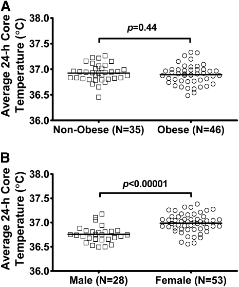
No difference was shown between obese and nonobese subjects when mean (±SE) daily core temperatures were compared (Figure 1A; 36.93 ± 0.03°C compared with 36.97 ± 0.03°C, P = 0.44). Similarly, when BMI was used as a continuous variable along with age, sex, race, and season in the regression analysis, the mean 24-h core temperature was not linearly related to BMI (β < 0.0002, P = 0.963). However, women, had an ≈0.23°C greater mean 24-h core body temperature than that of men (Figure 1B; 36.99 ± 0.0.03°C compared with 36.76 ± 0.03°C; P < 0.00001). Post hoc analyses that examined each sex separately showed no significant difference between nonobese women and obese women or men (P = 0.16 for obese compared with nonobese women; P = 0.50 for obese compared with nonobese men. There were no significant differences between white and black individuals overall (P = 0.44; adjusted for age, sex, BMI, and season), or between nonobese and obese individuals within white or black groups (adjusted for age, sex, and season).
FIGURE 1.
Mean daily core temperatures. A: Nonobese compared with obese subjects. No significant differences in the core body temperature between nonobese and obese individuals were detected. B: Male compared with female subjects. Women had an ≈0.23°C higher mean core temperature than that in men. The analysis was conducted by using Student's t test.
Circadian core temperature profiles in obese and nonobese adults
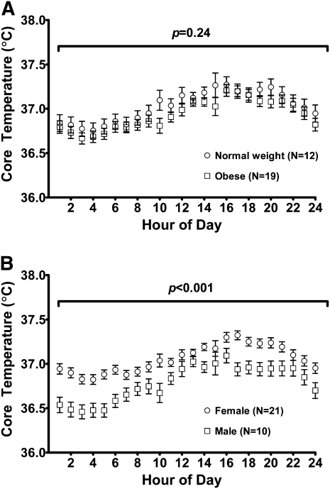
To examine the possibility that the calculation of the mean daily core temperature might have obscured differences in the circadian profiles of the core temperatures between nonobese and obese individuals, the diurnal pattern of the core temperature between normal-weight and obese individuals was examined by using 24-h temperature profiles constructed for the 31 subjects who participated in study 2 and, thus, had detailed information about activities available. In this analysis, obesity was not a significant predictor of the 24-h core temperature profile (Figure 2A; P = 0.244). When the 24-h core temperature profiles were analyzed by sex, a significant difference of ≈0.27°C was shown between men and women (Figure 2B), with women as a group having a higher temperature than that of men (P < 0.001). The use of fat mass as a covariate instead of BMI yielded similar results (P < 0.001 for the difference between men and women; P = 0.228 for total fat mass as covariate). Post hoc analyses that examined each sex separately showed no significant differences between normal-weight and obese individuals (P > 0.153).
FIGURE 2.
Twenty-four-hour core temperature profiles. A: Normal-weight compared with obese subjects. Obesity status was not a significant predictor of core temperature. B: Male compared with female subjects. Women had, on average, a 0.27°C higher core temperature than that in men throughout the day. The analysis was conducted by using ANCOVA with repeated measures.
DISCUSSION
In 2 analyses of core temperature data, we showed no evidence for differences in the core temperature between nonobese and obese individuals. There were no significant differences in the mean daily core temperature between nonobese and obese individuals or in circadian core temperature profiles between normal-weight and obese subjects.
To overcome the limitations of past studies that measured oral or tympanic temperatures to estimate the core body temperature in obesity (8–10), we directly measured the core temperature within the gastrointestinal tract with temperature-sensing capsules. The automated nature of this system allowed resting nighttime temperatures to be obtained without any intervention that might have interrupted sleep. In contrast to the wireless system used, the estimation of the core body temperature by oral or tympanic membrane temperatures has several potential limitations. Sources of error for oral temperature readings are mostly related to the variability in subjects' activities before measurement (14–19), including eating or drinking hot or cold substances (14, 16–18), smoking (14), mouth breathing (18), and tachypnea (15). Some previous studies of temperature in obesity requested that subjects not eat or drink for 15–30 min before measurements (8) but provided no data that indicated that subjects complied with this request. Because oral temperature measurements require subject participation to keep the thermometer properly placed, sleeping temperatures are not possible to obtain. In addition, oral and tympanic membrane temperatures have an imperfect concordance with the core temperature measured rectally (18, 19) and are not universally accepted to be as accurate as the rectal temperature (20).
We replicated previous observations that women have a greater core body temperature than that of men (20–23). Ovarian-derived sex hormones such as estradiol are possibly implicated in the greater temperatures observed in women because no temperature difference between men and women was observed in a sample of older (age >65y) adults in which postmenopausal women were studied (24). Our replication of the sexual dimorphism in the core body temperature observed in past studies served as a partial validation for the quality of data reported.
There were several potential limitations to the current study. The sample size was limited to a total of 81 participants and may have been insufficient to identify a small difference in the core temperature between lean and obese individuals. Nevertheless, we were easily able to replicate the known ≈0.2°C difference between male and female subjects. In addition, although our analyses controlled for factors such as sex, age, and race that might have affected the body temperature, it is possible that adjustment for these factors did not fully account for their potential to influence our measures. Another limitation is the absence of observations regarding the sleep and wakefulness of study subjects. Thus, it was not possible to study obesity-related differences in the core temperature specifically during sleep. Last, the core temperature–sensing capsules used did not always transmit data continuously, such that data points were occasionally dropped because of errors in transmission; however, the overlap between data collected on different days in study 2 allowed us to construct what we believe were representative 24-h core temperature profiles.
Although no general difference in the core body temperature was shown between normal-weight and obese individuals, these observations did not conclusively prove that the core temperature set point plays no role in the regulation of human body weight. Animal models supported the possibility that specific genetic defects that alter the core body temperature may lead to changes in body weight. The current data suggested that alterations in the core body temperature are likely infrequent or perhaps minor modulators of human body adiposity. A systematic search for, and study of, individuals with unusually low or high core body temperatures may conceivably reveal relations that could not be shown in an unselected sample. Because of the high prevalence of obesity in Pima Indians, the findings of Rising et al (11) that 10 Pima Indian men had lower sleeping core body temperatures than 10 whites matched for body weights and percentages of fat may have been preliminary evidence that alterations in body temperature could predispose to obesity. Studies of individuals with established function-altering mutations in core temperature–regulating genes are needed to critically test if differences in the core body temperature set point affect the regulation of human body weight.
Acknowledgments
We thank the volunteers who participated for their help in completing these studies.
The authors' responsibilities were as follows—AMG, DMS, HSE, JAY, KYC, and MCS: conception and design of the study; MJH and JAY: analysis and interpretation of data and drafting of the article; and all authors: contributed to the collection and assembly of data, provided critical revision of the article for content, and approved the final version of the manuscript. JAY and MCS are Commissioned Officers in the US Public Health Service, US Department of Health and Human Services. All authors reported that there were no disclosures or conflicts of interest relevant to this publication.
REFERENCES
- 1.Kurz A. Physiology of thermoregulation. Best Pract Res Clin Anaesthesiol 2008;22:627–44 [DOI] [PubMed] [Google Scholar]
- 2.Rosenbaum M, Hirsch J, Gallagher DA, Leibel RL. Long-term persistence of adaptive thermogenesis in subjects who have maintained a reduced body weight. Am J Clin Nutr 2008;88:906–12 [DOI] [PubMed] [Google Scholar]
- 3.Landsberg L, Young JB, Leonard WR, Linsenmeier RA, Turek FW. Is obesity associated with lower body temperatures? Core temperature: a forgotten variable in energy balance. Metabolism 2009;58:871–6 [DOI] [PubMed] [Google Scholar]
- 4.Trayhurn P, James WP. Thermoregulation and non-shivering thermogenesis in the genetically obese (ob/ob) mouse. Pflugers Arch 1978;373:189–93 [DOI] [PubMed] [Google Scholar]
- 5.Jurgens HS, Schurmann A, Kluge R, et al. Hyperphagia, lower body temperature, and reduced running wheel activity precede development of morbid obesity in New Zealand obese mice. Physiol Genomics 2006;25:234–41 [DOI] [PubMed] [Google Scholar]
- 6.Conti B, Sanchez-Alavez M, Winsky-Sommerer R, et al. Transgenic mice with a reduced core body temperature have an increased life span. Science 2006;314:825–8 [DOI] [PubMed] [Google Scholar]
- 7.Refinetti R. The circadian rhythm of body temperature. Front Biosci 2010;15:564–94 [DOI] [PubMed] [Google Scholar]
- 8.Adam K. Human body temperature is inversely correlated with body mass. Eur J Appl Physiol Occup Physiol 1989;58:471–5 [DOI] [PubMed] [Google Scholar]
- 9.Kim H, Richardson C, Roberts J, Gren L, Lyon JL. Cold hands, warm heart. Lancet 1998;351:1492. [DOI] [PubMed] [Google Scholar]
- 10.Eriksson H, Svardsudd K, Larsson B, Welin L, Ohlson LO, Wilhelmsen L. Body temperature in general population samples. The study of men born in 1913 and 1923. Acta Med Scand 1985;217:347–52 [PubMed] [Google Scholar]
- 11.Rising R, Fontvieille AM, Larson DE, Spraul M, Bogardus C, Ravussin E. Racial difference in body core temperature between Pima Indian and Caucasian men. Int J Obes Relat Metab Disord 1995;19:1–5 [PubMed] [Google Scholar]
- 12.Savastano DM, Gorbach AM, Eden HS, Brady SM, Reynolds JC, Yanovski JA. Adiposity and human regional body temperature. Am J Clin Nutr 2009;90:1124–31 [DOI] [PMC free article] [PubMed] [Google Scholar]
- 13.Gallagher D, Albu J, He Q, et al. Small organs with a high metabolic rate explain lower resting energy expenditure in African American than in white adults. Am J Clin Nutr 2006;83:1062–7 [DOI] [PMC free article] [PubMed] [Google Scholar]
- 14.Bickley LS, Szilagyi PG, Bates B. Bates' guide to physical examination and history taking. 9th ed. Philadelphia, PA: Lippincott Williams & Wilkins, 2007 [Google Scholar]
- 15.Tandberg D, Sklar D. Effect of tachypnea on the estimation of body temperature by an oral thermometer. N Engl J Med 1983;308:945–6 [DOI] [PubMed] [Google Scholar]
- 16.Newman BH, Martin CA. The effect of hot beverages, cold beverages, and chewing gum on oral temperature. Transfusion 2001;41:1241–3 [DOI] [PubMed] [Google Scholar]
- 17.Terndrup TE, Allegra JR, Kealy JA. A comparison of oral, rectal, and tympanic membrane-derived temperature changes after ingestion of liquids and smoking. Am J Emerg Med 1989;7:150–4 [DOI] [PubMed] [Google Scholar]
- 18.Varney SM, Manthey DE, Culpepper VE, Creedon JF., Jr A comparison of oral, tympanic, and rectal temperature measurement in the elderly. J Emerg Med 2002;22:153–7 [DOI] [PubMed] [Google Scholar]
- 19.Craig JV, Lancaster GA, Taylor S, Williamson PR, Smyth RL. Infrared ear thermometry compared with rectal thermometry in children: a systematic review. Lancet 2002;360:603–9 [DOI] [PubMed] [Google Scholar]
- 20.Sund-Levander M, Forsberg C, Wahren LK. Normal oral, rectal, tympanic and axillary body temperature in adult men and women: a systematic literature review. Scand J Caring Sci 2002;16:122–8 [DOI] [PubMed] [Google Scholar]
- 21.Horne JA, Coyne I. Seasonal changes in the circadian variation of oral temperature during wakefulness. Experientia 1975;31:1296–8 [DOI] [PubMed] [Google Scholar]
- 22.Mackowiak PA, Wasserman SS, Levine MM. A critical appraisal of 98.6 degrees F, the upper limit of the normal body temperature, and other legacies of Carl Reinhold August Wunderlich. JAMA 1992;268:1578–80 [PubMed] [Google Scholar]
- 23.Chamberlain JM, Terndrup TE, Alexander DT, et al. Determination of normal ear temperature with an infrared emission detection thermometer. Ann Emerg Med 1995;25:15–20 [DOI] [PubMed] [Google Scholar]
- 24.McGann KP, Marion GS, Camp L, Spangler JG. The influence of gender and race on mean body temperature in a population of healthy older adults. Arch Fam Med 1993;2:1265–7 [DOI] [PubMed] [Google Scholar]


