Abstract
The mechanosensitive (MS) channels MscS and MscL are essential for the survival of hypoosmotic shock by Escherichia coli cells. We demonstrate that MscS and MscL are induced by osmotic stress and by entry into stationary phase. Reduced levels of MS proteins and reduced expression of mscL– and mscS–LacZ fusions in an rpoS mutant strain suggested that the RNA polymerase holoenzyme containing σS is responsible, at least in part, for regulating production of MS channel proteins. Consistent with the model that the effect of σS is direct, the MscS and MscL promoters both use RNA polymerase containing σS in vitro. Conversely, clpP or rssB mutations, which cause enhanced levels of σS, show increased MS channel protein synthesis. RpoS null mutants are sensitive to hypoosmotic shock upon entry into stationary phase. These data suggest that MscS and MscL are components of the RpoS regulon and play an important role in ensuring structural integrity in stationary phase bacteria.
Keywords: MscS, MscL, Escherichia coli, osmoregulation
Mechanosensitive (MS) channels are gated by tension in the cell membrane. They have been identified in all of the major living domains, including Archaea, several Prokaryote species, and Eukaryotes, including fungi, plants, and mammals (1–4). However, MS channels are best characterized in Escherichia coli, where three activities have been recorded by using the patch-clamp technique on giant protoplasts (5). On the basis of their conductance, the MS channel activities are classified into MscM (0.3 nS), MscS (1 nS), and MscL (2–3 nS) (6). Each channel exhibits a characteristic pressure threshold for activation, with progressively greater pressure required to open the larger channels. MscL has been extensively analyzed at both gene and protein level and key residues involved in channel gating have been identified (7–10). MscS activity arises from two channels, MscS and MscK (KefA) (11, 12). The crystal structure of MscS has recently been solved and reveals a homoheptameric protein that has little in common with MscL (13). Genetic analysis supports key conclusions arising from the structure (14). Thus, we have two molecular paradigms for MS channel activity.
The major role for MS channels in cell physiology is protection of cell integrity when the cell is exposed to a dilute environment (hypoosmotic shock). During hypoosmotic shock, water rapidly enters the cell down the osmotic gradient. The water inflow causes a sufficiently rapid increase in turgor pressure that cells lacking MscS and MscL lyse (11). Lysis only occurs in a mscS mscL mutant when an osmotic pressure drop equivalent to >0.2 M NaCl is imposed irrespective of the initial and final osmolarity (ref. 11 and S.C. and I.R.B., unpublished data). Because commonly used growth media have an osmolarity of ≈200 mOsm (equivalent to ≈0.1 M NaCl), E. coli cells growing in such media cannot encounter a large enough hypoosmotic shock to threaten their structural integrity. However, when cells growing at higher osmolarity are exposed to hypoosmotic shock, MS channels must be activated on a ms time scale to prevent damage to cell integrity. De novo gene expression cannot modulate the levels of MS channel proteins on this time scale, suggesting that MS channel expression might be induced when cells are exposed to high osmolarity to prepare for the eventuality of hypoosmotic stress conditions.
Here we show that E. coli cells express a basal level of MS channel protein that is augmented by new expression upon growth at high osmolarity and upon entry into stationary phase. We demonstrate that the synthesis of the stress sigma factor, RpoS (σS), is required to effect the increased synthesis of MS channels and that RNA polymerase holoenzyme containing σS transcribes the mscS and mscL promoters in vitro. An rpoS null mutant exhibits osmotic sensitivity leading to cell lysis upon hypoosmotic shock.
Experimental Procedures
Bacterial Strains and Plasmids. All strains are E. coli K-12 derivatives and are listed, with the plasmids used in this study, in Table 1. Cells were grown aerobically in flasks at 37°C in citrate-phosphate medium (pH 7; ≈220 mOsm), prepared as described (11), or in LB medium with ampicillin (Sigma) at 100 μg/ml. Cell culture and membrane preparation were as described (14).
Table 1. Strains and plasmids used in this study.
| Genotype/characteristics | Ref./Source | |
|---|---|---|
| Strain | ||
| Frag1 | F–rha thi gal lacZ | 47 |
| MJF372 | Frag1 rpoS::Tn10 | 48 |
| MJF405 | Frag1 clpP::Tn9 | 48 |
| MJF455 | Frag1 ΔmscS mscl::Cm | 11 |
| MJF507 | Frag1 ΔlacU169 | This study* |
| MJF513 | Frag1 ΔlacU169 rpoS::Tn10 | This study* |
| MJF530 | Frag1 ΔlacU169 ompR::Tn10 | This study* |
| MJF541 | Frag1 rssB::Tn10 | This study* |
| JM109 | endA1 recA1 gyrA96 thi hsdR17 [rK–,mK+] relA1 supE44 Δ[lac-proAB] [F′ traD36proAB lacl ZΔM15] | 49 |
| Plasmid | ||
| pMC1871 | pBR322 copy-number fusion vector | Amersham Pharmacia |
| pHSG575 | pSC101 copy-number cloning vector | 16 |
| pMCS | pMC1871 derivative carrying mscS promoter–lacZ cassette | This study |
| pMCL | pMC1871 derivative carrying mscL promoter–lacZ cassette | This study |
| pRLG770 | pBR322 derivative carrying transcription terminators | 17 |
| pRLG6984 | pRLG770 carrying the promoter region of mscL | This study |
| pRLG6985 | pRLG770 carrying the promoter region of mscS | This study |
| pHSGL | pHSG575 derivative carrying mscL promoter–lacZ cassette | This study |
| pHSGS | pHSG575 derivative carrying mscS promoter–lacZ cassette | This study |
| pHSGlacZ | pHSG575 derivative carrying promoter-less lacZ cassette from pMC1871 | This study |
Created by P1 transduction
Construction of Reporter Plasmids. A 325-bp fragment of the mscS promoter, with the first 9 codons of the coding sequence, was amplified by using primers SPF (5′-TAGATGCCCGGGAATTGCCTGATGCGCTAC-3′) and SPR (5′-TAGATGCCCGGGGCTATCGACAACATTCAA-3′), by using standard PCR. A 321-bp fragment of the mscL promoter, including the first 11 codons, was amplified with primers LPF (5′-TAGATGCCCGGGGGAACGATTATTGGAGCG-3′) and LPR (5′-TAGATGCCCGGGCGCAAATTTCGCGAAATTCC-3′). Primers were designed to include SmaI sites (underlined). PCR products were cloned into the SmaI site of pMC1871, by using standard techniques (15), to create a translational fusion to the lacZ gene. Positive JM109 transformants were screened on LB agar containing 70 μg/ml 5-bromo-4-chloro-3-indoylyl-β-d-galactopyranoside (X-gal) and 1 mM isopropyl β-d-thiogalactopyranoside (IPTG). Putative recombinant plasmids were isolated (Qiaprep Spin Miniprep kit, Qiagen, Valencia, CA), PCR screened by using the vector-specific primer PMCF (5′-CAACGTTGTTGCCATTGC-3′) and either SPR or LPR, and confirmed by SmaI digestion. One clone of each type was sequenced, on both strands, by using the ABI PRISM BigDye Terminator Cycle Sequencing kit (Applied Biosystems) with primers SPF/SPR or LPF/LPR as appropriate. The fusions (complete with promoters) were then subcloned into the SalI site of the low copy number vector, pHSG575 (16), to create plasmids pHSGS and pHSGL. lacZ was subcloned from pMC1871 into pHSG575 to create the promoter-less control plasmid, pHSGlacZ. Promoter derivatives lacking the translation start site were used for mapping the transcription start site and were generated by PCR from plasmids containing a wild-type promoter. Primers for PCR were designed to include an EcoRI site at the upstream junction of the promoter sequence and a HindIII site at the downstream junction. The PCR products were cleaved with EcoRI and HindIII, then ligated into the multicopy plasmid pRLG770 (17) to construct pRLG6984 (mscL) and pRLG6985 (mscS). Plasmids were maintained in E. coli strain CAG1574.
Transcript Mapping. For in vivo mapping, strains containing pRLG6984 (mscL) and pRLG6985 (mscS) were grown at 37°C in LB to early stationary phase (A600 ≈ 1.0), RNA was extracted by using a boiling lysis procedure, and reverse transcription was performed as described (18, 19). For in vitro mapping, transcripts were synthesized from pRLG6984 (mscL) and pRLG6985 (mscS) by using purified RNA polymerase containing either σ70 or σS, (Eσ70 and EσS, respectively), which were prepared as described (20). Because saturating amounts of σ (either σS or σ70) were added to the same amount of core enzyme, the two holoenzymes have the same specific activity. Primer extension was performed essentially as described (21), except that the reaction volume was 50 μl, the salt was 100 mM KCl, and the reactions were carried out at 30°C.
β-Galactosidase Assays. Transformants carrying pHSGS, pHSGL, and pHSGlacZ were grown overnight in citrate-phosphate medium (22) with limiting glucose (0.04% wt/vol) and supplemented the following morning with glucose to 0.2%. After one doubling, the culture was diluted 20-fold into identical medium and incubated to mid-exponential phase (OD650 ≈0.4), before being diluted into the assay medium, giving a starting OD650 ≈0.05. At the times indicated, samples were removed for assays of β-galactosidase activity (23) and protein concentration determination (24). Activity is expressed as Miller units per microgram total cell protein (units/μg protein).
Antibodies and Immunoblots. The MscS-specific antiserum was generated against a synthetic peptide as described (14). The anti-MscL antibodies were kindly provided by S. Sukharev (University of Maryland, College Park). Western blots on total membrane from strain Frag1 (MscS+), MJF451 (ΔmscS), MJF455 (mscL::Cm, ΔmscS), and MJF455/pTrcMscS, induced with 1 mM IPTG for 60 min, were performed to verify the specificity of the antisera (14). Total membrane fractions were prepared essentially as described (14), and 30 μg of membrane protein was separated by SDS/PAGE on 12% acrylamide gels. Western blots were performed as described (14, 25). Signal intensities on developed film were measured (Bio-Rad GS-700 Imaging Densitometer), quantified by using molecular analysis software (Bio-Rad), and are displayed (in arbitrary units) below each blot image. By using a dilution series of membrane protein, prepared from IPTG-induced MJF455/pTrcMscS, the intensity of the signal was observed to be linear, with MscS protein up to values ≤9 (data not shown).
Survival Assays. Survival assays were performed essentially as described (11). Where appropriate, the overnight culture contained ampicillin, and thereafter, the remainder of the assay was undertaken in the absence of this antibiotic. Transformants retained the plasmid, despite multiple generations in the absence of the antibiotic (data not shown).
Chemicals. DNA polymerases and restriction endonucleases were obtained from Roche Molecular Diagnostics. Sigma-Genosys (Cambridge, U.K.) prepared the oligonucleotides and antiserum. All other reagents were purchased from Sigma.
Results
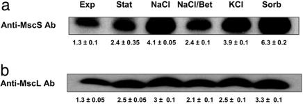
Growth Phase and Osmolarity Dependence of MscS and MscL Expression. The expression level of the MS channel proteins was investigated by probing membrane preparations from cells grown under different regimes with antibodies specific to MscS and MscL. Both channels were expressed in Frag1 (MscS+, MscL+) during exponential phase (Fig. 1), but expression increased upon entry into stationary phase (Fig. 1). Steady state growth at high osmolarity (0.5 M NaCl) also increased expression, and comparable results were observed with 0.5 M KCl or 0.8 M sorbitol (Fig. 1), indicating that expression was not salt-specific. Chloramphenicol and tetracycline were found to prevent the increased expression of the channel genes in response to 0.5 M NaCl (data not shown). Incubation with the osmoprotectant glycine betaine (1 mM) reduced expression in the presence of 0.5 M NaCl (Fig. 1). The reversal of induction by betaine is characteristic of many osmoregulated genes (26, 27). These data clearly point to osmotic induction of the MS channels. MscS was induced more than MscL, but both systems showed consistently increased expression at high osmolarity and in stationary phase.
Fig. 1.
Effect of growth phase and osmolarity on channel protein levels in membranes. Cells of Frag1 were cultured in citrate-phosphate medium (Exp, exponential; Stat, stationary phase) or in medium with the indicated supplements (NaCl, 0.5M NaCl; NaCl/Bet, 0.5 M NaCl plus 1 mM betaine; KCl, 0.5 M KCl; Sorb, 0.8 M sorbitol) and harvested during exponential growth (OD650 = 0.4), except for stationary phase cells, which were harvested after growth for 22 h. Membrane fractions were prepared as described (14), and Western blots were performed by using antisera specific for MscS (a) and MscL (b). Blots were developed by ECL, and the signal intensity was determined by scanning laser densitometry. Replicate measurements were made on the same gel to determine the reproducibility of the analysis and the data are the mean and error of three separate blots. We and others have previously demonstrated the specificity of the antisera (14, 46). Blots were replicated from at least two independent membrane preparations.
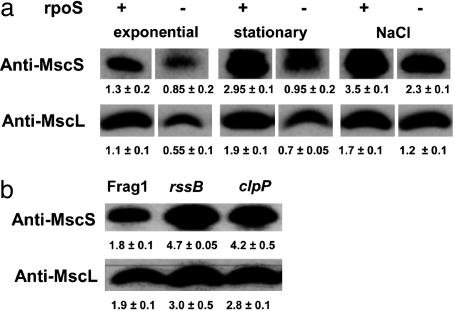
RpoS Is Required for Osmotic Induction of MS Channel Genes. Many different mechanisms could account for induction at high osmolarity (28, 29). Because σS levels increase at high osmolarity (30, 32) we tested whether cells lacking RpoS (rpoS::Tn10) exhibited altered accumulation of MscS and MscL during growth at low and high osmolarity (Fig. 2a). Inactivation of RpoS reduced E. coli MS channel expression; membranes harvested from MJF372 (Frag1, rpoS::Tn10) in exponential phase (OD650 = 0.3–0.4), stationary phase, and during exponential growth in the presence of 0.3 M NaCl exhibited lowered levels of both MscS and MscL (Fig. 2a). (Note that 0.3 M NaCl concentration was used in these experiments because rpoS null mutants fail to grow at very high osmolarity; ref. 27.)
Fig. 2.
RpoS levels affect MS channel protein expression. Cells of Frag1 (+) and MJF372 (rpoS::Tn10)(-)(a), and Frag1, MJF541 (rssB::Tn10), and MJF405 (clpP::Tn9) (b) were grown in McIlvaine's medium as described for Fig. 1. (a) Cells were harvested during exponential phase, after attainment of the stationary phase (22 h after inoculation) and in exponential phase in the presence of 0.3 M NaCl. (b) Cells were harvested from exponential phase. Membrane protein blots were probed with antisera to MscS or MscL as indicated. Other details were as in Fig. 1.
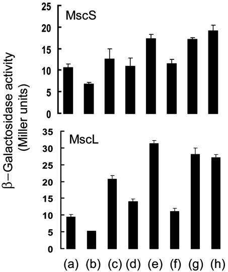
Gene expression is often investigated by the creation of gene fusions between the gene of interest and a reporter. Increased osmolarity (0.3–0.5 M NaCl or KCl or 0.8 M sorbitol) resulted in higher levels of β-galactosidase from the MscS–LacZ and MscL–LacZ gene fusions and this effect was reversed by the inclusion of betaine (1 mM) in the growth medium (Fig. 3). The fusions also exhibited increased expression upon entry into stationary phase; β-galactosidase increased ≈2- to 3-fold ≈2 h after exponential growth ceased (data not shown). An rpoS null mutation reduced expression of the MscL–LacZ gene fusion during exponential phase in low osmolarity medium and in the presence of 0.3 M NaCl (Fig. 3). For the MscS–LacZ fusion, the data were less clear-cut: expression was reduced in exponential phase cells at low osmolarity, but in the presence of 0.3 M salt, the effect of an rpoS mutation was not significant.
Fig. 3.
Expression of MscS–LacZ and MscL–lacZ fusions. Strains MJF507 (Δlac) (a, c, and e–h) and MJF513 (Δlac, rpoS::Tn10)(b and d) were transformed with pHSGS (MscS) or pHSGL (MscL) and grown in citrate-phosphate medium. Different supplements were added to the medium: a and b, none; c and d, 0.3 M NaCl; e, 0.5 M NaCl; f, 0.5 M NaCl plus 1 mM betaine; g, 0.5 M KCl; h, 0.8 M sorbitol. Aliquots of cells were harvested in mid exponential phase (OD650 ≈ 0.4) and assayed for β-galactosidase activity and total cell protein, as described in Experimental Procedures. Data represent means from three independent experiments and error bars display the SD. Cells transformed with pHSGLacZ did not exhibit β-galactosidase activity. Similar osmotic induction of the fusions was observed with cells transformed with pMCS and pMCL (data not shown).
Two types of mutation, clpP and rssB, are known to increase the stability of RpoS protein leading to its accumulation during exponential phase (27, 30). Expression of both MscS and MscL increased to levels equivalent to those seen at high osmolarity in strains carrying rssB or clpP mutations (Fig. 2b). These data confirm the involvement of RpoS in expression of the MS channels. RpoS is also required for the transcription of a MscS paralogue, YbiO (F786), (31) and for expression of the potassium channel homologue Kch (P.L., H.D.M., R.L.G., and I.R.B., unpublished data), which suggests that this σ factor integrates the expression of a range of channels that are potential components of the osmoregulatory system of E. coli.
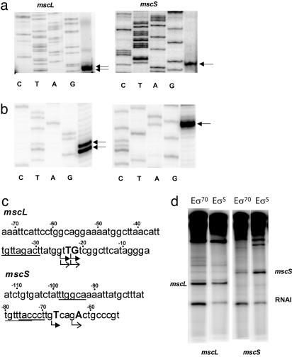
Transcription of mscS and mscL Promoters by both σS and σ70. RpoS could act either by driving transcription of the mscS and mscL genes directly or via its effect on the expression of another protein that is itself required for expression of the channel genes. The transcription start sites of the mscS and mscL genes were identified by primer extension (see Experimental Procedures). This analysis indicated that the mscL promoter initiates at two nucleotides (-24T and -23G, relative to the translation start site), whereas the mscS promoter initiates at a single nucleotide (-68T, relative to the translation start site) (Fig. 4a). Consistent with these results, the sequence upstream of the mscL transcription start site contained a good match to the consensus sequence recognized by both EσS and Eσ70 (TGtTAGAA/CT) (20). There was no discernible -35 hexamer, but it is known that TG immediately upstream of the -10 can obviate the need for a strong -35 sequence (32) (Fig. 4c). Potential matches to the -10 and -35 hexamers upstream of the mscS start sites were much less obvious than for mscL (Fig. 4c).
Fig. 4.
Mapping mscL and mscS transcripts in vivo and in vitro. (a) RNA was extracted from strains expressing pRLG6984 (mscL) and pRLG6985 (mscS) and reverse transcribed as described in Experimental Procedures. Transcript start site(s) are indicated by arrows. (b) RNA was synthesized in vitro from pRLG6984 (mscL) and pRLG6985 (mscS) by using EσS and reverse transcribed as described in Experimental Procedures. Transcript start site(s) are indicated by arrows. (c) DNA sequences surrounding the transcription start sites of each promoter. In vivo initiation sites are indicated by solid arrowheads, in vitro initiation sites are indicated by open arrowheads, and putative -10 and -35 hexamers are underlined. (d) In vitro transcription of pRLG6984 (mscL) and pRLG6985 (mscS) using Eσ70 or EσS. The plasmid-derived RNA interference (RNAI) transcript is also indicated. The RNAI promoter was shown previously to be transcribed better by Eσ70 than by EσS (20).
It was possible to compare transcription from the same promoters by purified EσS or Eσ70 in vitro because the reconstituted holoenzymes had equivalent catalytic activity (see Experimental Procedures). The mscL promoter was recognized by both EσS and Eσ70 (Fig. 4d) and initiated at the same position in vitro as it used in vivo (Fig. 4 a–c). In contrast, the mscS promoter was recognized much better by EσS than Eσ70 (Fig. 4d), and transcription initiated in vitro 4 nt downstream from where it initiated in vivo (Fig. 4 a–c; see Discussion). Taken together with the results from Fig. 2, these data are consistent with the model that, in vivo, both promoters are transcribed directly by EσS.
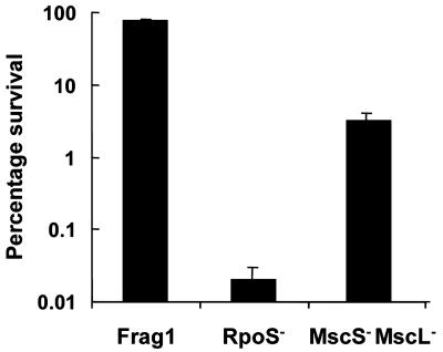
Stationary Phase Cultures of rpoS Null Mutants Lyse upon Hypoosmotic Shock. MS channels are required for E. coli cells to survive hypoosmotic shock (11). Because RpoS appears to be a significant factor regulating expression of the MS channels, we investigated survival of an rpoS null mutant during growth at high osmolarity and after entry into stationary phase. During exponential phase, the RpoS mutant strain MJF372 (Frag1, rpoS::Tn10) was not sensitive to hypoosmotic shock; survival was equivalent to the parent Frag1 (survival 90 ± 10%) (data not shown). However, cultures of the rpoS mutant that had entered stationary phase (18–22 h cultures) lysed almost completely when transferred to low osmolarity; survival was usually <0.01% (Fig. 5) and the culture turbidity was significantly reduced (data not shown). These changes were much greater than those seen with the mscL mscS double mutant exposed to the same hypoosmotic shock (Fig. 5; ref. 11). Expression of either MscS or MscL from an IPTG-inducible promoter did not increase survival of the rpoS mutant during osmotic shock.
Fig. 5.
Absence of RpoS causes sensitivity to hypoosmotic shock. Cells of MJF372 (rpoS::Tn10) were grown into stationary phase (18–22 h) in McIlvaine's minimal medium containing 0.3 M NaCl. The cells were then diluted 20-fold into sterile distilled water. After 30 min, the cells were serially diluted and 5-μl spots (four spots per dilution) were placed on LB plates and the plates were incubated at 37°C for 24 h before counting the colonies. For comparison with the effect of the rpoS mutation, hypoosmotic shock survival data from MJF455 (mscL mscS) is also shown. For the control incubation (used to establish 100% survival), McIlvaine's medium containing 0.3 M NaCl was used for the 20-fold dilution and the serial dilutions, and recovery of the colonies was on LB containing 0.3 M NaCl.
Discussion
In this study, we have demonstrated that expression of the MscS and MscL channel proteins is regulated by osmotic stress and upon entry into stationary phase, and we suggest this phenomenon is under the direct control of EσS. Mutants lacking RpoS exhibited reduced expression of MscS and MscL proteins, expression of MscS–LacZ and MscL–LacZ gene fusions is diminished, and the rpoS mutant exhibits enhanced sensitivity to osmotic down-shock after entry into stationary phase. RpoS protein is known to accumulate during growth at high osmolarity and upon entry into stationary phase, leading to altered patterns of transcription. Growth in the presence of betaine lowers the level of RpoS protein (27) and was found in this study to lower the osmotic enhancement of the MS channel proteins. Both MscL and MscS continue to be made at reduced levels during rapid exponential growth, when RpoS levels are lower, and in rpoS null mutants. The in vitro transcription data suggest that this is a result of transcription by Eσ70 (Fig. 4), which is a common observation for σS-dependent promoters (33). The transcript start site for mscS differs by four nucleotides in vivo versus in vitro (Fig. 4). One explanation is the potential participation of a transcription activator that interacts with RNA polymerase, thereby positioning it on -10/-35 hexamers that are not as favored as those used by RNA polymerase in the absence of the factor. Alternatively, a difference in template topology in vivo versus in vitro could alter the stability of RNA polymerase on the promoter, favoring the presence of different transcription intermediates, allowing utilization of a new start site, and/or preventing the utilization of the in vitro start site. Identifying the potential transcription factor or testing potential effects of altered DNA conformation on start site utilization are beyond the scope of this paper.
The central role of σS is supported by the effects of rssB and clpP mutations, which lead to increased expression of both MscS and MscL (Fig. 2). RssB regulates the activity of the ClpP protease (34), which is the primary determinant of σS half-life. In exponential phase, the σS protein has a short half-life, but under conditions of osmotic stress and after entry into stationary phase, the protein is stabilized because of physiological factors that control RssB activity (27). In rssB and clpP null mutants, σS levels are elevated and the high levels of expression of genes under the control of this sigma factor are observed (27, 34). The observations made here for MscL and MscS proteins follow the predictions for genes that can be expressed by RNAPσS holoenzyme. MscS and MscL are most necessary to cell survival during transfer from high to low osmolarity (11). Dilution of cells growing in conventional growth media (usually ≈200 mOsm) into water is insufficient to lyse a mscS mscL mutant, consistent with the failure of the MscS and MscL channels to fire under these conditions (11). However, increasing the osmolarity of cells by the addition of 100 mM NaCl imposes a requirement for these channels (C.S. and I.R.B., unpublished data). Very low levels of MscS protein, equivalent to the basal level of expression seen in exponential phase at low osmolarity, are insufficient to provide the required protection (14). Thus, transcriptional control by σS ensures increased expression during the specific growth conditions where the cells have the greatest potential need for high levels of MS channel activity.
The increase in the level of MS channels upon entry into stationary phase is, at first sight, less simply explained. This stage of the life cycle of the organism represents a period of overall diminished protein synthesis (35), but considerable biochemical and morphological changes occur in the membrane and wall during this period (36), which may require extra MS channels for protection against increased stress on the cell wall. We have demonstrated that entry into stationary phase imposes mechanical stress on these cell components, because cells of an rpoS mutant from stationary phase, but not from exponential phase, lyse upon hypoosmotic shock. The stability of the cell is determined by the balance between the turgor pressure, tending to cause cell lysis, and the resistance provided by the cell wall. Failure to regulate either or both of these properties may lead to cell lysis. From our data we infer that, during stationary phase, the mechanical strength of the wall diminishes unless new gene expression takes place and that RpoS controls the expression of the relevant gene products. In parallel, RpoS also controls the expression of MS channels, which potentiates survival.
RpoS has been implicated in modulating the structure of the cell wall. Penicillin-binding proteins (PBP) are regulated by RpoS (37). It has been shown that PBP3, which is involved in septum formation, is down-regulated by RpoS upon entry into stationary phase. RpoS is also implicated in remodeling of crosslinks in the cell wall of stationary phase E. coli cells (38). The frequency of direct crosslinks between two diaminopimelate residues on adjacent peptidoglycan strands increases 6-fold in stationary phase, a change paralleled by a decrease in d-Ala–d-Ala crosslinks (39). RpoS is required for the recovery of the d-Ala–d-Ala dipeptide because it controls the expression of both the peptidase and the transport system (39). Control over the expression of cell wall remodeling enzymes is critical for cell integrity (40–42). Finally, the increased synthesis of MS channels upon entry into stationary phase may represent a component of the coordinated modification of the cytoplasmic membrane. Cyclopropane fatty acid synthesis is under the control of RpoS, and the increased accumulation of this modified phospholipid has been suggested to affect stress tolerance (43). Because MS channels sense the tension within the bilayer, it is conceivable that their increased synthesis partially compensates for altered membrane fluidity.
There is a further aspect to the regulated expression of MS channels. Electrophysiological analysis suggests that there are few MS channel proteins per cell. Upper estimates suggest that there are four to five MscL and 20–30 MscS channels per cell, which equates to ≈20–30 copies of MscL protein and 140–210 copies of MscS. The MscM and MscK channels are of even lower abundance, possibly one to two functional channels per cell (12, 44). At such low protein abundance there will be a stochastic distribution of functional channels in the cell population (45) that will lead to some cells possessing insufficient channels to survive changes in either wall integrity or external osmolarity. Activated expression boosts the numbers of channels per cell and will diminish the effects of stochastic distribution of low abundance proteins, thereby enhancing cell survival. Thus, regulation by RpoS may be essential to ensure that the channels are expressed at significant levels during osmotic stress and stationary phase, two conditions that increase the potential demand for MS channels.
Acknowledgments
We thank H. Bell, Ulrike Schumann, and P. Carter (University of Aberdeen) for technical assistance and DNA sequencing, and S. Sukharev (University of Maryland, College Park) and C. Kung (University of Wisconsin, Madison) for provision of the anti-MscL antibody. We thank Paul Blount (University of Texas Southwestern Medical Center, Dallas) for helpful suggestions. N.R.S. was the recipient of a Biotechnology and Biological Sciences Research Council Food Directorate Studentship, P.L. was funded by an European Union Marie Curie Fellowship, C.S. was a Wellcome Trust Travel Fellow, S.M. is supported by a Wellcome Trust Program grant, W.B. is funded by European Union Contract QLK3-CT-2000-00640 (Hypersolutes), and I.R.B. is a Wellcome Trust Research Leave Fellow. H.D.M. was supported by a National Institutes of Health Genetics predoctoral training grant and National Institutes of Health Grant RO1 GM37048 (to R.L.G.).
Abbreviations: MS, mechanosensitive; IPTG, isopropyl β-d-thiogalactopyranoside.
References
- 1.Pivetti, C. D., Yen, M. R., Miller, S., Busch, W., Tseng, Y. H., Booth, I. R. & Saier, M. H. (2003) Microbiol. Mol. Biol. Rev. 67, 66-85. [DOI] [PMC free article] [PubMed] [Google Scholar]
- 2.Tavernarakis, N. & Driscoll, M. (2001) Cell Biochem. Biophys. 35, 1-18. [DOI] [PubMed] [Google Scholar]
- 3.Duggan, A., Garcia-Anoveros, J. & Corey, D. P. (2000) Curr. Biol. 10, R384-R387. [DOI] [PubMed] [Google Scholar]
- 4.Gustin, M. C., Zhou, X.-L., Martinac, B. & Kung, C. (1998) Science 242, 762-765. [DOI] [PubMed] [Google Scholar]
- 5.Strop, P., Bass, R. & Rees, D. C. (2003) in Membrane Proteins, ed. Rees, D. C. (Academic, New York), Vol. 63, pp. 177-209. [Google Scholar]
- 6.Booth, I. R. & Louis, P. (1999) Curr. Opin. Microbiol. 2, 166-169. [DOI] [PubMed] [Google Scholar]
- 7.Sukharev, S. I., Blount, P., Martinac, B., Blattner, F. R. & Kung, C. (1994) Nature 368, 265-268. [DOI] [PubMed] [Google Scholar]
- 8.Blount, P., Sukharev, S. I., Schroeder, M. J., Nagle, S. K. & Kung, C. (1996) Proc. Natl. Acad. Sci. USA 93, 11652-11657. [DOI] [PMC free article] [PubMed] [Google Scholar]
- 9.Ou, X., Blount, P., Hoffman, R. J. & Kung, C. (1998) Proc. Natl. Acad. Sci. USA 95, 11471-11475. [DOI] [PMC free article] [PubMed] [Google Scholar]
- 10.Chang, G., Spencer, R. H., Lee, A. T., Barclay, M. T. & Rees, D. C. (1998) Science 282, 2220-2226. [DOI] [PubMed] [Google Scholar]
- 11.Levina, N., Totemeyer, S., Stokes, N. R., Louis, P., Jones, M. A. & Booth, I. R. (1999) EMBO J. 18, 1730-1737. [DOI] [PMC free article] [PubMed] [Google Scholar]
- 12.Li, Y. Z., Moe, P. C., Chandrasekaran, S., Booth, I. R. & Blount, P. (2002) EMBO J. 21, 5323-5330. [DOI] [PMC free article] [PubMed] [Google Scholar]
- 13.Bass, R. B., Strop, P., Barclay, M. & Rees, D. C. (2002) Science 298, 1582-1587. [DOI] [PubMed] [Google Scholar]
- 14.Miller, S., Bartlett, W., Chandrasekaran, S., Simpson, S., Edwards, M. & Booth, I. R. (2003) EMBO J. 22, 36-46. [DOI] [PMC free article] [PubMed] [Google Scholar]
- 15.Sambrook, J., Fritsch, E. & Maniatis, T. (1989) Molecular Cloning: A Laboratory Manual (Cold Spring Harbor Lab. Press, Plainview, NY)
- 16.Takeshita, S., Sato, M., Toba, M., Masahashi, W. & Hashimotogotoh, T. (1987) Gene 61, 63-74. [DOI] [PubMed] [Google Scholar]
- 17.Ross, W., Thompson, J. F., Newlands, J. T. & Gourse, R. L. (1990) EMBO J. 9, 3733-3742. [DOI] [PMC free article] [PubMed] [Google Scholar]
- 18.Josaitis, C. A., Gaal, T. & Gourse, R. L. (1995) Proc. Natl. Acad. Sci. USA 92, 1117-1121. [DOI] [PMC free article] [PubMed] [Google Scholar]
- 19.Schneider, D. A., Gaal, T. & Gourse, R. L. (2002) Proc. Natl. Acad. Sci. USA 99, 8602-8607. [DOI] [PMC free article] [PubMed] [Google Scholar]
- 20.Gaal, T., Ross, W., Estrem, S. T., Nguyen, L. H., Burgess, R. R. & Gourse, R. L. (2001) Mol. Microbiol. 42, 939-954. [DOI] [PubMed] [Google Scholar]
- 21.Aiyar, S. E., Gaal, T. & Gourse, R. L. (2002) J. Bacteriol. 184, 1349-1358. [DOI] [PMC free article] [PubMed] [Google Scholar]
- 22.Jordan, S. L., Glover, J., Malcolm, L., Thomson-Carter, F. M., Booth, I. R. & Park, S. F. (1999) Appl. Environ. Microbiol. 65, 1308-1311. [DOI] [PMC free article] [PubMed] [Google Scholar]
- 23.Miller, J. H. (1972) Experiments in Molecular Genetics (Cold Spring Harbor Lab. Press, Plainview, NY).
- 24.Lowry, O., Rosebrough, N., Farr, A. & Randall, R. (1951) J. Biol. Chem. 193, 265-275. [PubMed] [Google Scholar]
- 25.Towbin, H., Staehelin, T. & Gordon, J. (1979) Proc. Natl. Acad. Sci. USA 76, 4350-4354. [DOI] [PMC free article] [PubMed] [Google Scholar]
- 26.Cairney, J., Booth, I. R. & Higgins, C. F. (1985) J. Bacteriol. 164, 1224-1232. [DOI] [PMC free article] [PubMed] [Google Scholar]
- 27.Muffler, A., Traulsen, D. D., Lange, R. & Hengge-Aronis, R. (1996) J. Bacteriol. 178, 1607-1613. [DOI] [PMC free article] [PubMed] [Google Scholar]
- 28.Hengge-Aronis, R. (1996) Mol. Microbiol. 21, 887-893. [DOI] [PubMed] [Google Scholar]
- 29.Hengge-Aronis, R. (1999) Curr. Opin. Microbiol. 2, 148-152. [DOI] [PubMed] [Google Scholar]
- 30.Schweder, T., Lee, K. H., Lomovskaya, O. & Matin, A. (1996) J. Bacteriol. 178, 470-476. [DOI] [PMC free article] [PubMed] [Google Scholar]
- 31.Schellhorn, H. E., Audia, J. P., Wei, L. I. C. & Chang, L. (1998) J. Bacteriol. 180, 6283-6291. [DOI] [PMC free article] [PubMed] [Google Scholar]
- 32.Ponnambalam, S., Webster, C., Bingham, A. & Busby, S. (1986) J. Biol. Chem. 261, 6043-6048. [PubMed] [Google Scholar]
- 33.Nguyen, L. H. & Burgess, R. R. (1997) Biochemistry 36, 1748-1754. [DOI] [PubMed] [Google Scholar]
- 34.Muffler, A., Fischer, D., Altuvia, S., Storz, G. & Hengge-Aronis, R. (1996) EMBO J. 15, 1333-1339. [PMC free article] [PubMed] [Google Scholar]
- 35.Kolter, R., Siegele, D. A. & Tormo, A. (1993) Annu. Rev. Microbiol. 47855-874. [DOI] [PubMed]
- 36.Huisman, G. W., Siegele, D. A., Zambrano, M. M. & Kolter, R. (1996) in Escherichia coli and Salmonella: Cellular and Molecular Biology, eds. Neidhardt, F. C., Curtis, R., III, Ingraham, J. L., Lin, E. C. C., Magasanik, B., Reznikoff, W. S., Rilwey, M., Scaechter, M. & Umbarger, H. E. (Am. Soc. Microbiol. Press, Washington, DC), Vol. 2, pp. 1672-1682. [Google Scholar]
- 37.Dougherty, T. J. & Pucci, M. J. (1994) Antimicrob. Agents Chemother. 38, 205-210. [DOI] [PMC free article] [PubMed] [Google Scholar]
- 38.Lessard, I. A. D., Pratt, S. D., McCafferty, D. G., Bussiere, D. E., Hutchins, C., Wanner, B. L., Katz, L. & Walsh, C. T. (1998) Chem. Biol. 5, 489-504. [DOI] [PubMed] [Google Scholar]
- 39.Lessard, I. A. D. & Walsh, C. T. (1999) Proc. Natl. Acad. Sci. USA 96, 11028-11032. [DOI] [PMC free article] [PubMed] [Google Scholar]
- 40.McCafferty, D. G., Lessard, I. A. D. & Walsh, C. T. (1997) Biochemistry 36, 10498-10505. [DOI] [PubMed] [Google Scholar]
- 41.Lazar, S. W., Almiron, M., Tormo, A. & Kolter, R. (1998) J. Bacteriol. 180, 5704-5711. [DOI] [PMC free article] [PubMed] [Google Scholar]
- 42.Morosini, M. I., Ayala, J. A., Baquero, F., Martinez, J. L. & Blazquez, J. (2000) Antimicrob. Agents Chemother. 44, 3137-3143. [DOI] [PMC free article] [PubMed] [Google Scholar]
- 43.Wang, A.-Y. & Cronan, J. E., Jr. (1994) Mol. Microbiol. 11, 1009-1017. [DOI] [PubMed] [Google Scholar]
- 44.Blount, P., Sukharev, S. I., Moe, P. C., Martinac, B. & Kung, C. (1999) Methods Enzymol. 294, 458-482. [DOI] [PubMed] [Google Scholar]
- 45.Booth, I. R. (2002) Int. J. Food Microbiol. 78, 19-30. [DOI] [PubMed] [Google Scholar]
- 46.Blount, P., Sukharev, S. I., Moe, P. C., Schroeder, M. J., Guy, H. R. & Kung, C. (1996) EMBO J. 15, 4798-4805. [PMC free article] [PubMed] [Google Scholar]
- 47.Epstein, W. & Kim, B. S. (1971) J. Bacteriol. 108, 639-644. [DOI] [PMC free article] [PubMed] [Google Scholar]
- 48.Ferguson, G. P., Creighton, R. I., Nikolaev, Y. & Booth, I. R. (1998) J. Bacteriol. 180, 1030-1036. [DOI] [PMC free article] [PubMed] [Google Scholar]
- 49.Yanischperron, C., Vieira, J. & Messing, J. (1985) Gene 33, 103-111. [DOI] [PubMed] [Google Scholar]


