ABSTRACT
Purpose: The purpose of this case study is to describe the management of a female patient with chronic left low back pain and sacroiliac joint pain (LBP/SIJP) using unique unilateral exercises developed by the Postural Restoration Institute (PRI) to address pelvic asymmetry and left hip capsule restriction, which is consistent with a Right Handed and Left Anterior Interior Chain pattern of postural asymmetry.
Client Description: The client was 65-year-old woman with a 10-month history of constant left LBP/SIJP and leg pain.
Intervention: The patient was seen six times to correct pelvic position/posture and left hip posterior capsule restriction via (1) muscle activation (left hamstrings, adductor magnus, and anterior gluteus medius) and (2) left hip adduction to lengthen the left posterior capsule/ischiofemoral ligament. Stabilization exercises included bilateral hamstrings, gluteus maximus, adductors, and abdominals to maintain pelvic position/posture.
Measures and Outcome: Left Ober's test (initially positive) was negative at discharge. Pain as measured on the Numeric Pain Rating Scale (initially 1/10 at best and 8/10 at worst) was 0/10–0/10 at discharge. Oswestry Disability Index score (initially 20%) was 0% at discharge. The patient no longer had numbness in her left leg, and sexual intercourse had become pain free.
Implications: Interventions to restore and maintain the optimal position of pelvis and hip (femoral head in the acetabulum) may be beneficial for treating patients with chronic LBP/SIJP. The patient's pain was eliminated 13 days after she first performed three exercises to reposition the pelvis and restore left posterior hip capsule extensibility and internal rotation.
Key Words: asymmetry, muscle inhibition, position, Postural Restoration, posture, sacroiliac joint
RÉSUMÉ
Objectif : L'objectif de cette étude de cas est de décrire la gestion d'une lombalgie chronique à gauche (LLBP) et de douleurs chroniques à l'articulation sacro-iliaque (SIJP) chez une femme, par le recours à des exercices unilatéraux uniques élaborés par le Postural Restoration Institute (PRI) pour la prise en charge de l'asymétrie pelvienne et de restriction au mouvement de la capsule de la hanche gauche, ce qui est cohérent avec le une personne droitière et un modèle d'asymétrie posturale de la chaîne antérieure gauche.
Description de la cliente : La cliente était une femme de 65 ans avec antécédents, depuis 10 mois, de lombalgie, de douleurs chroniques à l'articulation sacro-iliaque et de douleurs à la jambe.
Intervention : La patiente a été vue six fois pour corriger sa position, sa posture pelvienne et sa restriction au mouvement de la capsule de la hanche gauche par (1) une activation musculaire (ischiojambiers gauches, grand adducteur et fessier moyen antérieur) et (2) une adduction de la hanche gauche en vue d'allonger la capsule postérieure gauche / le ligament ischiofémoral. Les exercices de stabilisation ont notamment sollicité les ischiojambiers bilatéraux, le grand fessier et les adducteurs. Ils visaient le maintien de la posture et de la position pelvienne.
Mesures et résultat : Le test d'Ober à gauche (positif au départ) a été négatif une fois que la patiente a obtenu son congé. La douleur mesurée à l'échelle numérique (EN) d'évaluation de la douleur (au départ 1/10 au mieux et 8/10 au pire) était de 0/10-0/10 au congé. L'évaluation de l'incapacité fonctionnelle d'Oswestry (initialement de 20 %) était de 0 % au congé. La patiente ne ressentait plus d'engourdissement à la jambe gauche, et les relations sexuelles ne provoquaient plus de douleur chez elle.
Conséquences : Les interventions qui visent à restaurer et à maintenir la position optimale du bassin et de la hanche (tête du fémur dans l'acétabule) peuvent être bénéfiques pour traiter les patients qui souffrent de LBP et de SIJP chroniques. La douleur chronique a été éliminée 13 jours après la première exécution de ces trois exercices de repositionnement du bassin et de restauration de l'extensibilité et de la rotation interne de la capsule postérieure de la hanche.
Mots clés : articulation sacro-iliaque, asymétrie, inhibition musculaire, posture, rééducation posturale
INTRODUCTION
Both low back pain (LBP) and/or sacroiliac joint pain (SIJP) are common: an episode of LBP alone is thought to occur in approximately 80% of the general population at some point in their lives,1 while SIJP is thought to occur in between 13% and 30% of patients seen in outpatient orthopaedic clinics.2,3 Standard physical therapy (PT) interventions may include repetitive exercises;4 manual joint mobilization/manipulation;5–9 bracing;6,8,10,11 treatment modalities such as heat, ultrasound, or massage;6,8,12–14 patient education;6,8,9,15 aerobic conditioning;9,14,15 or general therapeutic exercise.8,9,14,15 Therapeutic exercises for the lumbar spine and/or SIJs have included, but are not limited to, hamstring stretches, erector spinae and/or abdominal strengthening, and core stabilization (transverse abdominis activation).8,16–18
The intervention used in this case did not include traditional techniques; the therapeutic exercises that were used are unique. Therapeutic exercises to restore postural position of the lumbar-pelvic-femoral complex are not commonly addressed in the literature,19 and identification of a restricted left hip posterior capsule/ischiofemoral ligament as it relates to LBP/SIJP and/or postural asymmetry is rarely discussed.19,20 Neither therapeutic exercises to activate the ischiocondylar portion of the adductor magnus (IC AM) to achieve a desired hip position (left hip internal rotation), strengthening, and lengthening of the posterior capsule for management of patients with SIJP nor strengthening of the anterior gluteus medius to facilitate femoral internal rotation (IR) (to help keep an externally rotated femoral head better seated in the acetabulum), especially for single-leg stance control, is discussed in the literature.
If a patient's LBP/SIJP is associated with an asymmetrical postural pattern, such as a Right Handed pattern21 (attributed to right hand dominance) or a Left Anterior Interior Chain (Left AIC) pattern20 (attributed to anatomical asymmetry), specific interventions to address unilateral impairments may be important. These patterns include common asymmetrical findings in individuals that are related to bony and soft tissue position—that is, muscle that may be relatively long/weak or overactive (strong/short). For example, Hruska, 22 Ebmeier and Hruska, 23 and Boyle20 describe the Left AIC pattern, which is named for a polyarticular chain of muscles that become overactive. The pattern of pelvic position includes an anterior tilt (sagittal plane) and forward rotation (transverse plane) of the left innominate (hemipelvis). An individual with this pelvic position would have his or her centre of gravity shifted to the right and would therefore have a tendency to be shifted over the right hip, that is, to have the right hip in IR and adduction and the left hip in concomitant abduction and oriented inward; compensation of the left hip into external rotation (ER) is commonly seen to realign the left femur to the midline.20 With the left hip in ER, the left hip posterior capsule is in a shortened position, and over time it may become restricted. This restriction will then limit weight-shifting ability to the left side, which requires left hip adduction and IR, whereby the left acetabulum (A) moves over the femoral head (F) (left AF IR). This pelvic position places the left hamstrings and IC AM in a lengthened position, which may contribute to weakness.20,22 With the right hip in IR, the right gluteus maximus would be in a lengthened position and possibly weaker (for transverse plane ER) than the left.20–22
To correct for this triplanar asymmetry, the left hamstrings could restore the anteriorly tilted pelvic position in the sagittal plane by providing muscular opposition into hip extension or posterior pelvic tilt. Activation of the left adductors and/or right hip abductors could help return the pelvic girdle to a more neutral state in the frontal plane. Lastly, the right gluteus maximus (hip ER) and left gluteus medius and IC AM (hip IR) could help restore position in the transverse plane.20,22,24 The purpose of this case study is to describe the management of a female patient with chronic left LBP/SIJP using unique unilateral exercises developed by the Postural Restoration Institute (PRI)20,22,23,25 to address pelvic asymmetry and left hip capsule restriction consistent with a Right Handed and Left AIC pattern of postural asymmetry.
CASE DESCRIPTION
History
The patient was a 65-year-old retired female nurse experiencing a 10-month episode of constant Left LBP, Left SIJ region pain, pain with intercourse and numbness down her left posterior thigh to the knee. The patient reported a 13-year history of intermittent LBP and 10 months of radicular pain in her left buttock and lower extremity (LE). She also reported three traumatic falls on her buttock that occurred 7 years before the onset of her left-buttock and LE radicular complaints. Using the Numeric Pain Rating Scale,26 the patient rated her current pain level as 1/10 at best and 8/10 at worst. She complained of pain with active left hip abduction, as well as with hip flexion, and described her left iliac crest as tender when touched. She reported that her pain and left posterior thigh numbness were worse in the afternoon and evening and that her pain and symptoms were better in the morning and in supine lying. Functional limitations included pain in the low back during sexual intercourse and when lying on her left side, standing, walking uphill, or gardening. Radiographs of the lumbar spine were positive for degenerative joint disease. Magnetic resonance imaging was not performed. The patient's medical history included a hysterectomy in 1972. The patient stated that her physician had prescribed ibuprofen for her left LBP/SIJP about 2 weeks after the onset of the current episode and that she had stopped taking it after a month because it did not seem to provide any relief. She had received no other treatment.
Findings
The patient's gait was unremarkable on a level surface. Standing posture observation revealed anterior pelvic tilt, forward left pelvis, such that the left anterior superior iliac spine was more anterior than the right and the right hip in relative IR. This pelvic position would include relative left hip flexion, ER, and abduction and concomitant right hip extension, IR, and adduction.27(p.370) The patient presented with moderate tenderness during palpation of the left buttock, posterior superior iliac spine, inferior lateral angle of sacrum, and lateral iliac crest. Ober's test21,28 was positive on the left (thigh well above parallel) but negative on the right. Ober's test, which has historically been used to assess iliotibial band length,21 has more recently been used to reflect pelvic position (the relative position of the femoral head in the acetabulum), which, if not in neutral (e.g., a pelvis in an anteriorly tilted or hip-flexed position), may not allow for the femur to adduct fully on the acetabulum.19,20,29 If the acetabulum is neutral over the femoral head, it should allow full movement of the femur into the two phases of Ober's test without bony abutment. If, however, the acetabulum is flexed relative to the femur (anterior pelvic tilt), the bony relationships are different, which may account for the bony end feel perceived during the test. Ober's test allows the examiner to determine whether or not the patient can (a) extend the femur and (b) adduct the femur.
The Thomas test21,28 was negative bilaterally. The Thomas test is used to determine, when Ober's test is positive, whether the patient can or cannot extend the hip; a negative result indicates joint laxity. Even with hip flexor tone perceived by the therapist during Ober's test in a side-lying position, the weight of the patient's leg, combined with gravity and laxity of the anterior capsule, may contribute to the leg's extending down to the level of the plinth. In this case, the combination of a positive Ober's test and negative Thomas test on the left was interpreted as left anterior hip joint capsule laxity. During Ober's test, laxity in the anterior hip capsule would allow the femoral head to move more anteriorly in the acetabulum during the abduction/extension phase of the test; however, if the pelvis was not in neutral relative to the femoral head (i.e., anteriorly tilted/forwardly rotated), the femoral neck could still abut on the posterior cotyloid rim of the acetabulum.
SIJ compression and distraction tests were positive on the left.30 Passive range of motion (PROM)31 of the patient's hips measured in sitting position was as follows: right IR: 46°; right ER: 25°; left IR: 40°; left ER: 25°. Active and passive hip flexion was 120° and pain-free on the right but 95° and painful, with a bony end feel, on the left. The patient was unable to shift her right femur forward (i.e., to move her left acetabulum over her left femur) in a sitting position. Manual muscle tests for hip internal rotators were 3/5 on the left and 4/5 on the right; for hip external rotators, 4/5 on the left and 3/5 on the right.21 Dermatomes for bilateral L1–S2 (light touch sensation) were intact. Deep tendon reflexes for bilateral quadriceps and Achilles tendon were 2+ (normal). The Oswestry Disability Index (ODI) score was 20%, equivalent to mild disability.32
The patient presented with pelvic asymmetry, based on visual observation, bony palpation of pelvic landmarks, passive hip IR 6° less for the left than for the right, 25° less hip flexion (HF) on the left and a bony end feel (perhaps due to an anterior pelvic tilt (HF)/forward rotation position more on the left side, which may result in bony abutment of the femur on the acetabulum), positive Ober's test on the left side only, and the patient's inability to shift as far to the left as to the right (i.e., to move her left acetabulum over her left femur in sitting). The patient also presented with a left posterior hip capsule/ligament restriction (less passive hip IR on left and inability to shift to the left side as easily as to the right), and she was therefore a good candidate for active therapeutic exercises to restore position and left hip ROM. The patient had equal limitation in PROM for hip ER. If the pelvis is in a neutral position statically and the femurs are lined up parallel to each other and facing the clinician straight on, then the femoral head is neutral in the acetabulum in the frontal and transverse planes. If, however, the pelvis is not neutral, as in the Left AIC pattern (left innominate anteriorly tilted and forwardly rotated), then in order for the femurs to be neutral in the acetabula, the femurs need to be shifted to the patient's right rather than lined up straight facing the clinician. Because standard procedures for taking hip PROM goniometric measurements include positioning the femurs parallel and straight in front of the clinician, the femoral heads would then not be in a neutral position within the acetabula: if the acetabula are facing to the right, the right hip would be passively positioned in relative adduction/IR, and the left hip would be passively positioned in relative abduction/ER. With a typical Left AIC pattern, it is common to see less hip ER on the right than on the left,22 and in this case the left and right appeared equally limited, perhaps as a result of measurement error. The treatment goals were to achieve a minimal clinically significant difference (MCSD)33 in both the patient's ODI score (change score of 20%) and her worst pain rating by discharge (improvement of 2.5 points).
INTERVENTION
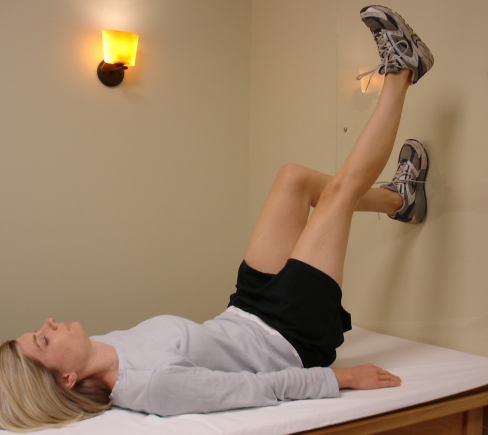
The patient was seen 6 times over 8 weeks by one physical therapist and instructed in 8 exercises. The first four unilateral exercises (Figures 1–4) were developed by the PRI to restore optimal triplanar pelvic/hip position and maintain it during functional movement. Three traditional stabilization exercises (bridging, adductor ball squeeze, and abdominal marching) and one stabilization exercise (reverse curl-ups) developed by the PRI were prescribed to help maintain and stabilize the lumbar–pelvic–femoral complex. During all exercise instruction, the therapist asked the patient where she felt her muscles working to ensure that the correct muscles were activated. The therapist reviewed and progressed the patient's home programme on each follow-up visit. On the first visit, the therapist instructed the patient in an exercise called a 90/90 left hemibridge with a left hip shift34 (see Figure 1). This exercise was prescribed by the treating therapist to activate the left hamstrings and left IC AM to address the identified pelvic asymmetry (left pelvis anteriorly tilted (sagittal plane) and forwardly rotated (transverse plane)) and restricted left posterior hip capsule/ligament. Muscle activation of the left hamstring would move the left hemipelvis into posterior rotation, which is also hip extension (acetabular movement on the femur), restoring position in the sagittal plane by opposing the anterior pelvic tilt. Muscle activation of the IC AM would help to achieve left AF IR (i.e., left hip movement (acetabulum over the femur) into IR and adduction) and lengthen the posterior left hip capsule/ligament.
Figure 1.
Instructions:
- Lie on your back with your feet flat on a wall and your knees and hips bent at a 90° angle.
- Inhale through your nose and exhale through your mouth while pressing your heels down toward the mat (you should feel the muscles in the back of your legs engage), and your tailbone will lift up slightly off the mat. Keep your back flat on the mat.
- Move (shift) your left knee down lower than the right knee.
- Maintain your hip lift with your left leg on the wall and straighten your right leg.
- Continue your breathing for three breaths, then slowly lower back to the mat and repeat.
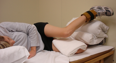
Figure 4.
Instructions:
- Lie on your right side with your toes on a wall, ankles and knees together and your back rounded.
- Place your right arm under your head and your left hand on the floor in front of you to stabilize your trunk.
- Place a pillow between your ankles and a folded towel between your knees.
- Place a 3–5 lb ankle weight around your left ankle.
- Slide and guide your left hip backward as far as you can without arching your back.
- Push your right toes into the wall
- Raise your left ankle up while turning your left thigh “in” so that your left ankle is higher than your left hip. Now lift your left knee and ankle simultaneously towards the ceiling. You should feel your left outer hip engage.
- Hold this position while taking four or five deep breaths in through your nose and out through your mouth.
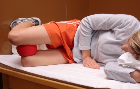
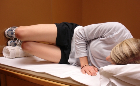
On the second visit (5 days after the initial exam), the therapist reviewed the 90/90 hemibridge and instructed the patient in two new exercises in left side-lying position: a scissor slide34 (see Figure 2) and a knee-to-knee34 (see Figure 3). The scissor-slide technique was prescribed to promote movement of the left acetabulum over the femur and to facilitate elongation of the patient's left posterior hip capsule/ligament to ensure that the left femoral head was seated properly in the left acetabulum and to gain passive left hip IR. The knee-to-knee34 was prescribed to activate the right gluteus maximus (GM), left IC AM, and abdominals to help hold the left acetabulum over the left femur. The left adductors were shortened first via a left pelvic shift (acetabulum moving over the femur) and then shortened further with the concentric contraction against gravity into hip IR and adduction. This position effectively strengthens a weak and long muscle by placing it in a shortened position before activating it against gravity. The right GM was activated to oppose right hip IR (a concomitant position secondary to forward rotation of the left hemipelvis)27 via the GM's action to move the hip into ER.
Figure 2.
Instructions:
- Lie on your left side with your knees bent and place a ball between your knees.
- Press your left toes slightly into the wall.
- Inhale through your nose and gently slide your right leg forward without letting your trunk rotate forward.
- Exhale and gently push your left leg up into the ball.
- Inhale again and slide your right leg forward further.
- Exhale and squeeze your left leg into the ball.
- Repeat this sequence until you have taken a total of three breaths in and out.
Figure 3.
Instructions:
- Lie on your left side with your toes on the wall, knees together and back rounded.
- Place a bolster underneath your ankles.
- Push your left toes into the wall.
- Shift your right knee forward.
- Lift up or turn “out” your right thigh.
- Then lift up or turn “in” your lower thigh. You should feel your left inner thigh engage.
- Hold your legs together while you take four or five deep breaths in through your nose and out through your mouth.
On the third visit (9 days after initial exam), the patient was instructed in a basic bridge9 exercise and bilateral adductor ball squeezes. Bridges were prescribed by the therapist for muscle activation of bilateral GM and hamstrings. This exercise was done in supine hook-lying position, with activation of GM and abdominals before lifting the pelvis up off the plinth. The patient was instructed not to use her back muscles (paraspinals) to lift her pelvis. The position was then held for 5 seconds before the pelvis was lowered back to the plinth. The adductor ball squeezes were prescribed in order to activate the patient's bilateral adductors, pelvic floor musculature, and abdominals for stabilization. This exercise was also done in supine hook-lying position, using a ball 15.2 cm in diameter. The patient was instructed to activate pelvic floor muscles (as in a Kegel exercise) and abdominals and to squeeze the ball with both legs, holding for 5 seconds.
During the fourth visit (11 days after initial exam), the therapist instructed the patient in a right side-lying left anterior gluteus medius34 exercise (see Figure 4). The clinical reasoning for this exercise was that the IR action of the anterior gluteus medius would further help to train the patient to keep her left femoral head seated optimally in the acetabulum, rather than rotating out into ER, which would decrease the surface area of the femoral head in the acetabulum. Left anterior gluteus medius activation/strengthening was needed because of the anterior hip capsule laxity inferred from the negative Thomas test on the left. The anterior gluteus medius is an internal rotator35 and thereby acts to rotate the femoral head inward, which is important if the anterior hip capsule/ligaments are loose. The anterior hip ligaments serve to check excessive hip ER (iliofemoral ligament) and hip hyperextension (iliofemoral and pubofemoral ligaments).36(p.668) If the anterior hip ligaments are loose, however, they will allow excessive femoral head ER, requiring more hip internal rotator muscle strength/activation to keep the femoral head rotated inward.
On the fifth visit (36 days after initial exam), the therapist prescribed abdominal marching (lower-abdominal exercise with unilateral hip flexion) for activation of bilateral transverse abdominus, obliques, and rectus abdominis;17 this exercise has been recommended for patients to stabilize the trunk.14 The patient was instructed to lie supine hook-lying and to activate her abdominal muscles by drawing her navel up and in (posterior). The patient was instructed to hold the abdominal contraction while lifting one leg at a time off the plinth, then lowering the leg and lifting the other leg.
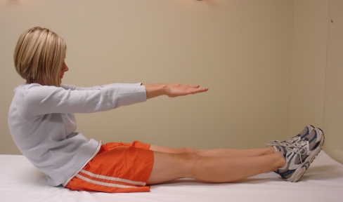
At the discharge (sixth) visit (55 days after initial exam), the therapist instructed the patient in long sitting reverse curl-ups37 (see Figure 5). This technique was prescribed for muscle activation of abdominal obliques (both eccentric and concentric) in the sagittal and transverse planes (right and left trunk rotation) during exhalation and muscle inhibition of paraspinals (to avoid an anterior pelvic tilt), again to stabilize the pelvis/trunk. Dosage details of the patient's home programme are given in Table 1.
Figure 5.
Instructions:
- With knees straight in a long seated position, breathe in through your nose.
- Lower your back to a firm surface slowly, while curling your back (trunk flexion), reaching with extended arms and blowing slowly through curled lips.
- Your low back will touch the surface first, followed by your midback and then your head.
- Inhale through your nose.
- Turn to one side and blow out again as your trunk curls up.
- Repeat eight times, alternating curl-ups from one side to the other.
Table 1.
Home Programme Treatment Parameters for a Female Patient with Left Low Back Pain and Left Sacroiliac Joint Pain
| Exercise* | Prescription (volume, frequency) |
|---|---|
| 90/90 hemibridge with left shift | 5 reps×1 set, 3×/day |
| Scissor slides | 10 reps×1 set, 2×/day |
| Left side-lying knee-to-knee | 10 reps×1 set, 2×/day |
| Basic bridges | 10 reps×1 set with 5 s hold, 1×/day |
| Adductor ball squeezes | 10 reps×1 set with 5 s hold, 1×/day |
| Right side-lying left anterior gluteus medius | 8 reps×2 sets with 5 s hold 1×/day |
| Abdominal marching | 10 reps×1–2 sets, 1×/day |
| Reverse curl-ups | 8 reps×2 sets, 1×/day |
Stabilization exercises in italics
reps=repetitions; s=seconds
OUTCOMEs
Both subjective and objective outcome measures over the course of the 8 weeks of physical therapy management (6 visits) are given in Table 2. On the patient's second visit, her Ober's test was positive initially but was negative immediately after she performed her 90/90 left hemibridge exercises. During the Ober's test, her left thigh dropped well below horizontal, and she no longer had bony end feels at the ER and adduction phases of the test. On her third visit, she stated that she was able to have pain-free sexual intercourse and that her low back and SIJ were 100% pain free (and had been for 6 days prior to her third visit); she no longer had left LE numbness, and she rated her pain 0/10 at best and 0/10 at worst (0–0/10). This represented a change of 8 points, far exceeding the MCSD of 2.5 points for chronic pain.33 The therapist palpated the patient over her original areas of tenderness, and she reported no tenderness. On this third visit the patient's passive left hip IR was measured at 45°.
Table 2.
Findings from Initial and Discharge Examinations for a Female Patient with Left Low Back Pain and Sacroiliac Joint Pain
| Test/Measure | Initial— First Visit |
Second Visit | Third Visit | Fourth Visit | Fifth Visit | Discharge— Sixth Visit |
|---|---|---|---|---|---|---|
| Days since initial visit | 0 | 5 | 19 | 21 | 47 | 61 |
| Passive left hip flexion | 95° with bony end feel |
NT | NT | NT | NT | 120° with normal end feel |
| Passive left hip IR | 40° | NT | 45° | NT | NT | 45° |
| Ober's test | (+) L | (+) L start of session, (−) L after 90/90 hemibridge |
(−) L start of session |
(−) L start of session |
(−) L start of session |
(−) L |
| Palpation over left buttock and inferior lateral angle of sacrum |
Tender | NT | Non-tender | NT | NT | Non-tender |
| MMT hip internal rotators | L 3/5 R 4/5 |
NT | NT | NT | NT | L 4/5 R 4/5 |
| MMT hip external rotators | L 4/5 R 3/5 |
NT | NT | NT | NT | L 4/5 R 4/5 |
| Left-leg numbness | Yes | Yes | No | No | No | No |
| Pain level | 1–8/10 | 1–5/10 | 0/10 | 0/10 | 0/10 | 0–0/10 |
| ODI | 20% | NT | NT | NT | NT | 0% |
(+)=positive; (−) negative; NT=not tested; MMT=manual muscle test; IR=internal rotation; L=left; R=right; ODI=Oswestry Disability Index
During the fourth and fifth visits, Ober's test remained negative; no other testing was done on these visits. On the sixth visit, the patient's passive left hip flexion was measured at 120°, while her left hip IR remained at 45°—increases of 15° and 5°, respectively, from the initial examination. Manual muscle tests for both bilateral hip internal rotators and bilateral hip external rotators were 4/5. Finally, the patient completed another ODI, which the therapist scored as 0%, equivalent to no disability. This improvement in function also met the MCSD for the ODI of 20%.33
DISCUSSION
The purpose of this case report was to describe unique therapeutic exercises that address pelvic–hip asymmetry/impairments without manual techniques, bracing, or treatment modalities such as heat, ultrasound, or massage. The intervention strategies used to reposition the patient's pelvis/femur and increase extensibility of her left posterior hip capsule appeared to eliminate her chronic LBP and SIJP. If the patient was compensating for a left anterior tilted/forwardly rotated pelvis by externally rotating her left hip (femoral ER), this could eventually lead to shortening of the left posterior hip capsule/ligament and contribute to laxity of the left anterior hip ligaments.20 Left hip ER is a common finding of physical therapists trained by the PRI.20 They believe it is a compensation for the Left Anterior Interior Chain (Left AIC) pattern, to help realign the left femur, which is oriented inward with an anteriorly tilted and forwardly rotated pelvis toward the midline or forward sagittal position.20 The clinical reasoning would therefore be to provide interventions to lengthen the left posterior hip capsule/ligaments to allow the femoral head to be seated back in the acetabulum and to provide neuromuscular re-education of internal rotator musculature (i.e., anterior gluteus medius, IC AM) to achieve femoral IR instead of compensatory femoral ER. The initial exercises prescribed in this case were based on this clinical reasoning.
For this case, the therapist hypothesized that activation of the patient's left hamstrings (left hip extension—acetabulum moving on femur) would help to realign the pelvis. The therapist also hypothesized that moving the left hip into adduction and IR would increase the extensibility of the posterior hip capsule and ischiofemoral ligament to increase the acceptance of the femoral head within the acetabulum. The scissor-slide technique was prescribed to help the patient regain left posterior hip capsule flexibility and move her left hip into IR and adduction.
The knee-to-knee technique was prescribed to help activate/recruit the right gluteus maximus, left AM, and abdominals to help hold the left acetabulum over the left femoral head. Recruiting the adductors in combination with internal obliques can help to increase pelvic stability.38 The right side-lying left anterior gluteus medius technique was prescribed to help recruit the left gluteus medius for hip IR (to maintain femoral head position within the acetabulum), to rest tender left buttock muscles, and to avoid possible compensatory left hip ER.
A previous study reported a similar case in which a female patient was managed for right LBP and sciatica.19 Although the case study was of an older female patient, the impairments were similar: pelvic asymmetry (left anterior tilt and forward rotation), decreased passive left hip IR relative to the right, positive left Ober's test, restricted left posterior capsule/ligament, and relative weakness in right hip external rotators and left hip internal rotators.
Ober's test was used in both cases, and in both cases changed from a positive to a negative value after hamstring activation. Ober's test has traditionally been used to assess tensor fascia latae and iliotibial band flexibility;21,39 more recently, however, it has been used to reflect pelvic position, because the position of the acetabulum over the femoral head may influence the test results.19 If the pelvis is in a neutral position, the acetabulum is maximally covering the femoral head, but if the pelvis is in an anterior tilt or forward rotation, then the position of the acetabulum over the femur is not in anatomical neutral. The bony blocks felt by the therapist during the test may have been the impact of the posterior inferior femoral neck on the posterior inferior rim of the acetabulum (during the abduction/ER phase of the test) and the impact of the inferior femoral neck on the inferior rim of the acetabulum (during the adduction phase). This test was repeated at each visit to assess whether or not the effect seen with hamstring activation (90/90 left hemibridge with left shift) was maintained. There is no known literature reporting on the validity, sensitivity, or specificity of Ober's test;40(p.269) however, its reliability has been found to be 0.94 ICC.41
In the two cases, the therapists addressed patient impairments by prescribing similar but different exercises. Both patients did a 90/90 hemibridge, left side-lying knee-to-knee, and right side-lying left anterior gluteus medius exercise. In both cases the left posterior capsule was lengthened, yet the method of doing so was different. In the earlier case, a balloon blow was added to the 90/90 exercise to further activate the abdominals and encourage rib depression/internal rotation.19 The outcomes in that case were remarkable, with a change in ODI score from moderate disability (40%) to no disability (0%).19 The ODI has high specificity, moderate to low sensitivity, and good reliability.42 No other literature was found correlating the above pattern of impairments with LBP, nor did the literature search reveal any studies discussing activation of hamstrings, adductors, and anterior gluteus medius or stretching left posterior capsule for management of patients with LBP.
The literature does discuss passive hip ROM asymmetry in patients with SIJP. Cibulka43 found that patients with SIJP had more external than internal rotation ROM on one side. The patient in the present case had less ER than IR on both sides, but her left IR was less than her right IR; the two equalized with intervention. No literature on management of patients with SIJP was found that discussed activation of hamstrings or adductors to correct for an anteriorly tilted/forwardly rotated innominate/pelvis, a posterior capsule stretch to allow the femoral head to seat into the acetabulum, or anterior gluteus medius activation to help train the femur to rotate in to remain in the acetabulum.
When the patient in this case reported that she was pain free and had a negative Ober's test and normal hip PROM, general bilateral stabilization exercises were prescribed by the therapist. The therapist believed that gaining stabilization strength would help the patient to maintain her pelvic position and thus help prevent future painful episodes. Stabilization exercises are well supported in the literature as an intervention strategy for LBP and SIJ region pain.44–46 Based on the patient's reported outcomes (a 100% reduction in disability), the therapeutic exercises seem to have assisted in addressing the patient's target problem, reducing her pain and disability. The patient stated that she did her unilateral exercises on average 2×/day and the stabilization exercises 1×/day and that she was motivated to be compliant because she felt better as a result of doing the exercises.
This is the second case report describing a postural pattern of asymmetry for an older female patient in which the left innominate was anteriorly tilted/forwardly rotated, passive hip IR was less on the left than on the right, left posterior capsule was restricted, hip external rotators were weaker on the right than on the left, and hip internal rotators were weaker on the left than the right. This case study contributes to the literature on unique unilateral therapeutic exercises (developed by the PRI), which appeared to eliminate pain and improve function for this 65-year-old woman with LBP, SIJ region pain, and LE radicular complaints on the left side. Thirteen days after initiation of the home programme, the patient was able to resume sexual intercourse without pain and had no more LBP, SIJP, or left LE pain. The techniques described above targeted muscle activation for left hamstrings, left IC AM, right GM, left anterior gluteus medius, and abdominals to correct lumbar–pelvic–femoral position and improve extensibility of the left hip posterior capsule/ischiofemoral ligament. Bilateral stabilization strengthening exercises were prescribed to maintain pelvic position and prevent future episodes of pain. At discharge, the patient scored 0% (no disability) on the ODI, a 100% reduction. Common interventions for LBP/SIJ region pain, such as heat, ultrasound, electrical stimulation modalities, massage, lumbar or SIJ bracing, repetitive lumbar flexion or extension exercises, or manual therapy, were not prescribed by the therapist for this patient.
IMPLICATIONS AND FUTURE DIRECTIONS
Pelvic asymmetry and posterior hip capsule restriction may contribute to symptoms of LBP and/or SIJP. Further outcomes-based research is needed to investigate the efficacy of the exercises discussed in this case for individuals with similar pelvic and hip impairments and the efficacy of the management approach developed by the PRI.
KEY MESSAGES
What Is Already Known on This Topic
One previous case report describes similar impairments (left anteriorly tilted and forwardly rotated innominate, less passive hip internal rotation on the left than on the right, restriction of left posterior hip capsule/ligament, hip external rotators weaker on the right than on the left and left hip internal rotators weaker on the left than on the right) and interventions for a female patient with right LBP and sciatica. No other literature describes this pattern of asymmetry/impairments or describes interventions to address the impairments as it relates to patients with LBP and/or SIJP.
What This Study Adds
Pelvic asymmetry (innominate position, passive hip ROM, hip rotator strength, and posterior hip capsule extensibility) appears to be associated with LBP and SIJP and may be important to identify. Management with active unilateral exercises (developed by the Postural Restoration Institute) to restore optimal lumbar–pelvic–femoral position may be beneficial in improving function and decreasing pain.
Boyle KL. Managing a female patient with left low back pain and sacroiliac joint pain with therapeutic exercise: a case report. Physiother Can. 2010;preprint. doi:10.3138/ptc.2009-37
References
- 1.Waddell G. A new clinical model for the treatment of low back pain. Spine. 1987;12:632–41. doi: 10.1097/00007632-198709000-00002. doi: 10.1097/00007632-198709000-00002. [DOI] [PubMed] [Google Scholar]
- 2.Maigne JY, Aivaliklis A, Pfefer F. Results of sacroiliac joint double block and value of sacroiliac pain provocation tests in 54 patients with low back pain. Spine. 1996;21:1889–92. doi: 10.1097/00007632-199608150-00012. doi: 10.1097/00007632-199608150-00012. [DOI] [PubMed] [Google Scholar]
- 3.O'Sullivan PB. Altered motor control strategies in subjects with sacroiliac joint pain during the active straight-leg-raise test. Spine. 2002;27(1):E1–8. doi: 10.1097/00007632-200201010-00015. doi: 10.1097/00007632-200201010-00015. [DOI] [PubMed] [Google Scholar]
- 4.McKenzie R May S. The lumbar spine: mechanical diagnosis and therapy. 2nd ed. Waikanae, New Zealand: Spinal Publications New Zealand; 2003. [Google Scholar]
- 5.Maitland G. Vertebral manipulation. 5th ed. Boston: Butterworth-Heinemann; 1986. [Google Scholar]
- 6.Solomon J, Gregory L, Cooke P, Gage T. Discogenic low back pain. Phys Med Rehabil Clin N Am. 2004;16:177–210. doi: 10.1615/CritRevPhysRehabilMed.v16.i3.20. [Google Scholar]
- 7.Aure O, Hoel NJ, Vasseljen L. Manual therapy and exercise therapy in patients with chronic low back pain: a randomized, controlled trial with 1-year follow-up. Spine. 2003;28:525–31. doi: 10.1097/01.BRS.0000049921.04200.A6. doi: 10.1097/00007632-200303150-00002. [DOI] [PubMed] [Google Scholar]
- 8.Porterfield J, DeRosa C. Mechanical low back pain: perspectives in functional anatomy. 2nd ed. Philadelphia: W.B. Saunders; 1998. [Google Scholar]
- 9.Kisner C, Colby LA. Therapeutic exercise foundations and techniques. 4th ed. Philadelphia: F.A. Davis Co.; 2002. [Google Scholar]
- 10.Lee D. The pelvic girdle. 3rd ed. New York: Churchill Livingstone; 2004. [Google Scholar]
- 11.Roncarati A, McMullen W. Correlates of low back pain in a general population sample: a multidisciplinary perspective. J Manip Physiol Ther. 1988;11(3):158–64. [PubMed] [Google Scholar]
- 12.Nadler S, Steiner DJ, Erasala GN, Hengehold DA, Abeln SB, Weingand KW. Continuous low-level heatwrap therapy for treating acute nonspecific low back pain. Arch Phys Med Rehabil. 2003;84:329–34. doi: 10.1053/apmr.2003.50102. doi: 10.1053/apmr.2003.50102. [DOI] [PubMed] [Google Scholar]
- 13.Young J, Press JM, Hweeinf A. The disc at risk in athletes: perspectives on operative and nonoperative care. Med Sci Sport Exerc. 1997;29(Suppl.7):S222–32. doi: 10.1097/00005768-199707001-00004. doi: 10.1097/00005768-199707001-00004. [DOI] [PubMed] [Google Scholar]
- 14.Rittenberg J, Ross AE. Functional rehabilitation for degenerative lumbar spinal stenosis. Phys Med Rehabil Clin N Am. 2003;14:111–20. doi: 10.1016/s1047-9651(02)00082-7. doi: 10.1016/S1047-9651(02)00082-7. [DOI] [PubMed] [Google Scholar]
- 15.Bodack M, Monteiro M. Therapeutic exercise in the treatment of patients with lumbar spinal stenosis. Clin Orthop Relat Res. 2001;384:144–52. doi: 10.1097/00003086-200103000-00017. doi: 10.1097/00003086-200103000-00017. [DOI] [PubMed] [Google Scholar]
- 16.Kisner C, Colby LA. Therapeutic exercise foundations and techniques. 4th ed. Philadelphia: F.A. Davis Co.; 2002. The spine; pp. 418–79. [Google Scholar]
- 17.Sahrmann SA. Diagnosis and treatment of movement impairment syndromes. St. Louis (MO): Mosby; 2002. [DOI] [PMC free article] [PubMed] [Google Scholar]
- 18.Damen L, Spoor CW, Snijders CJ, Stam HJ. Does a pelvic belt influence sacroiliac joint laxity? Clin Biomech. 2002;17:495–8. doi: 10.1016/s0268-0033(02)00045-1. doi: 10.1016/S0268-0033(02)00045-1. [DOI] [PubMed] [Google Scholar]
- 19.Boyle K, Demske J. Management of a female with chronic sciatica and low back pain: a case report. Physiother Theory Pract. 2009;25(1):44–54. doi: 10.1080/09593980802622677. doi: 10.1080/09593980802622677. [DOI] [PubMed] [Google Scholar]
- 20.Boyle K. Ethnography of the Postural Restoration subculture: a posture based approach to patient/client management [dissertation] Fort Lauderdale (FL): Nova Southeastern University; 2006. [Google Scholar]
- 21.Kendall FP, McCreary EK, Provance PG, Rodgers MM, Romani WA. Muscles: testing and function, with posture and pain. 5th ed. Philadelphia: Lippincott Williams & Wilkins; 2005. [Google Scholar]
- 22.Hruska R. Myokinematic restoration: an integrated approach to treatment of patterned lumbo-pelvic-femoral pathomechanics [course manual] Elon (NC): Postural Restoration Institute; 2005. [Google Scholar]
- 23.Ebmeier J, Hruska R. Postural Restoration Institute [Internet] Lincoln (NE): The Institute; 2007. [updated 2008; cited 2007 Dec 28]. Available from: http://www.posturalrestoration.com/ [Google Scholar]
- 24.Masek J. Soccer hip impingement as it relates to postural restoration, part III: management considerations. Performance Soccer Conditioning. 2007. pp. 1–4.
- 25.Hruska R. Advanced integration assessment and treatment of patterned pathomechanics [course manual] Lincoln (NE): Postural Restoration Institute; 2005. [Google Scholar]
- 26.O'Sullivan S, Schmitz T. Physical rehabilitation: assessment and treatment. Philadelphia: F.A. Davis Co.; 2001. [Google Scholar]
- 27.Levangie P. The hip complex. In: Levangie P, Norkin C, editors. Joint structure and function: a comprehensive analysis. Philadelphia: F.A. Davis Co.; 2005. pp. 355–91. [Google Scholar]
- 28.Magee D. Orthopedic physical assessment. Philadelphia: W.B. Saunders; 1997. [Google Scholar]
- 29.Boyle K, Albarran I. The influence of hamstring activation on the outcome of a positive Ober's test; APTA Annual Conference San Antonio, TX; 2008 Feb. [Google Scholar]
- 30.Magee DJ. Orthopedic physical assessment. 4th ed. Philadelphia: W.B. Saunders; 1997. Pelvis; pp. 567–605. [Google Scholar]
- 31.Norkin C, White DJ. Measurement of joint motion: a guide to goniometry. Philadelphia: F.A. Davis Co.; 1995. [Google Scholar]
- 32.Fairbank J, Davies JB, Couper J, O'Brien JP. The Oswestry Low Back Pain Disability Questionnaire. Physiotherapy. 1980;66(8):271–3. [PubMed] [Google Scholar]
- 33.Ostelo RWJG, DeVet H. Clinically important outcomes in low back pain. Best Pract Res Clin Rheumatol. 2005;19:593–607. doi: 10.1016/j.berh.2005.03.003. doi: 10.1016/j.berh.2005.03.003. [DOI] [PubMed] [Google Scholar]
- 34.Hruska R. Myokinematic restoration—an integrated approach to treatment of lower half musculoskeletal dysfunction [course manual] Lincoln (NE): Postural Restoration Institute; 2003. [Google Scholar]
- 35.Moore KL, Dalley AF. Clinically oriented anatomy. 5th ed. Philadelphia: Lippincott Williams & Wilkins; 2006. [Google Scholar]
- 36.Oatis CA. Kinesiology: the mechanics and pathomechanics of human movement. Philadelphia: Lippincott Williams & Wilkins; 2004. Structure and function of the bones and noncontractile elements of the hip; pp. 661–708. [Google Scholar]
- 37.Hruska R. Influences of dysfunctional respiratory mechanics on orofacial pain. Dental Clin N Am. 1997;41:211–27. [PubMed] [Google Scholar]
- 38.Hungerford B, Gileard W, Hodges P. Evidence of altered lumbopelvic muscle recruitment in the presence of sacroiliac joint pain. Spine. 2003;28:1593–600. doi: 10.1097/00007632-200307150-00022. [PubMed] [Google Scholar]
- 39.Kendall FP, McCreary EK. Muscles: testing and function. 3rd ed. Baltimore: Williams & Wilkins; 1983. [Google Scholar]
- 40.Cook C, Hegedus E. Orthopedic physical examination tests: an evidence-based approach. Upper Saddle River (NJ): Pearson Prentice Hall; 2008. Physical examination tests for the hip; pp. 259–77. [Google Scholar]
- 41.Melchione W, Sullivan S. Reliability of measurements obtained by use of an instrument designed to measure iliotibial band length indirectly. J Orthop Sport Phys Ther. 1993;18:511–5. doi: 10.2519/jospt.1993.18.3.511. [DOI] [PubMed] [Google Scholar]
- 42.Fardon DF, Milette P. Nomenclature and classification of lumbar disc pathology: recommendations from the Combined Task Forces of the North American Spine Society, American Society of Spine Radiology, and American Society of Neuroradiology. Spine. 2001;26(5):E93–113. doi: 10.1097/00007632-200103010-00006. doi: 10.1097/00007632-200103010-00007. [DOI] [PubMed] [Google Scholar]
- 43.Cibulka MT. Unilateral hip rotation range of motion asymmetry in patients with sacroiliac joint regional pain. Spine. 1998;23:1009–15. doi: 10.1097/00007632-199805010-00009. doi: 10.1097/00007632-199805010-00009. [DOI] [PubMed] [Google Scholar]
- 44.Kasai R. Current trends in exercise management for chronic low back pain: comparison between strengthening exercise and spinal segmental stabilization exercise. J Phys Ther Sci. 2006;18:97–105. doi: 10.1589/jpts.18.97. [Google Scholar]
- 45.O'Sullivan P, Phyty GD, Twomey LT, Allison GT. Evaluation of specific stabilizing exercise in the treatment of chronic low back pain with radiologic diagnosis of spondylolysis or spondylolisthesis. Spine. 1997;22:2959–67. doi: 10.1097/00007632-199712150-00020. doi: 10.1097/00007632-199712150-00020. [DOI] [PubMed] [Google Scholar]
- 46.Richardson CA, Jull GA, Hodges PW, Hides JA. Therapeutic exercise for spinal segmental stabilization in low back pain: scientific basis and clinical approach. Edinburgh: Churchill Livingstone; 1999. [Google Scholar]


