Abstract
Evaluating the effect of Histoacryl on the outcome and recurrence rate after excision and primary closure of sacrococcygeal pilonidal disease. Forty patients with sacrococcygeal pilonidal sinus were randomly divided into 2 equal groups through computer randomization program. Group I was operated by complete excision of sinus with wound closure using Histoacryl. Group II was operated with primary wound closure by interrupted inverting sutures. Mean operative time was 31.5 ± 5.6 minutes in group I and 35.9 ± 5.1 minutes in group II. Mean healing time was 13.4 ± 2.7 days in group I and 18.0 ± 8.9 days in group II. Wound infection occurred in 2 patients (10%) in Group II. Delayed wound healing occurred in 3 patients (15%) in group I and 4 patients (20%) in group II. Recurrence occurred in 1 patient (5%) in group I and 3 patients (15%) in group II. Histoacryl improves outcome (significantly decreases operative and healing times and increases patient satisfaction score, insignificantly decreases rates of complications and recurrence) after excision and primary closure of sacrococcygeal pilonidal disease.
Keywords: Histoacryl, Pilonidal sinus, Midline operation
Introduction
Sacrococcygeal pilonidal disease is a common problem. In 1833 Herbert described a hair-containing sinus [1]. In 1880 Hodge suggested the term “pilonidal” [2] (Latin; pilus: hair and nidus: nest), to indicate a disease consisting of hair-containing sinus in the sacrococcygeal area.
There were controversies about the etiology and management of pilonidal sinus disease. It was thought to be of congenital origin secondary to a remnant of an epithelial-lined tract from postcoccygeal epidermal cell rests or vestigial scent cells. Pilonidal disease is now widely accepted as an acquired disorder based on the observations that congenital tracts do not contain hair and are lined by cuboidal epithelium. The recurrence after complete excision of the diseased tissue down to the sacrococcygeal fascia and the high incidence of chronic pilonidal sinus disease in hirsute patients further support the acquired theory of pathogenesis. Karydakis suggested three main factors interacting to produce the disease, namely hair, force and vulnerability [3, 4].
Many procedures have been described for the management of symptomatic pilonidal sinus, none of which, judged by the yardsticks of primary healing and recurrence of disease, is perfect [5].
On review of the literature, recurrence rates ranged from 20% to 40% and sometimes more regardless of the technique used [6, 7]. Causes of recurrence are thought to be due to unrecognized sinus at the time of initial excision, repeated infections of the scar or intergluteal cleft anatomy promoting the accumulation of perspiration, friction, and the tendency for hair to grow into the scar [3].
The study of physical and chemical properties of tissue adhesives during the past decades proved to be valuable for surgery. They are now frequently used in various surgical operations [8–10]. Tissue adhesives have advantages over the classic methods of wound closure: they diminish operation time, do not affect tissue blood supply, and are atraumatic and wound infections are reduced [11]. Moreover, they have proven to have negligible histotoxicity as they form a strong bond at the wound edges and provide long-term cosmesis [12]
The aim of this study is to evaluate the effect of skin glue (Histoacryl) on the outcome and recurrence rate after excision and primary closure of sacrococcygeal pilonidal disease.
Patients and Methods
This single blind trial was performed in the General Surgery Department, Ghodran General Hospital, KSA during the period from may 2004 to July 2008 on 40 patients with primary (non recurrent) pilonidal sinus in the lower back. Asymptomatic patients and patients having pilonidal abscess were excluded from this study. Patients having any degree of inflammation were properly treated with suitable antibiotics before being scheduled for operation. Full explanation of procedures and possible complications and patient consent were assured before inclusion in the research. The study protocol was approved by the Ethics Committee of Ghodran General Hospital, KSA
Patients were randomly divided into 2 groups through computer randomization program (www.randomization.com) before the onset of research. Group I included 20 patients operated by complete excision of sinus though midline elliptical incision including all puncti with a core of tissue down to the sacrococcygeal fascia. Meticulous hemostasis was assured with insertion of closed suction drain. The skin and about 1 cm of subcutaneous tissue were released from the rest of subcutaneous tissue in both sides of wound. Deep interrupted sutures were used to close the depth of wound. The superficial part of subcutaneous tissue was sutured separately using inverted interrupted sutures. Wound was closed using n-Butyl-2 Cyanoacrylate (Histoacryl- TissueSeal, B. Braun Corporation, Ann Arbor, MI, USA).
Group II included 20 patients operated by complete excision of sinus though midline elliptical incision including all puncti with a core of tissue down to the sacrococcygeal fascia. Meticulous hemostasis was assured with insertion of closed suction drain through a separate stab away from the wound. The skin and about 1 cm of subcutaneous tissue were released from the rest of subcutaneous tissue in both sides of wound. Deep interrupted sutures were used to close the depth of wound. The superficial part of subcutaneous tissue was sutured separately using inverted interrupted sutures. Wound was primarily closed using monocryl (Ethicon) interrupted inverting sutures. All operative maneuvers in both groups were done by the same surgeon (article author).
All patients were discharged on the first postoperative day with oral Diclofinac analgesia for the first 3 days. Instructions on discharge included improving of local hygiene and regular removal of hairs by depilatory creams monthly. Closed suction drain was removed when 24-hour drainage was minimal, usually by the fourth postoperative day. In Group I patients the wound was left without dressing after the second postoperative day. Histoacryl was allowed to slough separately with showering after 10 days. In Group II patients sutures were removed after complete wound healing.
All patients were followed up in visits at 3 days interval until complete wound healing was assured, then every month for 6 months. Telephone follow up was made every 6 months to complete 2 year follow up. Patients were encouraged to visit the clinic at any time if they have any problem. The mean length of follow up was 21.30 ± 5.23 months in Group I patients and 20.85 ± 5.54 months in Group II patients.
Pain was evaluated using pain visual analog scale [13] before and after treatment. Healing was defined as complete re-epithelization of the wound. Wound sepsis was diagnosed clinically. Wound cultures were taken for bacteriological confirmation. Recurrence was defined by re-existence of sinus openings in anatomic exploration whether associated with symptoms or not. Recurrence was confirmed by siograms. Patient satisfaction score was designed by asking patients to express their satisfaction in a numerical score from 0 to 10 one month after surgery. Results were evaluated by auther.
Statistical Analysis
Quantitative variables were expressed as mean ± Standard Deviation. Qualitative variables were expressed as frequency and percent. Quantitative parametric variables were compared between the two groups using unpaired student t-test, quantitative non-parametric variables were compared using Mann-Whitney test. Qualitative variables were compared using Chi-square test or Fisher exact test (when the criteria for using Chi-square were not sufficient. The power used was 0.80 while the level of significance was 5%.
Results
Forty patients were included in our study, 33 males (82.50%) and 7 females (17.50%). The age of patients ranged from 16 to 37 years with mean age of 23.61 ± 6.08 years.
Variable degrees of Low back pain was the main presenting symptom in 37 patients (92.50%). Single or multiple (up to 5) puncti in the natal clefts intermittently discharging pus were present in all patients (Table 1).
Table 1.
Clinical data of patients
| Group I | Group II | Total | |
|---|---|---|---|
| Age | 22.9 ± 5.27 | 23.2 ± 4.35 | 23.61 ± 6.08 |
| Sex | |||
| Males | 15(37.5%) | 18(45%) | 33 (82.5%) |
| Females | 5(12.5%) | 2(5%) | 7 (17.5%) |
| Presentation | |||
| Low back pain | 18(45%) | 19(47.5%) | 37 (92.5%) |
| Single punctum | 12(30%) | 15(37.5%) | 27 (67.5%) |
| Multiple puncti | 8(20%) | 5(12.5%) | 13 (32.5%) |
| Total | 20 | 20 | 40 |
Operative Time
In group I, operative time ranged from 24–45 minutes. The mean operative time was 31.5 ± 5.6 minutes. In group II, operative time ranged from 30–50 minutes. The mean operative time was 35.9 ± 5.1 minutes. The difference between the two groups was proved to be statistically significant (p: 0.014).
Time of Complete Healing
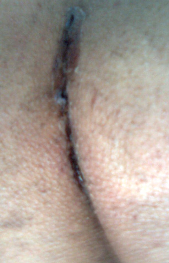
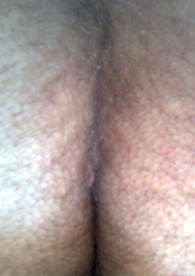
Time elapsed from end of surgery till complete wound epithelialization without any residual open segments was calculated. In group I, healing time ranged from 12–21 days. The mean healing time was 13.4 ± 2.7 days. In group II, healing time ranged from 12–45 days. The mean healing time was 18.0 ± 8.9 days. The difference between the two groups was proved to be statistically significant (p: 0.033) (Table 2). Figure 1 shows midline sacrococcygeal wound sealed with Histoacryl 9 days after operation and Fig. 2 shows the same wound 21 days after operation.
Table 2.
Time of complete healing
| Time of healing | Group I | Group II |
|---|---|---|
| 0–12 days | 14 | 10 |
| 13–18 days | 3 | 6 |
| 19–24 days | 1 | 0 |
| 25–30 days | 1 | 2 |
| More than 30 days | 1 | 2 |
| Mean Time of healing | 13.4 ± 2.7 | 18.0 ± 8.9 |
| p-value | 0.03 | |
Fig. 1.

Midline sacrococcygeal wound sealed with Histoacryl 9 days after operation
Fig. 2.

Midline sacrococcygeal wound sealed with Histoacryl 21 days after operation
Complications
One patient in each group (5%) suffered from severe pain in the first postoperative day. Early wound infection occurred before complete wound healing in 2 patients (10%) in Group II. Delayed wound healing (more than 18 days) occurred in 3 patients (15%) in group I and 4 patients (20%) in group II. There was no statistical significant difference between the two groups regarding complications (p: 1.00, 0.49 and 0.49 respectively) (Table 3).
Table 3.
Complications
| Complications | Group I | Group II | p-value |
|---|---|---|---|
| Severe postoperative pain | 1 (5%) | 1 (5%) | 1.00 |
| Early wound infection | 0 | 2 (10%) | 0.49 |
| Delayed wound healing | 3 (15%) | 4 (20%) | 0.49 |
Recurrence
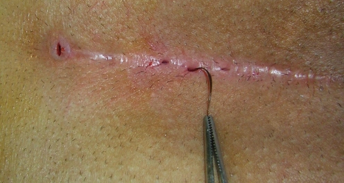
Recurrence after complete healing occurred in 2 patients (10%) in group II 5 and 8 months after operation. These 2 patients are the patients who suffered from early wound infection. One patient (5%) in group I and 1 patient (5%) in group II suffered from late recurrences after 19 and 23 months respectively. Second excision with Rhomboid flap repair solved their problems. There was no statistical significant difference between the two groups regarding recurrence (p:0.61) (Table 4). Figure 3 shows midline sacrococcygeal wound with recurrence 8 months after operation.
Table 4.
Time of recurrence
| Time of Recurrence | Group I | Group II | p-value |
|---|---|---|---|
| 0–12 months | 0 | 2 (10%) | 0.49 |
| 12–24 months | 1 (5%) | 1 (5%) | 1.00 |
| Total | 1 (5%) | 3 (15%) | 0.61 |
Fig. 3.
Midline sacrococcygeal wound showing recurrence 8 months after operation
Patient Satisfaction Score
The mean patient satisfaction score for Group I patients was 9.1 ± 1.02 versus 7.9 ± 2.15 for Group II patients. The difference between the two groups was proved to be statistically significant (p: 030).
Discussion
Surgical treatment of chronic pilonidal sinus by excision of the diseased tissue down to the sacrococcygeal fascia is generally accepted but management of the remaining defect is still a matter of debate. Many methods have been described such as open excision; excision with primary closure and excision with flap closure. Open excision and healing by secondary intention technique is associated with long hospitalization, frequent wound dressing, increased postoperative morbidity, loss of work days and poor cosmetic outcome due to wide unacceptable scars [14]. Primary closure of the wound is a simple technique but it may have a high recurrence rate due to continuing deep natal cleft [15]. Excision with local flap procedures have a low recurrence rate but they are more technically demanding and their use is generally restricted to recurrent complex pilonidal sinus [16].
Many authors believe that complete excision of the pilonidal sinus with primary closure yields good results in terms of healing, morbidity, early return to work and recurrence rate and can be considered the treatment of choice for pilonidal sinus. More demanding flap techniques and plasties should be reserved for complicated pilonidal sinus, which requires a wider excision [17–19].
In the present study, the use of Cyanoacrylate significantly decreased operative and healing times, significantly increased patient satisfaction and insignificantly decreased rates of complications and recurrence. We think that the decrease in early postoperative infection that happened in wounds sealed with Cyanoacrylate is the reason for decreased rate of recurrence in those patients.
In the present study, the patients who suffered from early wound infection were the patients having early recurrence. Søndena et al. [20] tried to find out whether failure of primary wound healing after excision and primary suture for chronic pilonidal sinus predicts recurrence. They followed a total of 197 consecutive patients operated for chronic pilonidal sinus; 52 patients in the prospective group were given cloxacillin perioperatively and 145 patients randomized to have either a single preoperative dose of cefoxitin 2 g intravenously (73 patients) or no prophylaxis (72 patients). Patients were followed for 7 years. In the prospective group there were 10 recurrences (19%). In the randomized study 6 patients (8%) with antibiotic prophylaxis had a recurrence compared with 14 patients (19%) without prophylaxis. In both groups, failure of primary healing was significantly associated with early recurrence. The use of antibiotics did not have significant influence on the incidence of recurrence. Most recurrences occurred within the first year. They concluded that wound complications significantly influenced the recurrence rate whereas antibiotics did not.
Cyanoacrylate tissue adhesive precludes the need for foreign bodies (sutures) to close wounds. It also has an in vitro antimicrobial effect. To determine whether contaminated wounds closed with cyanoacrylate tissue adhesive will have a lower infection rate compared with wounds closed with 5-0 monofilament sutures, Quinn et al. [21] designed a randomized, blinded, experimental animal study. Two contaminated incisions were made on 20 albino guinea pigs and randomly assigned to be closed with either topical octylcyanoacrylate tissue adhesive or percutaneous 5-0 polypropylene suture. 25% in the tissue adhesive group were sterile on day 5, whereas all sutured wounds had positive cultures. Fewer wounds in the tissue adhesive group were determined to be infected by histologic and clinical criteria. They concluded that contaminated wounds closed with sutures had higher infection rates compared with those reported with topical tissue adhesive.
Howell et al. [22] studied the effects of closing lacerations with suture or cyanoacrylate tissue adhesive on staphylococcal counts in inoculated guinea pig lacerations. Wounds closed with adhesive alone had lower counts than wounds containing suture material. The results of a time-kill study were consistent with a bacteriostatic adhesive effect of the adhesive against Staphylococcus aureus.
We conclude that Histoacryl improves outcome (significantly decreases operative and healing times and increases patient satisfaction, insignificantly decreases rates of complications and recurrence) after excision and primary closure of sacrococcygeal pilonidal disease.
References
- 1.Mayo OH. Observations on injuries and disease of rectum. Burgess and Hill, London, pp 45–46 (Quoted from Da Silva JH (2000) Pilonidal cyst: cause and treatment. Dis Colon Rectum. 1833;43:1146–1156. doi: 10.1007/BF02236564. [DOI] [PubMed] [Google Scholar]
- 2.Hodge RM. Pilonidal sinus. Poston Med Surg J 103:485–6, 493, 544 (Quoted from Da Silva JH (2000) Pilonidal cyst: cause and treatment. Dis Colon Rectum. 1880;43:1146–1156. doi: 10.1007/BF02236564. [DOI] [PubMed] [Google Scholar]
- 3.Caestecker J, Mann BD, Castellanos AE, Straus J (2006) Pilonidal Disease. http://emedicine.medscape.com/article/192668
- 4.Karydakis GE. Easy and successful treatment of pilonidal sinus after explanation of its causative process. Aust NZJ Surg. 1992;62:385–389. doi: 10.1111/j.1445-2197.1992.tb07208.x. [DOI] [PubMed] [Google Scholar]
- 5.Senapati A, Cripps NP, Thompson MR. Bascom’s operation in the day-surgical management of symptomatic pilonidal sinus. Br J Surg. 2000;87:1067–1070. doi: 10.1046/j.1365-2168.2000.01472.x. [DOI] [PubMed] [Google Scholar]
- 6.Ringelheim R, Silverberg MA, Villanueva NJJ (2006) Pilonidal Cyst and Sinus; http://emedicine.medscape.com/article/788127
- 7.Berger A, Frileux P. Pilonidal sinus. Ann Chir. 1995;49:889–901. [PubMed] [Google Scholar]
- 8.Binmoeller KF, Soehendra N. New haemostatic techniques: histoacryl injection, banding/endoloop ligation and haemoclipping. Baillieres Best Pract Res Clin Gastroenterol. 1999;13:85–96. doi: 10.1053/bega.1999.0010. [DOI] [PubMed] [Google Scholar]
- 9.Oppell UO, Zilla P. Tissue adhesives in cardiovascular surgery. J Long Term Eff Med Implants. 1998;8:87–101. [PubMed] [Google Scholar]
- 10.Skeist I. Handbook of adhesives. York: Reinhold Publishing corp; 1962. [Google Scholar]
- 11.Zografos GC, Marti KC, Morris DL. Laser Doppler flowmetry in evaluation of cutaneous wound blood flow using various suturing techniques. Ann Surg. 1992;215:266–268. doi: 10.1097/00000658-199203000-00011. [DOI] [PMC free article] [PubMed] [Google Scholar]
- 12.Fotiadis C, Leventis I, Adamis S, Gorgoulis V, Domeyer P, Zografos G, et al. The use of isobutylcyanoacrylate as a tissue adhesive in abdominal surgery. Acta Chir Belg. 2005;105:392–396. doi: 10.1080/00015458.2005.11679743. [DOI] [PubMed] [Google Scholar]
- 13.Huskisson EC. Measurement of pain. Lancet. 1974;2(7889):1127–1131. doi: 10.1016/S0140-6736(74)90884-8. [DOI] [PubMed] [Google Scholar]
- 14.Fuzun M, Bakir H, Soylu M, Tansug T, Kaymak E, Harmancioglu O. Which technique for pilonidal sinus—open or closed? Dis Colon Rectum. 1994;37:1148–1150. doi: 10.1007/BF02049819. [DOI] [PubMed] [Google Scholar]
- 15.Iesalnieks I, Furst A, Rentsch M, Jauch KW. Primary midline closure after excision of a pilonidal sinus is associated with a high recurrence rate. Chirurg. 2003;74:461–468. doi: 10.1007/s00104-003-0616-8. [DOI] [PubMed] [Google Scholar]
- 16.Khatri V, Espinosa MH, Amin AK. Management of recurrent pilonidal sinus by simple V-Y fasciocutaneous flap. Dis Colon Rectum. 1994;37:1232–1235. doi: 10.1007/BF02257787. [DOI] [PubMed] [Google Scholar]
- 17.Toccaceli S, Persico Stella L, Diana M, Dandolo R, Negro P. Treatment of pilonidal sinus with primary closure. A twenty-year experience. Chir Ital. 2008;60:433–438. [PubMed] [Google Scholar]
- 18.Tocchi A, Mazzoni G, Bononi M, Fornasari V, Miccini M, Drumo A, et al. Outcome of chronic pilonidal disease treatment after ambulatory plain midline excision and primary suture. Am J Surg. 2008;196:28–33. doi: 10.1016/j.amjsurg.2007.05.051. [DOI] [PubMed] [Google Scholar]
- 19.Al-Salamah SM, Hussain MI, Mirza SM. Excision with or without primary closure for pilonidal sinus disease. J Pak Med Assoc. 2007;57:388–391. [PubMed] [Google Scholar]
- 20.Søndenaa K, Diab R, Nesvik I, Petter Gullaksen F, Magne Kristiansen R, Sæbø A, Kørner H. Influence of failure of primary wound healing on subsequent recurrence of pilonidal sinus. Combined prospective study and randomized controlled trial. Eur J Surg. 2002;168:614–618. doi: 10.1080/11024150201680007. [DOI] [PubMed] [Google Scholar]
- 21.Quinn J, Maw J, Ramotar K, Wenckebach G, Wells G. Octylcyanoacrylate tissue adhesive versus suture wound repair in a contaminated wound model. Surgery. 1997;122:69–72. doi: 10.1016/S0039-6060(97)90266-X. [DOI] [PubMed] [Google Scholar]
- 22.Howell JM, Bresnahan KA, Stair TO, Dhindsa HS, Edwards BA. Comparison of effects of suture and cyanoacrylate tissue adhesive on bacterial counts in contaminated lacerations. Antimicrob Agents Chemother. 1995;39:559–560. doi: 10.1128/aac.39.2.559. [DOI] [PMC free article] [PubMed] [Google Scholar]


