Human Shugoshin 1 (Sgo1) protects centromeric sister-chromatid cohesion during mitosis. Heterochromatin protein 1 (HP1) has been proposed to recruit Sgo1 to mitotic centromeres. We show that the molecular interaction targeting HP1 to mitotic centromeres is incompatible with HP1 further recruiting Sgo1. Our results clarify the role of centromeric HP1 in chromosome segregation.
Abstract
Human Shugoshin 1 (Sgo1) protects centromeric sister-chromatid cohesion during prophase and prevents premature sister-chromatid separation. Heterochromatin protein 1 (HP1) has been proposed to protect centromeric sister-chromatid cohesion by directly targeting Sgo1 to centromeres in mitosis. Here we show that HP1α is targeted to mitotic centromeres by INCENP, a subunit of the chromosome passenger complex (CPC). Biochemical and structural studies show that both HP1–INCENP and HP1–Sgo1 interactions require the binding of the HP1 chromo shadow domain to PXVXL/I motifs in INCENP or Sgo1, suggesting that the INCENP-bound, centromeric HP1α is incapable of recruiting Sgo1. Consistently, a Sgo1 mutant deficient in HP1 binding is functional in centromeric cohesion protection and localizes normally to centromeres in mitosis. By contrast, INCENP or Sgo1 mutants deficient in HP1 binding fail to localize to centromeres in interphase. Therefore, our results suggest that HP1 binding by INCENP or Sgo1 is dispensable for centromeric cohesion protection during mitosis of human cells, but might regulate yet uncharacterized interphase functions of CPC or Sgo1 at the centromeres.
INTRODUCTION
Faithful chromosome segregation is essential for the genomic integrity of eukaryotic cells and requires the proper establishment and resolution of sister-chromatid cohesion (Nasmyth, 2002). The cohesin complex is required for sister-chromatid cohesion and is loaded to chromosomes in telophase and modified during DNA replication to establish functional cohesion (Onn et al., 2008; Peters et al., 2008; Nasmyth and Haering, 2009). Cohesin removal is a prerequisite for sister-chromatid separation during mitosis and occurs in two steps in vertebrates (Waizenegger et al., 2000). In prophase, most cohesin along chromosome arms is phosphorylated by the mitotic kinase Plk1 (Sumara et al., 2002; Hauf et al., 2005), and subsequently removed by Wapl (Losada et al., 2005; Kueng et al., 2006; Shintomi and Hirano, 2009). Cohesin at the centromeres (the term “centromere” in this article refers to both the core centromere and the pericentric heterochromatin) is, however, protected from the prophase pathway by the Shugoshin 1 (Sgo1)–PP2A complex (Kitajima et al., 2006; Riedel et al., 2006; Tang et al., 2006). The centromeric pool of cohesin ensures the biorientation of sister chromatids and is cleaved by separase at the metaphase–anaphase transition (Hauf et al., 2001; Onn et al., 2008; Peters et al., 2008; Nasmyth and Haering, 2009).
Three mechanisms have been suggested to target Sgo1 to centromeres during mitosis of human cells. First, the mitotic kinase Bub1 phosphorylates centromeric histone H2A at its C-terminal tail (Kawashima et al., 2010). This phospho-histone mark is required to target Sgo1 to centromeres. Second, PP2A prevents Plk1-dependent removal of Sgo1 from chromosomes (Tang et al., 2006). Third, Sgo1 binds to heterochromatin protein 1 (HP1), and this interaction has been proposed to promote Sgo1 centromeric localization in mitosis (Yamagishi et al., 2008).
Human cells contain three HP1 proteins: HP1α, HP1β, and HP1γ (Li et al., 2002; Maison and Almouzni, 2004). Each HP1 protein has an N-terminal chromo domain (CD) that binds to di-/trimethylated histone H3 lysine 9 (H3-K9me2/3), a flexible hinge region, and a C-terminal chromo shadow domain (CSD) that interacts with PXVXL/I motifs in a diverse set of proteins (Smothers and Henikoff, 2000; Li et al., 2002; Maison and Almouzni, 2004; Thiru et al., 2004; Nozawa et al., 2010). In interphase, HP1 is targeted to centromeric heterochromatin through the CD–H3-K9me2/3 interaction and further recruits other proteins to centromeres through the CSD–PXVXL/I interaction. During mitosis, Aurora B in the chromosome passenger complex (CPC) phosphorylates histone H3 serine 10 (H3-pS10), disrupts the HP1 CD–H3-K9me2/3 interaction, and releases HP1 from chromatin (Fischle et al., 2005; Hirota et al., 2005; Ruchaud et al., 2007). In addition, inactivation of the Suv39h methyltransferases that write the H3-K9me2/3 marks does not cause gross sister-chromatid cohesion defects in mammalian cells (Koch et al., 2008). Therefore, inactivation of centromeric targeting of HP1 mediated by the CD–H3-K9me2/3 interaction does not appear to be required for centromeric cohesion protection. These findings have cast doubts about the proposed function of HP1 in recruiting Sgo1 to mitotic centromeres. Nonetheless, HP1α can be detected at mitotic centromeres in human cells (Hayakawa et al., 2003). Furthermore, the mitotic centromeric targeting of HP1α is independent of its CD (Hayakawa et al., 2003). Thus, it has been suggested that HP1 is recruited to mitotic centromeres through a mechanism distinct from that in interphase and that this pool of HP1 at mitotic centromeres contributes to Sgo1 targeting through a CSD-dependent interaction (Yamagishi et al., 2008).
To clarify the role of the Sgo1–HP1 interaction in centromeric cohesion protection in human cells, we have determined the mechanism by which HP1α is targeted to mitotic centromeres. Consistent with previous observations (Ainsztein et al., 1998; Nozawa et al., 2010), we show that HP1 binds to INCENP, a subunit of CPC, and that this interaction involves the binding of HP1 CSD and a PXVXL/I motif in INCENP. We further show that the HP1–INCENP interaction is required for the recruitment of HP1α to mitotic centromeres. Because the HP1–Sgo1 interaction also requires HP1 CSD and a PXVXL/I motif in Sgo1 (Yamagishi et al., 2008), the INCENP-bound centromeric HP1 is incapable of binding Sgo1. Consistently, an INCENP mutant deficient in HP1 binding fully rescues mitotic defects of INCENP RNAi cells. More importantly, a Sgo1 mutant deficient of HP1 binding is fully functional in sister-chromatid cohesion. Therefore, our results indicate that the Sgo1–HP1 interaction is dispensable for sister-chromatid cohesion. Both INCENP and Sgo1 mutants deficient of HP1 binding fail to enrich at interphase centromeres, suggesting that HP1 might regulate yet unidentified interphase functions of Sgo1 and CPC.
RESULTS
HP1 binds to INCENP through its CSD
To study the potential functions of HP1 in chromosome segregation, we constructed HeLa cell lines that stably expressed HP1-CFP and examined HP1 localization in different cell-cycle stages. Consistent with previous reports (Hayakawa et al., 2003), we observed centromeric localization of HP1α, HP1β, and HP1γ during interphase. Most HP1 proteins were released from chromatin in mitosis, but HP1α still localized to the centromeres during mitosis (Supplemental Figure S1A). Ectopically expressed HP1β and HP1γ did not enrich at the mitotic centromeres (Supplemental Figure S1B). In mitotic chromosome spread, the endogenous HP1α localized at the inner centromeres (Supplemental Figure S1C), whereas HP1β and HP1γ did not (unpublished data). Interestingly, a small pool of HP1α was also seen at the midbody during telophase (Supplemental Figure S1A).
Many regulators of cytokinesis, including the CPC, localize to the midbody (Ruchaud et al., 2007). CPC consists of four subunits, Aurora B, INCENP, survivin, and borealin (Ruchaud et al., 2007). It is critical for several mitotic processes, including chromosome alignment, the spindle checkpoint, and cytokinesis. Consistent with its multiple functions, CPC exhibits a dynamic localization pattern during mitosis. It localizes to centromeres and chromosome arms during prophase, to centromeres during metaphase, to the central spindle and the cleavage furrow in anaphase, and to the midbody during telophase. The midbody localization of HP1α suggested to us that HP1 might interact with CPC. Consistently, HP1α had previously been shown to interact with INCENP, a CPC subunit, although the function of such an interaction was unclear (Ainsztein et al., 1998). We thus decided to further investigate the function of the HP1α–INCENP interaction in mitosis.
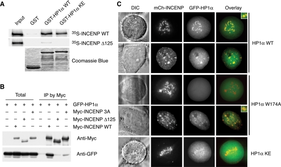
We first confirmed the interaction between HP1 and INCENP. Myc-INCENP bound to all three HA-HP1 proteins (Supplemental Figure S1D). The endogenous INCENP bound to HP1α during mitosis (Supplemental Figure S2A). Further domain-mapping experiments narrowed the HP1-binding region of INCENP to residues 124–248 (unpublished data). Deletion of this region (referred to as INCENP Δ125) greatly diminished the interaction between INCENP and HP1 (Figure 1, A and B, and Supplemental Figure S1D). A previous study showed that the C-terminal region of HP1α containing the hinge region and the CSD was sufficient to bind to INCENP (Hayakawa et al., 2003). We tested whether mutation of several conserved basic residues in the HP1α hinge region to glutamates (HP1 KE) affected INCENP binding. Mutation of the hinge region disrupted the nuclear localization of HP1α (unpublished data), suggesting that this region might contain a nuclear localization signal. GST-HP1α KE bound to INCENP slightly less efficiently, as compared with GST-HP1α WT (Figure 1A).
FIGURE 1:
INCENP binding and mitotic centromere localization of HP1 require the CSD. (A) Recombinant purified GST, GST-HP1α WT, or GST-HP1α hinge mutant (KE; K89E, R90E, and K91E) on glutathione-agarose beads were incubated with in vitro translated 35S-labeled Myc-INCENP WT or Δ125. Bound fractions were analyzed by SDS–PAGE followed by autoradiography and Coomassie blue staining. (B) HeLa tet-on cells were transfected with plasmids encoding GFP-HP1α and Myc-INCENP WT, Δ125, or the PXVXL/I motif mutant (3A; P167A, V169A, and I171A). Cell lysates and the α-Myc IP were blotted with α-Myc and α-GFP. (C) HeLa tet-on cells were transfected with plasmids encoding mCherry-INCENP and GFP-HP1α WT, W174A, or KE and monitored with live-cell imaging. mCherry-INCENP and GFP-HP1α signals are shown in red and green, respectively, in the overlay. DIC, differential interference contrast.
We next showed that recombinant HP1 CSD alone bound to INCENP efficiently in vitro (unpublished data). HP1 CSD binds to peptide motifs with the consensus of PXVXL/I. The HP1-binding domain of INCENP contains one such motif at residues 167–171. An INCENP mutant with this PVVEI motif mutated to AVAEA (referred to as INCENP 3A) bound to HP1α much more weakly than INCENP WT did (Figure 1B). These results indicate that the HP1α–INCENP interaction is mainly mediated through HP1 CSD, with the HP1 hinge region playing an auxiliary role.
The HP1α–INCENP interaction is required for targeting HP1α to mitotic centromeres
Centromeric localization of HP1α in mitosis was previously shown to be mediated through its C-terminal region, including the hinge region and CSD (Hayakawa et al., 2003). We examined the localization of the HP1α hinge mutant (KE) and an HP1α CSD mutant (W174A) that lost its ability to bind PXVXL/I ligands. As shown in Figure 1C, green fluorescent protein (GFP)-HP1α WT and mCherry-INCENP colocalized to centromeres in both interphase and mitosis. GFP-HP1α W174A localized normally to centromeres in interphase but failed to localize to centromeres in mitosis. By contrast, GFP-HP1α KE still localized to mitotic centromeres. Therefore, HP1α CSD is largely responsible for its centromeric targeting in mitosis. Different mechanisms mediate HP1 centromere targeting during interphase and mitosis.
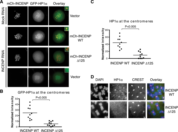
We then tested whether the INCENP–HP1α interaction regulated each other’s centromeric localization. Both mCherry-INCENP WT and Δ125 localized at the centromeres during mitosis (Figure 2A), indicating that the INCENP–HP1α interaction was not critical for the centromeric localization of INCENP. Consistent with previous reports, depletion of INCENP caused enhanced chromosome arm localization of GFP-HP1α (Figure 2A and Supplemental Figure S2B). Ectopic expression of mCherry-INCENP WT restored the centromeric localization of GFP-HP1α in mitosis. By contrast, expression of INCENP Δ125 (which was deficient in HP1α binding) did not restore the centromeric localization of GFP-HP1α (Figure 2, A and B) or the endogenous HP1α (Figure 2, C and D). Therefore, INCENP is critical for HP1α centromeric localization in mitosis. Because the mitotic centromeric localization of HP1α also requires the ligand-binding activity of its CSD, our results collectively indicate that HP1α centromeric targeting in mitosis is mediated by an interaction between HP1α CSD and the PVVEI motif in INCENP.
FIGURE 2:
INCENP recruits HP1α to mitotic centromeres. (A) HeLa tet-on cells were first cotransfected with mCherry-INCENP and GFP-HP1α for 6 h and then either mock transfected or transfected with INCENP siRNA for another 48 h. Cells were examined with live-cell imaging. mCherry-INCENP and GFP-HP1α signals are shown in red and green, respectively, in the overlay. (B) Quantification of the mitotic centromeric signals of GFP-HP1α of cells (N = 10 cells) in (A). (C) HeLa tet-on cells that stably express mCherry-INCENP WT or Δ125 were transfected with INCENP siRNA for 48 h. Metaphase chromosome spread was prepared from these cells and stained with DAPI, CREST, and α-HP1α. Staining intensities of HP1α at the centromeres were quantified (N = 10 cells). (D) Representative images of the metaphase chromosome spread described in (C). DAPI, HP1α staining, and CREST staining are colored blue, green, and red, respectively, in the overlay.
We tested whether the binding of HP1α CSD to INCENP was required for the functions of CPC in cytokinesis and the spindle checkpoint. To do so, we depleted endogenous INCENP by RNAi from HeLa cells and ectopically expressed siRNA-resistant mCherry-INCENP WT or Δ125 and analyzed these cells with flow cytometry (fluorescence-activated cell sorting [FACS]) (Supplemental Figure S3). In INCENP RNAi cells, due to cytokinesis failure, the population of cells with 2N DNA content decreased greatly, whereas the population of polyploid, 4N cells increased (Supplemental Figure S3B). Ectopic expression of either mCherry-INCENP WT or Δ125 largely restored the 2N population, indicating that INCENP Δ125 was not grossly defective in its cytokinesis function. Both INCENP WT and Δ125 also significantly rescued the deficiency of INCENP RNAi cells to undergo Taxol-triggered mitotic arrest (Supplemental Figure S3C), indicating that the INCENP–HP1α interaction is not required for the spindle checkpoint.
The INCENP–HP1 interaction is not required for sister-chromatid cohesion
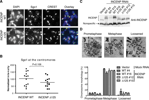
Because HP1α has been implicated in the centromeric recruitment of Sgo1 in human cells (Yamagishi et al., 2008), we next examined whether Sgo1 localization at centromeres was affected in cells expressing the HP1-binding-deficient INCENP mutant, INCENP Δ125. For this purpose, we generated HeLa cell lines that stably expressed RNAi-resistant mCherry-INCENP WT or Δ125. Depletion of INCENP led to increased chromosome arm localization of Sgo1 (Supplemental Figure S4). Ectopic expression of either INCENP WT or Δ125 restored the centromeric localization of Sgo1 (Figure 3, A and B). These HeLa cell lines expressed mCherry-INCENP WT or Δ125 at levels comparable to that of the endogenous INCENP (Figure 3C). This finding indicates that HP1α at the mitotic centromeres is not required for Sgo1 localization at centromeres.
FIGURE 3:
INCENP Δ125 supports Sgo1 localization and sister-chromatid cohesion. (A) HeLa tet-on cells that stably express mCherry-INCENP WT or Δ125 were transfected with INCENP siRNA for 48 h. Metaphase chromosome spread was prepared from these cells and stained with DAPI, CREST, and α-Sgo1. DAPI, Sgo1 staining, and CREST staining are colored blue, green, and red, respectively, in the overlay. (B) Quantification of Sgo1 staining intensities at the centromeres in the chromosome spread described in (A) (N = 13 cells). (C) Different clones of HeLa tet-on cell lines stably expressing mCherry-INCENP WT or Δ125 were isolated and transfected with INCENP siRNA for 48 h. The total cell lysates were blotted with α-INCENP. A nonspecific band is used as the loading control. mCherry-INCENP Δ125 migrated at the same position as the endogenous INCENP did. (D) Giemsa-stained mitotic chromosome spread of cells described in (C). Representative images (top panel) and percentages (bottom panel) of different chromosome morphology are shown (mean ± SD, N = 100 cells). Cells that exhibited more than five pairs of separated sister-chromatids were counted as the “loosened” phenotype.
We then tested whether sister-chromatid cohesion was affected by the INCENP Δ125 mutation. The same HeLa cell lines expressing mCherry-INCENP WT or Δ125 were depleted of the endogenous INCENP and treated briefly with nocodazole to enrich for mitotic cells. Neither cell line exhibited gross premature sister-chromatid separation (Figure 3D). Similar results were obtained for two different clones of each cell line. Therefore, centromeric localization of HP1α in mitosis is not required for sister-chromatid cohesion, consistent with normal Sgo1 centromeric localization in these cells.
Using live-cell imaging, we further monitored the mitotic progression of cells expressing mCherry-INCENP WT or Δ125 that had been depleted of endogenous INCENP. We did not observe significant differences in mitotic timing between these two cell lines (Supplemental Figure S5). Furthermore the mitotic localization patterns of mCherry-INCENP WT and Δ125 were indistinguishable (Supplemental Figure S5C). Therefore, these data strongly suggest that centromeric localization of HP1α is dispensable for mitotic progression. Because defects in sister-chromatid cohesion trigger spindle checkpoint-dependent mitotic delays, these results confirm that centromeric HP1α in mitosis is not required for sister-chromatid cohesion.
There was, however, a striking difference in the localization patterns of mCherry-INCENP WT and Δ125 during interphase (Supplemental Figure S5C). Whereas INCENP WT was enriched at centromeres in interphase, INCENP Δ125 localized to nucleoli and failed to localize to centromeres. Similar nucleolus localization was observed for the INCENP 3A mutant (unpublished data). Therefore the INCENP–HP1 interaction is not required for proper mitotic progression or sister-chromatid cohesion, but regulates the centromeric localization of INCENP and possibly CPC in interphase.
Structural basis for HP1 CSD binding to the PXVXL/I motif in Sgo1
HP1α has been suggested to contribute to centromeric targeting of Sgo1 in mitosis through a direct interaction between HP1α CSD and a PXVXL/I motif in Sgo1 in human cells (Yamagishi et al., 2008). In contrast, our results described earlier in the text show that the centromeric targeting of HP1α in mitosis requires an interaction between HP1α CSD and a PXVXL/I motif in INCENP. The centromeric pool of HP1α is thus bound to INCENP and is incapable of simultaneously binding to Sgo1. We therefore decided to further characterize the Sgo1–HP1 interaction.
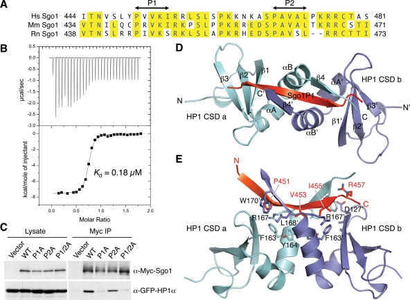
Human Sgo1 contains two PXVXL/I motifs that can potentially bind to HP1 CSD (Figure 4A). We synthesized two peptides (Sgo1P1 and Sgo1P2) containing either motif and tested their binding to HP1β CSD by isothermal titration calorimetry (ITC). Sgo1P1 bound to HP1β CSD with a dissociation constant Kd of 0.18 μM (Figure 4B), whereas no detectable binding was observed between HP1β CSD and Sgo1P2. Mutation of the P1 motif in Sgo1 (P1A) disrupted the Myc-HP1α–GFP-Sgo1 interaction in cells, whereas mutation of the P2 motif had no effect (Figure 4C). Therefore the P1 motif is necessary and sufficient for HP1 binding.
FIGURE 4:
Binding Sgo1 to HP1 involves HP1 CSD and a PXVXL/I motif in Sgo1. (A) Sequence alignment of the HP1-binding region of human (Hs), mouse (Mm), and rat (Rn) Sgo1. The two PXVXL/I motifs are labeled P1 and P2. (B) ITC measurement of the binding between HP1β CSD and Sgo1P1. (C) HeLa tet-on cells were transfected with the GFP-HP1 plasmid together with plasmids encoding Myc-Sgo1 WT, P1A (P451A, V453A, and I455A), P2A (P469A, V471A, and L473A), or P1/2A (P451A, V453A, I455A, P469A, V471A, and L473A) for 24 h and then treated with nocodazole for another 18 h. Cell lysates and Myc IP were blotted with α-Myc and α-GFP. (D) Ribbon drawing of the structure of HP1β CSD–Sgo1P1. Sgo1P1 is colored red, and the two CSD monomers are colored cyan and blue, respectively. (E) Ribbon drawing of the structure of HP1β CSD–Sgo1P1 in an orientation different from (C) and with key binding residues shown in sticks and labeled.
We then determined the crystal structure of HP1β in complex with Sgo1P1. As expected, HP1β CSD forms a dimer (Figure 4D). One CSD dimer binds to one Sgo1P1 peptide. Each CSD monomer has a mixed α/β fold, in which a three-stranded antiparallel β sheet (β1–3) packs against an N-terminal 310 helix and two α helices (αA and αB). αA and αB form the dimer interface. Sgo1P1 is sandwiched between the β4 strands of the two CSD monomers. The N-terminal segment of Sgo1P1 (residues 448–452) forms antiparallel β sheet interactions with β4 of one CSD monomer, whereas the C-terminal segment of Sgo1P1 (residues 453–456) forms parallel β sheet interactions with β4 of the other CSD monomer (Figure 4D). P451, V453, and I455 of the PXVXL/I motif in Sgo1P1 form extensive hydrophobic interactions with CSD (Figure 4E). R457 packs against CSD W170 (equivalent to W174 in HP1α) and also forms an electrostatic interaction with CSD D127. These interactions were similar to those observed in the solution structure of mouse HP1β CSD bound to the PXVXL/I motif in chromatin assembly factor-1 (Thiru et al., 2004). Therefore, our biochemical and structural analyses confirm that the Sgo1–HP1 interaction adopts a canonical binding mode between HP1 CSD and the PXVXL/I motif in Sgo1.
The Sgo1–HP1 interaction is not required for sister-chromatid cohesion
Our results so far have established that HP1α is recruited to mitotic centromeres by INCENP through the CSD–PXVXL/I interaction. Binding of Sgo1 to HP1 requires a similar molecular interaction. The centromeric pool of HP1α is thus not expected to bind Sgo1 and is indeed not required for mitotic centromeric localization of Sgo1 or centromeric sister-chromatid cohesion. It has, however, been argued that Sgo1 needs to be recruited to centromeres in interphase to promote centromeric cohesion protection in mitosis (Perera and Taylor, 2010). HP1 might contribute to Sgo1 recruitment to centromeres in interphase. Furthermore, it is possible that the cytoplasmic pool of HP1 might indirectly regulate Sgo1 function at centromeres in mitosis.
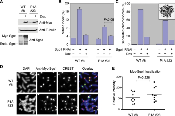
To directly examine the functions of the Sgo1–HP1 interaction, we created HeLa cell lines that stably expressed Myc-Sgo1 WT or the Sgo1 P1A mutant deficient for HP1 binding driven by the tetracycline-inducible promoter (Figure 5A) and depleted the endogenous Sgo1 with RNAi from these cells. In the absence of doxycycline and ectopic Myc-Sgo1 WT expression, Sgo1 depletion resulted in premature sister-chromatid separation and mitotic arrest (Figure 5, B and C). Ectopic expression of Myc-Sgo1 WT induced by doxycycline effectively rescued these phenotypes. Surprisingly, doxycycline-induced expression of Myc-Sgo1 P1A also rescued the mitotic phenotypes caused by Sgo1 RNAi. In fact, even the leaky expression of Myc-Sgo1 P1A in the absence of doxycycline greatly reduced the mitotic index and chromosome missegregation caused by Sgo1 RNAi. Two other Myc-Sgo1 P1A-expressing clones exhibited similar phenotypes (Supplemental Figure S6). Therefore, the Sgo1 P1A mutant deficient in HP1 binding is functional in centromeric cohesion protection. Consistently, both Myc-Sgo1 WT and P1A localized properly to centromeres in mitosis (Figure 5, D and E).
FIGURE 5:
The Sgo1–HP1 interaction is dispensable for Sgo1 localization and sister-chromatid cohesion. (A) HeLa tet-on cells that stably express Myc-Sgo1 WT (clone #8) or P1A (clone #23) under the control of doxycycline were cultured in the absence (–) or presence (+) of doxycycline (Dox) and transfected with Sgo1 siRNA for 24 h. Cell lysates were blotted with α-Myc, α-tubulin, and α-Sgo1. (B) The mitotic index of cells in (A). Cells were stained with propidium iodide and α-H3-pS10 and analyzed by FACS. Ten thousand events were counted for each sample. Mitotic cells have 4N DNA content and are H3-pS10-positive. The average and SD of three experiments are shown. (C) The extent of sister-chromatid separation of cells in (A) as determined by metaphase spread with Giemsa staining (N = 100 cells). A representative image of a cell with separated chromosomes is shown in inset. (D) Metaphase spread of cells in (A) was stained with DAPI (blue in overlay), Myc (green in overlay), and CREST (red in overlay). (E) Quantification of Myc-Sgo1 staining intensities at the centromeres for cells in (D) (N = 10 cells).
Finally, we performed live-cell imaging on cells depleted of endogenous Sgo1 and transiently expressing GFP-Sgo1 WT or P1A (Supplemental Figure S7). GFP-Sgo1 P1A supported proper mitotic progression. Chromosome alignment and segregation occurred with normal timing in these cells. Moreover, the timing of GFP-Sgo1 P1A localization to the centromeres during mitosis was indistinguishable from that of GFP-Sgo1 WT. Taken together, these data indicate that the Sgo1–HP1 interaction is not critical for sister-chromatid cohesion or Sgo1 centromeric localization in mitosis.
The Sgo1–HP1 interaction is required for centromeric localization of Sgo1 in interphase
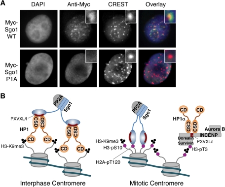
During the live-cell imaging experiment, we noticed that GFP-Sgo1 WT, but not GFP-Sgo1 P1A, localized to centromeres in G2-arrested cells (unpublished data). To confirm this finding, we stained cells expressing Myc-Sgo1 WT or P1A with anti-Myc. Myc-Sgo1 WT, but not Myc-Sgo1 P1A, localized to centromeres in interphase cells (Figure 6A). Therefore the Sgo1–HP1 interaction regulates the centromeric targeting of Sgo1 in interphase.
FIGURE 6:
The HP1–Sgo1 interaction targets Sgo1 to interphase centromeres. (A) HeLa tet-on cells that stably express Myc-Sgo1 WT or P1A were cultured in the presence of doxycycline, transfected with Sgo1 siRNA for 24 h, and stained with DAPI (blue in overlay), α-Myc (green in overlay), and CREST (red in overlay). (B) Model for the distinct binding modes and interactions of HP1 at the centromeres during interphase and mitosis. See Discussion for details.
DISCUSSION
Distinct mechanisms target HP1α to centromeres during interphase and mitosis
A previous study has shown that different regions of HP1α mediate its centromeric targeting in interphase and in mitosis (Hayakawa et al., 2003). The CD is required for targeting HP1α to centromeres in interphase, whereas the CSD mediates HP1α centromeric localization in mitosis. Consistent with this finding, Aurora B (as a subunit of CPC) phosphorylates H3-S10 during mitosis and disrupts the interaction between HP1α CD and H3-K9me2/3 (Fischle et al., 2005; Hirota et al., 2005). These earlier findings suggest that centromeric targeting of HP1α in mitosis likely involves the binding of its CSD with centromeric proteins containing PXVXL/I motifs. Intriguingly, two studies have shown that HP1α binds to the CPC subunit INCENP through its CSD (Ainsztein et al., 1998; Nozawa et al., 2010). In this study, we show that the HP1α–INCENP interaction is indeed required for the centromeric localization of HP1α in mitosis. Cells expressing an INCENP mutant deficient for HP1α binding fail to localize HP1α to centromeres in mitosis. Our study has thus identified the major mitotic centromeric receptor of HP1α.
Collectively, these findings establish that distinct mechanisms target HP1α to centromeres during interphase and mitosis, and support the following model for HP1 centromeric targeting during the cell cycle (Figure 6B). In interphase, HP1 CD binds to H3-K9me2/3 and recruits HP1 to centromeres in interphase. In mitosis, Aurora B phosphorylates H3-S10 and releases most HP1 from chromatin by disrupting the CD–H3-K9me2/3 interaction. HP1α CSD binds to a PXVXL/I motif in INCENP, maintaining a pool of HP1α at centromeres in mitosis.
Another recent study has shown that HP1α binds to the Mis14 (also known as Nsl1) subunit of the Mis12 kinetochore complex only in interphase, but not in mitosis (Kiyomitsu et al., 2010). The interphase HP1α–Mis14 interaction is nonetheless required for the centromeric localization of HP1 in mitosis. Interestingly, mutations of Mis14 that disrupt its binding to HP1α in interphase also cause defects in the centromeric localization of CPC (and presumably INCENP) in mitosis. Therefore, the defective HP1α mitotic centromeric localization in cells expressing these Mis14 mutants is likely an indirect consequence of the loss of INCENP localization at centromeres. How the HP1α–Mis14 interaction in interphase promotes the centromeric localization of CPC during mitosis is unclear.
What is the function of centromeric HP1α during mitosis?
As discussed earlier in the text, we have demonstrated in this study that INCENP directly recruits HP1α to centromeres in mitosis. Surprisingly, cells expressing an INCENP mutant deficient for HP1 binding undergo proper mitotic progression. They do not exhibit discernible defects in chromosome alignment, sister-chromatid cohesion, cytokinesis, or the spindle checkpoint. Therefore the INCENP–HP1α interaction appears to be dispensable for the mitotic processes examined. We cannot exclude the possibility that the residual, undetectable amount of HP1α at centromeres is still sufficient for its mitotic functions.
It has been proposed that centromeric HP1α directly recruits Sgo1 to centromeres in mitosis of human cells (Yamagishi et al., 2008). We have shown, however, that both Sgo1 binding and INCENP binding to HP1 involve the binding of HP1 CSD to canonical PXVXL/I motifs on Sgo1 or INCENP. The INCENP-bound HP1α cannot simultaneously bind to Sgo1, and vice versa (Figure 6B). Therefore, the centromeric pool of HP1α is unlikely to directly contribute to Sgo1 centromeric recruitment in mitosis. This notion is consistent with the lack of mitotic phenotypes in cells expressing HP1-binding-deficient mutants of INCENP.
What, then, is the function of centromeric HP1α in mitosis? We envision two possibilities. First, HP1-binding-deficient mutants of INCENP fail to localize to centromeres in interphase. HP1 binding of INCENP during mitosis may facilitate the return of INCENP and CPC to centromeres following mitotic exit and Aurora B inactivation. The function of INCENP at interphase centromeres, if any, is unclear at present. Second, a recent study has shown that the POGZ protein releases HP1α from mitotic chromosomes by disrupting HP1α binding to proteins containing PXVXL/I motifs (Nozawa et al., 2010). Depletion of POGZ from human cells causes mitotic phenotypes similar to those caused by Aurora B inactivation. That study suggests that HP1α negatively regulates the functions of CPC during mitosis, although the mechanism of such regulation is not understood. It is thus possible that the HP1α–INCENP interaction in mitosis attenuates Aurora B function and might be one of several redundant mechanisms contributing to CPC inactivation during mitotic exit.
What is the function of the HP1–Sgo1 interaction?
During meiosis in fission yeast, Swi6 (the HP1 orthologue) binds directly to Sgo1 (the meiotic form of shugoshin) and recruits Sgo1 to centromeres (Yamagishi et al., 2008). Sgo1 mutations that disrupt Swi6 binding also disrupt the centromeric localization of Sgo1. Therefore, the HP1–shugoshin interaction is clearly critical for meiotic progression in the fission yeast. The mitotic functions of HP1 in Sgo1 regulation and centromeric cohesion in mammalian cells have been murky, however. Yamagishi et al. first showed that human Sgo1 bound directly to HP1 through a CSD–PXVXL/I interaction (Yamagishi et al., 2008). They further showed that depletion of HP1α from human cells by RNAi weakened Sgo1 centromeric localization after a prolonged mitotic arrest and caused premature sister-chromatid separation. They concluded that HP1α directly recruited Sgo1 to mitotic centromeres and contributed to centromeric cohesion protection in human cells. In contrast, a later study reported that depletion of HP1α failed to produce mitotic defects in human cells (Serrano et al., 2009), casting doubts on the original report by Yamagishi et al. In addition, our results presented here suggest that the centromeric pool of HP1 bound to INCENP cannot physically interact with Sgo1. Our finding further questions the validity of the proposed, direct contribution of HP1α in Sgo1 centromeric targeting during mitosis of human cells.
To resolve this controversy, we further examined the effects of depleting HP1α in human cells. In our experiments, some, but not all, HP1α siRNAs that depleted HP1α caused loss of Sgo1 localization at centromeres and premature sister-chromatid separation (unpublished results). Ectopic expression of siRNA-resistant HP1α, however, failed to rescue the mitotic defects of HP1α RNAi cells, raising the possibility that the observed mitotic defects caused by certain HP1α siRNAs were due to off-target effects. Furthermore, human cells contain three HP1 isoforms (α, β, and γ). It is exceedingly difficult to deplete all three isoforms and perform functional rescue experiments to ascertain the specificity of RNAi-mediated depletion.
To avoid these complications, we have focused on the phenotypes of cells expressing a Sgo1 mutant deficient of HP1 binding in this study. Mutation of the sole, functional PXVXL/I motif in Sgo1 disrupts HP1 binding. Ectopic expression of this Sgo1 mutant fully restores sister-chromatid cohesion and Sgo1 centromeric localization in Sgo1 RNAi cells. This result indicates that the HP1–Sgo1 interaction is dispensable for mitotic progression and sister-chromatid cohesion during mitosis in human cells. We cannot, however, rule out the possibility that HP1 binding is one of several redundant mechanisms targeting Sgo1 to centromeres in mitosis.
The HP1α-binding-deficient mutant of Sgo1 does not localize to centromeres during interphase, indicating that, like the HP1–INCENP interaction, the HP1–Sgo1 interaction is required for the interphase centromeric localization of Sgo1. Consistent with our findings, inactivation of Suv39H also disrupted the centromeric targeting of Sgo1 in interphase (Perera and Taylor, 2010). In contrast, the interphase centromeric localization of Sgo1 does not appear to be critical for centromeric cohesion protection in mitosis, as the HP1α-binding-deficient mutant of Sgo1 is functional in sister-chromatid cohesion. Future studies are needed to address the functions of Sgo1 at the interphase centromeres.
CONCLUSION
In this study, we have shown that HP1α is targeted to mitotic centromeres through an interaction between its CSD and a PXVXL/I motif in INCENP. The Sgo1–HP1 interaction also requires the binding of HP1 CSD to a PXVXL/I motif in Sgo1. Therefore, the centromeric, INCENP-bound pool of HP1α cannot simultaneously engage Sgo1. Consistently, INCENP or Sgo1 mutants deficient for HP1 binding support proper mitotic progression and sister-chromatid cohesion. Therefore, our results strongly suggest that the HP1–Sgo1 interaction is dispensable for centromeric cohesion protection.
MATERIALS AND METHODS
Plasmids and siRNAs
Full-length human INCENP, HP1 isoforms, and Sgo1 were isolated from human fetal thymus cDNA (Clontech, Mountain View, CA) and cloned into the appropriate vectors. INCENP, HP1α, and Sgo1 mutants were generated using the QuikChange Site-directed Mutagenesis Kit (Stratagene, La Jolla, CA). siRNA oligonucleotides of INCENP (5′-AGAUCAACCCAGAUAACUA-3′) and Sgo1 (5′-CCUGCUCAGAACCAGGAAA-3′) were synthesized by Dharmacon (Lafayette, CO). siRNA-resistant INCENP plasmids were created by introducing the following silent mutations: bases 2451 (C to T), 2454 (C to T), 2457 (A to T), 2460 (T to C), and 2463 (C to T). siRNA-resistant Sgo1 plasmids were created by introducing the following silent mutations: bases 336 (T to A), 339 (G to A), 342 (C to T), and 345 (G to A).
Cell culture, transfection, and live cell imaging
HeLa tet-on (Clontech) cells were grown in DMEM (Invitrogen, Carlsbad, CA) supplemented with 10% fetal bovine serum. Transfections of siRNAs and plasmids were performed with Lipofectamine RNAiMAX (Invitrogen) and Effectene (Qiagen, Chatsworth, CA), respectively, according to manufacturers’ protocols. For transient rescue experiments of INCENP, HeLa tet-on cells were transfected first with RNAi-resistant pIRESpuro-mCherry-INCENP vectors. Six hours after transfection, the cells were transfected with INCENP siRNA and cultured for another 48 h. For the functional analysis of the spindle checkpoint, 24 h after transfection, the cells were incubated with 100 ng/ml nocodazole for another 16 h. Live cell imaging of HeLa tet-on cells expressing mCherry-INCENP or GFP-Sgo1 was performed using a DeltaVision deconvolution fluorescence microscope with a 40× objective (Applied Precision, Issaquah, WA). The images were processed and analyzed with ImageJ software.
Antibodies and immunoprecipitation (IP)
Production of antibodies against human Sgo1 was described previously (Tang et al., 2004). The following antibodies were purchased from the indicated commercial sources: α-INCENP antibody (Bethyl, Montgomery, TX), α-HP1α (clone 15.19s2; Upstate Biotechnology, Lake Placid, NY), CREST (ImmunoVision, Springdale, AR), α-Myc antibody (Roche, Basel, Switzerland), and α-H3-pS10 (Upstate Biotechnology). For coIP experiments, HeLa tet-on cells were lysed in lysis buffer (25 mM Tris, pH 7.5, 150 mM KCl, 0.2% NP-40, 1 mM dithiothreitol, 5 mM NaF, 5 mM β-glycerophosphate, 0.5 μM okadaic acid, protease inhibitors) by sonication. After centrifugation at 15,000 × g for 10 min at 4°C, the supernatant was incubated with Affi-Prep Protein A beads (Bio-Rad, Hercules, CA) and the appropriate antibodies for 2 h at 4°C. The beads were washed with the lysis buffer. The bound proteins were resolved by SDS–PAGE followed by immunoblotting.
Immunofluorescence (IF) and chromosome spread
For HP1α localization, HeLa tet-on cells were grown in six-well plates, trypsinized, resuspended in hypotonic solution (55 mM KCl), spun down onto slides by Shandon Cytospin 4 (Thermo Fisher, Waltham, MA), fixed in 4% paraformaldehyde for 10 min, permeabilized with the IF buffer (phosphate-buffered saline [PBS] containing 0.1% Triton X-100), and incubated with α-HP1α (1 μg/ml) in the IF buffer containing 3% bovine serum albumin (BSA) for 2 h at room temperature. The cells were then washed with the IF buffer, incubated with fluorescein isothiocyanate– or Cy5-conjugated secondary antibodies (4 μg/ml; Molecular Probes, Eugene, OR) in the IF buffer containing 3% BSA for 1 h, washed again with the IF buffer containing 1 μg/ml 4’,6-diamidino-2-phenylindole (DAPI), and analyzed using a DeltaVision deconvolution fluorescence microscope with a 60× objective. In other cases, IF was performed similarly, except that cells were extracted with the IF buffer before fixation. For quantitative analysis, the signal intensities of proteins in five randomly chosen centromeres of a given cell were averaged, subtracted by background, and normalized by the staining intensity of CREST. For chromosome spread, HeLa tet-on cells were grown in six-well plates, trypsinized, treated with hypotonic solution, fixed in the fixative solution (methanol/acetic acid = 3:1 [vol/vol]), dropped on slides, and stained with 5% Giemsa solution in PBS.
In vitro binding assay
For in vitro binding assays, ∼2 μg of purified GST-HP1α was incubated with 5 μl of glutathione beads in 50 μl of Q-A buffer (20 mM Tris, pH 7.7, 100 mM KCl, 1 mM MgCl2) for 1 h at room temperature. Beads were then incubated with 400 μl of blocking solution (25 mM Tris, pH 8.0, 150 mM NaCl, 2.5 mM KCl, 0.05% Tween-20, 5% dry milk) for 1 h. Ten microliters of in vitro translated 35S-labeled INCENP was added and incubated for 1 h. After washing with blocking solution without dry milk, the beads were boiled in SDS sample buffer and subjected to SDS–PAGE followed by autoradiography.
ITC
ITC was performed with a MicroCal Omega VP-ITC titration calorimeter (GE Life Sciences, Uppsala, Sweden) at 20ºC. Human HP1β CSD was expressed in bacteria and purified using affinity and size exclusion chromatography. The Sgo1P1 (residues 425–443) and Sgo1P2 (residues 443–458) were chemically synthesized. For each titration experiment, 2 ml of 50 μM HP1β CSD in a phosphate buffer was added to the calorimeter cell. Sgo1P1 or Sgo1P2 (0.4 mM) in the exact same buffer was injected with 28 steps of 10 μl each with an injection syringe. Binding parameters were analyzed as a single binding site model using the Origin 7.0 software package (OriginLab, Northampton, MA).
X-ray crystallography
HP1β CSD was concentrated to 14 mg/ml and mixed with SgoP1 at a molar ratio of 1:1.2. Crystals of the HP1β–Sgo1P1 complex were obtained by adding 1 μl of the above mixture to 1 μl of a crystallization buffer (0.1 M Bis-Tris, pH 6.6, 0.2 M NaCl, and 21% [wt/vol] PEG 3350) and incubating for 14 d. The crystals diffracted x-rays to a dmin of ∼1.9 Å. X-ray diffraction data were acquired from a single crystal on beamline 19-ID at the Structural Biology Center at Argonne National Laboratories. The data were indexed, integrated, and scaled using HKL2000 (Otwinowski and Minor, 1997). The data were further treated to correct negative intensities (French and Wilson, 1978). Phases for the structure were obtained by molecular replacement using the program Phaser (Read, 2001; Storoni et al., 2004). The HP1β CSD subunit from the crystal structure of the HP1β CSD–EMSY complex (PDB ID:2FMM) was used as the search model (Huang et al., 2006). The structure was refined using Phenix (Adams et al., 2010) and manually adjusted using Coot (Emsley and Cowtan, 2004). Parameters and statistics of the structure determination and refinement are shown in Supplemental Table S1. There were four HP1β CSD and two Sgo1P1 molecules in one asymmetric unit. Gel filtration chromatography, however, confirmed that the HP1β CSD–Sgo1P1 complex in solution contained two HP1β CSD and one Sgo1P1 molecules.
Supplementary Material
Acknowledgments
We thank Haydn Ball, the Protein Chemistry Technology Center at UT Southwestern Medical Center, for peptide synthesis, and Ross Warrington for technical support. Results shown in this report are derived from work performed at Argonne National Laboratory, Structural Biology Center at the Advanced Photon Source. Argonne is operated by University of Chicago Argonne, LLC, for the U.S. Department of Energy, Office of Biological and Environmental Research under contract DE-AC02–06CH11357. This work is supported by the National Institutes of Health (GM76481) and the Welch Foundation (I-1441). H.Y. is an Investigator at the Howard Hughes Medical Institute.
Abbreviations used:
- BSA
bovine serum albumin
- CD
chromo domain
- CPC
chromosome passenger complex
- CSD
chromo shadow domain
- DAPI
4′,6-diamidino-2-phenylindole
- FACS
fluorescence-activated cell sorting
- GFP
green fluorescent protein
- HP1
heterochromatin protein 1
- IF
immunofluorescence
- IP
immunoprecipitation
- ITC
isothermal titration calorimetry
- PBS
phosphate-buffered saline
- Sgo1
Shugoshin 1
Footnotes
This article was published online ahead of print in MBoC in Press (http://www.molbiolcell.org/cgi/doi/10.1091/mbc.E11-01-0009) on February 23, 2011.
REFERENCES
- Adams PD, et al. PHENIX: a comprehensive Python-based system for macromolecular structure solution. Acta Crystallogr D Biol Crystallogr. 2010;66(Part 2):213–221. doi: 10.1107/S0907444909052925. [DOI] [PMC free article] [PubMed] [Google Scholar]
- Ainsztein AM, Kandels-Lewis SE, Mackay AM, Earnshaw WC. INCENP centromere and spindle targeting: identification of essential conserved motifs and involvement of heterochromatin protein HP1. J Cell Biol. 1998;143:1763–1774. doi: 10.1083/jcb.143.7.1763. [DOI] [PMC free article] [PubMed] [Google Scholar]
- Chen VB, Arendall WB III, Headd JJ, Keedy DA, Immormino RM, Kapral GJ, Murray LW, Richardson JS, Richardson DC. MolProbity: all-atom structure validation for macromolecular crystallography. Acta Crystallogr D Biol Crystallogr. 2010;66(Part 1):12–21. doi: 10.1107/S0907444909042073. [DOI] [PMC free article] [PubMed] [Google Scholar]
- Emsley P, Cowtan K. Coot: model-building tools for molecular graphics. Acta Crystallogr D Biol Crystallogr. 2004;60(Pt 12 Pt 1):2126–2132. doi: 10.1107/S0907444904019158. [DOI] [PubMed] [Google Scholar]
- Fischle W, Tseng BS, Dormann HL, Ueberheide BM, Garcia BA, Shabanowitz J, Hunt DF, Funabiki H, Allis CD. Regulation of HP1-chromatin binding by histone H3 methylation and phosphorylation. Nature. 2005;438:1116–1122. doi: 10.1038/nature04219. [DOI] [PubMed] [Google Scholar]
- French S, Wilson K. On the treatment of negative intensity observations. Acta Crystallogr A. 1978;34:517–525. [Google Scholar]
- Hauf S, Roitinger E, Koch B, Dittrich CM, Mechtler K, Peters JM. Dissociation of cohesin from chromosome arms and loss of arm cohesion during early mitosis depends on phosphorylation of SA2. PLoS Biol. 2005;3:e69. doi: 10.1371/journal.pbio.0030069. [DOI] [PMC free article] [PubMed] [Google Scholar]
- Hauf S, Waizenegger IC, Peters JM. Cohesin cleavage by separase required for anaphase and cytokinesis in human cells. Science. 2001;293:1320–1323. doi: 10.1126/science.1061376. [DOI] [PubMed] [Google Scholar]
- Hayakawa T, Haraguchi T, Masumoto H, Hiraoka Y. Cell cycle behavior of human HP1 subtypes: distinct molecular domains of HP1 are required for their centromeric localization during interphase and metaphase. J Cell Sci. 2003;116:3327–3338. doi: 10.1242/jcs.00635. [DOI] [PubMed] [Google Scholar]
- Hirota T, Lipp JJ, Toh BH, Peters JM. Histone H3 serine 10 phosphorylation by Aurora B causes HP1 dissociation from heterochromatin. Nature. 2005;438:1176–1180. doi: 10.1038/nature04254. [DOI] [PubMed] [Google Scholar]
- Huang Y, Myers MP, Xu RM. Crystal structure of the HP1-EMSY complex reveals an unusual mode of HP1 binding. Structure. 2006;14:703–712. doi: 10.1016/j.str.2006.01.007. [DOI] [PubMed] [Google Scholar]
- Kawashima SA, Yamagishi Y, Honda T, Ishiguro K, Watanabe Y. Phosphorylation of H2A by Bub1 prevents chromosomal instability through localizing shugoshin. Science. 2010;327:172–177. doi: 10.1126/science.1180189. [DOI] [PubMed] [Google Scholar]
- Kitajima TS, Sakuno T, Ishiguro K, Iemura S, Natsume T, Kawashima SA, Watanabe Y. Shugoshin collaborates with protein phosphatase 2A to protect cohesin. Nature. 2006;441:46–52. doi: 10.1038/nature04663. [DOI] [PubMed] [Google Scholar]
- Kiyomitsu T, Iwasaki O, Obuse C, Yanagida M. Inner centromere formation requires hMis14, a trident kinetochore protein that specifically recruits HP1 to human chromosomes. J Cell Biol. 2010;188:791–807. doi: 10.1083/jcb.200908096. [DOI] [PMC free article] [PubMed] [Google Scholar]
- Koch B, Kueng S, Ruckenbauer C, Wendt KS, Peters JM. The Suv39h-HP1 histone methylation pathway is dispensable for enrichment and protection of cohesin at centromeres in mammalian cells. Chromosoma. 2008;117:199–210. doi: 10.1007/s00412-007-0139-z. [DOI] [PubMed] [Google Scholar]
- Kueng S, Hegemann B, Peters BH, Lipp JJ, Schleiffer A, Mechtler K, Peters JM. Wapl controls the dynamic association of cohesin with chromatin. Cell. 2006;127:955–967. doi: 10.1016/j.cell.2006.09.040. [DOI] [PubMed] [Google Scholar]
- Li Y, Kirschmann DA, Wallrath LL. Does heterochromatin protein 1 always follow code? Proc Natl Acad Sci USA. 2002;99:16462–16469. doi: 10.1073/pnas.162371699. [DOI] [PMC free article] [PubMed] [Google Scholar]
- Losada A, Yokochi T, Hirano T. Functional contribution of Pds5 to cohesin-mediated cohesion in human cells and Xenopus egg extracts. J Cell Sci. 2005;118:2133–2141. doi: 10.1242/jcs.02355. [DOI] [PubMed] [Google Scholar]
- Maison C, Almouzni G. HP1 and the dynamics of heterochromatin maintenance. Nat Rev Mol Cell Biol. 2004;5:296–304. doi: 10.1038/nrm1355. [DOI] [PubMed] [Google Scholar]
- Nasmyth K. Segregating sister genomes: the molecular biology of chromosome separation. Science. 2002;297:559–565. doi: 10.1126/science.1074757. [DOI] [PubMed] [Google Scholar]
- Nasmyth K, Haering CH. Cohesin: its roles and mechanisms. Annu Rev Genet. 2009;43:525–558. doi: 10.1146/annurev-genet-102108-134233. [DOI] [PubMed] [Google Scholar]
- Nozawa RS, Nagao K, Masuda HT, Iwasaki O, Hirota T, Nozaki N, Kimura H, Obuse C. Human POGZ modulates dissociation of HP1alpha from mitotic chromosome arms through Aurora B activation. Nat Cell Biol. 2010;12:719–727. doi: 10.1038/ncb2075. [DOI] [PubMed] [Google Scholar]
- Onn I, Heidinger-Pauli JM, Guacci V, Unal E, Koshland DE. Sister chromatid cohesion: a simple concept with a complex reality. Annu Rev Cell Dev Biol. 2008;24:105–129. doi: 10.1146/annurev.cellbio.24.110707.175350. [DOI] [PubMed] [Google Scholar]
- Otwinowski Z, Minor W. Processing X-ray diffraction data collected in oscillation mode. Methods Enzymol. 1997;276:307–326. doi: 10.1016/S0076-6879(97)76066-X. [DOI] [PubMed] [Google Scholar]
- Perera D, Taylor SS. Sgo1 establishes the centromeric cohesion protection mechanism in G2 before subsequent Bub1-dependent recruitment in mitosis. J Cell Sci. 2010;123:653–659. doi: 10.1242/jcs.059501. [DOI] [PubMed] [Google Scholar]
- Peters JM, Tedeschi A, Schmitz J. The cohesin complex and its roles in chromosome biology. Genes Dev. 2008;22:3089–3114. doi: 10.1101/gad.1724308. [DOI] [PubMed] [Google Scholar]
- Read RJ. Pushing the boundaries of molecular replacement with maximum likelihood. Acta Crystallogr D Biol Crystallogr. 2001;57:1373–1382. doi: 10.1107/s0907444901012471. [DOI] [PubMed] [Google Scholar]
- Riedel CG. Protein phosphatase 2A protects centromeric sister chromatid cohesion during meiosis I. Nature. 2006;441:53–61. doi: 10.1038/nature04664. [DOI] [PubMed] [Google Scholar]
- Ruchaud S, Carmena M, Earnshaw WC. Chromosomal passengers: conducting cell division. Nat Rev Mol Cell Biol. 2007;8:798–812. doi: 10.1038/nrm2257. [DOI] [PubMed] [Google Scholar]
- Serrano A, Rodriguez-Corsino M, Losada A. Heterochromatin protein 1 (HP1) proteins do not drive pericentromeric cohesin enrichment in human cells. PLoS ONE. 2009;4:e5118. doi: 10.1371/journal.pone.0005118. [DOI] [PMC free article] [PubMed] [Google Scholar]
- Shintomi K, Hirano T. Releasing cohesin from chromosome arms in early mitosis: opposing actions of Wapl-Pds5 and Sgo1. Genes Dev. 2009;23:2224–2236. doi: 10.1101/gad.1844309. [DOI] [PMC free article] [PubMed] [Google Scholar]
- Smothers JF, Henikoff S. The HP1 chromo shadow domain binds a consensus peptide pentamer. Curr Biol. 2000;10:27–30. doi: 10.1016/s0960-9822(99)00260-2. [DOI] [PubMed] [Google Scholar]
- Storoni LC, McCoy AJ, Read RJ. Likelihood-enhanced fast rotation functions. Acta Crystallogr D Biol Crystallogr. 2004;60:432–438. doi: 10.1107/S0907444903028956. [DOI] [PubMed] [Google Scholar]
- Sumara I, Vorlaufer E, Stukenberg PT, Kelm O, Redemann N, Nigg EA, Peters JM. The dissociation of cohesin from chromosomes in prophase is regulated by Polo-like kinase. Mol Cell. 2002;9:515–525. doi: 10.1016/s1097-2765(02)00473-2. [DOI] [PubMed] [Google Scholar]
- Tang Z, Shu H, Qi W, Mahmood NA, Mumby MC, Yu H. PP2A is required for centromeric localization of Sgo1 and proper chromosome segregation. Dev Cell. 2006;10:575–585. doi: 10.1016/j.devcel.2006.03.010. [DOI] [PubMed] [Google Scholar]
- Tang Z, Sun Y, Harley SE, Zou H, Yu H. Human Bub1 protects centromeric sister-chromatid cohesion through Shugoshin during mitosis. Proc Natl Acad Sci USA. 2004;101:18012–18017. doi: 10.1073/pnas.0408600102. [DOI] [PMC free article] [PubMed] [Google Scholar]
- Thiru A, Nietlispach D, Mott HR, Okuwaki M, Lyon D, Nielsen PR, Hirshberg M, Verreault A, Murzina NV, Laue ED. Structural basis of HP1/PXVXL motif peptide interactions and HP1 localisation to heterochromatin. EMBO J. 2004;23:489–499. doi: 10.1038/sj.emboj.7600088. [DOI] [PMC free article] [PubMed] [Google Scholar]
- Waizenegger IC, Hauf S, Meinke A, Peters JM. Two distinct pathways remove mammalian cohesin from chromosome arms in prophase and from centromeres in anaphase. Cell. 2000;103:399–410. doi: 10.1016/s0092-8674(00)00132-x. [DOI] [PubMed] [Google Scholar]
- Yamagishi Y, Sakuno T, Shimura M, Watanabe Y. Heterochromatin links to centromeric protection by recruiting shugoshin. Nature. 2008;455:251–255. doi: 10.1038/nature07217. [DOI] [PubMed] [Google Scholar]
Associated Data
This section collects any data citations, data availability statements, or supplementary materials included in this article.


