Abstract
Lingual osseous choristoma is an extremely rare condition, of which only 61 cases have been reported. Monserrat in 1913 was the first to report this bony lesion on the dorsum of the tongue and it was labelled as lingual osteoma, the term that normally describes neoplastic pathology. Krolls et al changed this term later to osseous choristoma, which means normal tissue in an abnormal location. The aetiology and pathogenesis of lingual osseous choristoma remain debatable. We report a case of lingual osseous choristoma and review the literature.
Keywords: Lingual osteoma, Osseous choristoma Torus
Case Presentation
A 14- years- old girl was referred to the ENT clinic at Harrogate District Hospital with a long history of lump on the tongue; she had this lump for as long as she could remember. It was slowly getting bigger, and started causing some gagging sensation but was never painful. Clinical examination revealed a very mobile white 1 cm pedunculated swelling on the midline of the posterior third of the tongue. It was difficult to reach and feel without causing considerable discomfort to the patient. The rest of the ENT examination was unremarkable.
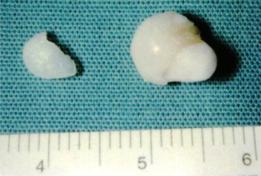
Neck ultrasound showed a normal thyroid gland in shape and position. She also had a normal thyroid function test. The swelling was excised under general anaesthetic, using an ordinary tonsillectomy approach, there was vascular pedicle connecting the swelling to the tongue, which required a suture tie and diathermy to achieve full haemostasis. The swelling was noticed to be bony hard, and enclosed by a thin membrane. On an attempt to section it, the membrane detached to leave a hard pearly central nodule (Figure 1).
Figure 1.
Excised lesion from tongue
The pathologist described it as a white polypoidal swelling of cortical bone covered with orthokeratinised squamous epithelium mucosa; the bone was viable bone with osteoblastic activity arranged as trabeculae with a small amount of fibrous marrow. Initially the pathologist described it as a torus, on further histological assessment it was described as a lingual osseous choristoma. Our patient had uneventful recovery, and became symptom free after the surgical excision.
Review Of The Literature
Method of the review: For this review we searched the English language literature in both the MEDLINE and the PUBMED from 1966 through 2006 with the following Medical Subject Heading (MsHe): “choristoma”; “osteoma”; “bone”; “osseous”; “benign neoplasm”; together with “tongue”; “lingual”; in different forms and combination. We also performed a manual search of all the available papers to us by going through the bibliography at the back of each citation. We used the following criteria to include in our review;
- Lesion clearly named as lingual osseous choristoma.
- If the lesion was not named by the author as osteoma.
- Clinically and histologically the lesion presented as tumor like fully developed osseous growth on the tongue dorsum.
Results of the review: The largest report was a series of nine patients with osseous choristoma that was reported by Krolls in 1971 [1]. He used the term Osseous Choristoma; as he noticed that these lesions are not osteogenic in origin and are not progressively enlarging like benign tumour and as result the term osteoma do not apply [2]. In his series, the age ranged from nine to seventy three years with five females and four males, most of the lesions were pedunculated. On detailed reviewing of the nine cases, it was found that one of them was not lingual and was located in the buccal mucosa [3], this make this series equal in size to Supiyaphun initial series of eight patients reported in 1998 [4]. Supiyaphun, added three more cases the following year and that made his series the largest although it was not reported together, the age ranged from nine to thirty five with female to male ratio of 7 to 1 [5] (Table 2). Cabbabe reported the youngest patient in 1986; he reported a pedunculated lingual osseous choristoma in five-year-old black female in the region of the circumvallate papillae [6]. Weitzner suggested that 80% of these lesions occur in women and in patients less than 40 years old when he reported three new cases and reviewed thirty-eight previously reported cases [7]. Lingual osseous choristoma is usually reported on the dorsal surface of the tongue; Wesley and Zielinski (1978) reported a case of osseous choristoma on the ventral surface of the tongue [8].
Table 2.
lingual osseous choristoma reported in literature
| Author | Year | LOC | OC | Total | RC |
|---|---|---|---|---|---|
| Krolls | 1971 | 8 | 1 | 9 | – |
| Mclendon | 1975 | 3 | – | 3 | – |
| Ohno | 1979 | 1 | – | – | – |
| Sato | 1981 | 1 | – | – | – |
| Sugita | 1981 | 1 | – | – | – |
| Wasserstein | 1983 | 1 | – | – | – |
| Shimono | 1984 | 2 | – | – | – |
| Sheridan | 1984 | 1 | 1 | 2 | 27 |
| Weitzner | 1986 | 3 | – | 3 | 38 |
| Cabbabe | 1986 | 1 | – | 1 | – |
| Markaki | 1987 | 1 | – | 1 | – |
| Ishikawa | 1993 | 2 | – | – | – |
| Negeow | 1996 | 1 | – | 1 | – |
| Manganaro | 1996 | 1 | – | 1 | 75 |
| Vered | 1998 | 2 | – | 2 | 38 |
| Lin | 1998 | 1 | 1 | 2 | – |
| Supiyaphun | 1998 | 8 | – | 8 | 50 |
| Supiyaphun | 1999 | 3 | – | 3 | – |
LOC, Oral Osseous Choristoma; OC, Other Choristoma of oral cavity; RC, reviewed cases
Discussion
Lingual osseous choristoma is a benign condition, which usually affects the dorsum of the tongue posterior to the circumvallate papillae. It affects females four times more than males, and its size can range from 0.3 to 2.5 cm [9]. The age at which it occurs can range from 5 to 73 years, with the majority of patients being in the second or third decades of life [9]. Lingual osseous choristomas can be pedunculated or sessile [10].
The osseous choristoma by definition is a normal bony tissue but in abnormal location, this can be in the skin (previously known as osteoma cutis), or in the oral cavity mucosa (previously known as osteoma mucosae), the last can affect the tongue and called Lingual Osseous Choristoma or the oral buccal mucosa.
Several theories tried to explain the pathogenesis of these lesions (Table 1). In general these theories are sub grouped in two categories; Developmental theory and Reactive (Posttraumatic) theory [2].
Table 1.
Theories implicated in etiology of lingual osseous choristoma
| Pathogenesis | |
|---|---|
| 1 | Developmental |
| 2 | Reactive |
Embryologically the tongue is very complicated structure. The anterior two thirds originate from the first branchial arch, the posterior one third originate from the third branchial arch, the union occurs at the site of the Foramen Cecum, both arches also give rise to normal bony structures such as the middle ear bony ossicles and the hyoid bone. It was suggested that pluripotential cells from these arches might give rise to the osseous choristoma lesions. Some explains formations of osseous choristoma on basis that Foramen Cecum is the site of the development and decent of the future thyroid gland in the neck, it was suggested that remnants of the undescended thyroid tissue might produce an osseous choristoma lesion especially noticing that these lesion occur around the puberty and adolescence time [2].
The posterior one third of the tongue is also the site of frequent and constant irritation by the different lingual activity such as swallowing and articulation, it is known that frequent trauma lead to local inflammation with the deposition of calcium and thickening of the tissue, all this might form the choristoma, these changes had been seen in other skeletal muscles and called “myositis ossificans”. This theory cannot explain the formation of osseous choristoma as these lesions contains fully developed bone with haversian system and not just calcifications [2].
Most of Lingual Osseous Choristomas present as an asymptomatic lump on the dorsum of the tongue (25.8%), but occasionally with dysphagia (6.9%), gagging sensation (5.1%), irritation (3.4%), and nausea (3.4%) [4].
Surgical excision remains the treatment of choice and recurrence of the lingual osseous choristoma has not been reported.
Histologically it is formed of mature lamellar bone with well developed Haversian system and bone marrow spaces covered with mucosa, but real osteoblastic and osteoclastic activity is absent.
Other Choristomas of oral cavity
During our methodological search of lingual osseous choristoma, we came cross several reports of other Choristomas occur in oral cavity [11] (Table 3)
Table 3.
Other Choristomas occur in oral cavity.
| 1 | Salivary gland choristoma |
| 2 | Cartilaginous Choristoma. |
| 3 | Oral Osseous Choristoma |
| 4 | Lingual Thyroid Choristoma. |
| 5 | Lingual Sebaceous Choristoma. |
| 6 | Glial choristoma |
References
- 1.Krolls SO, Jacoway JR, Alexander WN. Osseous Choristomas (Osteomas) of intra oral soft tissues. Oral Surg. 1971;32:588–595. doi: 10.1016/0030-4220(71)90324-0. [DOI] [PubMed] [Google Scholar]
- 2.Vered M, Lustig J, Buchner A. Lingual Osteoma: A debatable Entity. J Oral Maxillofac Sur. 1998;56:9–13. doi: 10.1016/s0278-2391(98)90906-5. [DOI] [PubMed] [Google Scholar]
- 3.Sheridan SM. Osseous choristoma: A report of two cases. Br J Oral Maxillofac Surg. 1984;22:99–102. doi: 10.1016/0266-4356(84)90021-4. [DOI] [PubMed] [Google Scholar]
- 4.Supiyaphun P, Sampatanakul P, Kerekhanjanarong V, Chawakitchareon P, Sastarasadhit V. Lingual Osseous Choristoma: A Study of Right Cases and Review of the Literature. Ear, Nose& Throat Journal. 1998;77(4):316–325. [PubMed] [Google Scholar]
- 5.Supiyaphun P, Sampatanakul P, Kerekhanjanarong V, Aeumjaturapat S, Sastarasadhit V. Lingual osseous choristoma:report of three cases. J Med Assoc Thai. 2000;83(5):564–8. [PubMed] [Google Scholar]
- 6.Cabbabe EB, Sotelo-Avila C, Mononey ST, Makhlouf MV. Osseous choristoma of the tongue. Annals of Plastic Surgery. 1986:150–152. doi: 10.1097/00000637-198602000-00013. [DOI] [PubMed] [Google Scholar]
- 7.Weitzner S. Osseous choristoma of the tongue. South Med J. 1986;79:69–70. doi: 10.1097/00007611-198601000-00020. [DOI] [PubMed] [Google Scholar]
- 8.Wesley R.K, Zielinski R.J. Osteocartilaginous choristoma of the tongue: clinical and histopathologic considerations. Journal of Oral Surgery. 1978;36:59–61. [PubMed] [Google Scholar]
- 9.Lin C, Chen C, Shen Y, Lin L. Osseous Choristoma of Oral Cavity. Kaohsiung J Med Sci. 1998;14(11):727–733. [PubMed] [Google Scholar]
- 10.Manganaro A. Lingual Osseous Choristoma. General Dentistry. 1996 Sep–Oct;44(5):430–431. [PubMed] [Google Scholar]
- 11.Chou L, Hansen LS, Daniels TE. Choristoma of the oral cavity: A review. Oral Surg Oral Med Oral Pathol. 1991;72:584–593. doi: 10.1016/0030-4220(91)90498-2. [DOI] [PubMed] [Google Scholar]
- 12.Monsarrat M. Osteome de la langue. Bulletin de la société d'anatomie. 1913;88:282–283. [Google Scholar]
- 13.Markaki S, Gearty J, Markakis P. Osteoma of the Tongue. British Journal of Oral and Maxillofacial surgery. 1987;25:79–82. doi: 10.1016/0266-4356(87)90161-6. [DOI] [PubMed] [Google Scholar]
- 14.McClendon EH. Lingual osseous choristoma: report of two cases. Oral Surg Oral Med Oral Pathol. 1975;39(1):39–44. doi: 10.1016/0030-4220(75)90393-x. [DOI] [PubMed] [Google Scholar]
- 15.Shimono M, Tsuji T, Iguchi Y, et al. Lingual osseous choristoma: report of two cases. Int J oral Surg. 1984;13:355–259. doi: 10.1016/s0300-9785(84)80045-9. [DOI] [PubMed] [Google Scholar]
- 16.Wasserstein MH, SunderRaj M, Jain R, et al. Lingual osseous choristoma. J Oral Med. 1983;38:87–89. [PubMed] [Google Scholar]
- 17.Ishikawa M, et al. Osseous choristoma of the tongue: report of two cases. Oral Surg Oral Med Oral Pathol. 1993;76:561–563. doi: 10.1016/0030-4220(93)90062-9. [DOI] [PubMed] [Google Scholar]
- 18.Sugita H, Yamamoto E, Sunakawa H, Matsubara T, Furuta I, Kohama G. Lingual osseous choristoma: report of a case. JpnJ Oral Maxillofac Surg. 1979;25:165–169. [Google Scholar]
- 19.Ohno T, Yanbe H, Morii E, Takahashi A, Miyazima H, Tanaka Y, Adachi F, Saito I, Kawahara H. Osseous choristoma situated on the dorsum of the root of the tongue: report of a case. Jpn J maxillofac surg. 1981;27:78–81. [Google Scholar]
- 20.Sato Y, Ozawa S, aria N, Toguchi I, Fukuda T, Ueda Y, Yoshimoto T. Osseous Choristoma of the tongue: report of a case. Jpn J Oral Maxillofac surg. 1981;27:93–95. [Google Scholar]


